Original Article
The role of capsulin in the morphogenesis and differentiation
of rat fetal gastric mucosa
MARIA ANDERSSON, ANDREW S. GIRAUD and MARY FAMILARI*
University of Melbourne, Deptartment of Medicine at Western Hospital, Footscray, Australia
ABSTRACT The signals that guide the morphogenesis and differentiation of rat fetal gastric mucosa remain largely unknown. We have investigated the role of capsulin in pit/gland formation and epithelial cell differentiation in cultured stomach tissue. Embryonic day 16.5 (E 16.5) stomach tissue cultured for three days in the presence of 1 µM hydrocortisone underwent dramatic transformation, from undifferentiated, stratified cells to differentiated epithelia composed of polarised columnar cells with mucous cells and pit/glands. In the presence of capsulin antisense oligonucleotides directed against capsulin mRNA, tissues do not undergo further development. Significantly, both mucous granules and pit/gland formation were inhibited compared to capsulin sense/scrambled oligonucleotide treated controls. However, in tissues treated with specific anti-rat HGF-antiserum to neutralise secreted HGF, pit/gland formation was inhibited, but the number of mucous granules remained unchanged compared to controls treated with non-specific antiserum (mouse monoclonal cytokeratin 8 antiserum). This data suggests that capsulin may have a role in the morphogenesis of pit/glands and mucin granule formation in the developing rat gastric mucosa. We discuss the possibility that this role of capsulin may be partly mediated through the actions of HGF.
KEY WORDS:
capsulin, HGF, fetal stomach, organ culture, antisense oligonucleotides
0214-6282/2001/$25.00
© UBC Press Printed in Spain
www.ijdb.ehu.es
*Address correspondence to: Dr. Mary Familari. Department of Zoology. University of Melbourne, Parkville, VIC 3010, Australia. Fax: +613--8344-7909. e-mail: m.familari@unimelb.edu.au
Abbreviations used in this paper: dNTPs, deoxynucleotide triphosphates; GAPDH, Glyceraldehyde-3-Phosphate Dehydrogenase; HCS, hydrocorti-sone; HGF, Hepatocyte Growth Factor.
Introduction
The signals that guide morphogenesis and differentiation in the fetal stomach remain largely unknown. However, Fukada et al., (1994) and Tsukada et al., (1998) have demonstrated that mesen-chymal tissue signals can regulate the expression of the gastric epithelial specific pepsinogen gene, and further that Gata5 may regulate this gene (Sakamoto et al., 2000). The mesenchyme plays an important role in the developmental fate of gastric epithelia in both the mice (Fukamachi et al., 1979) and chick (Yasugi et al., 1993). However, there are now many studies, which have demon-strated convincingly, that undifferentiated rat stomach endoderm when cultured without mesenchyme, can differentiate into mucus, parietal and even chief cells, under various experimental condi-tions, and using different substratum such as collagen gels (Terano
et al., 1982; Ishizuka-Oka and Mizuno 1984; Fukamachi et al.,
1994a; Matsuda et al., 1996; Ichinose et al., 1997; Tømmerås et al., 1997; Kong et al., 1998; Tsukada et al., 1998). Although differen-tiated cell types of the gastric epithelium can be induced under defined culture conditions, the formation of pit/glands in cultured fetal gastric epithelial cells is yet to be described.
Organ cultures - the maintenance of small pieces of explanted tissue in vitro under conditions in which embryonic development
can continue - are a useful way to dissect the signals that guide pit/ gland formation and differentiation of epithelial cell-types in the stomach. Human small intestine was first successfully cultured in chemically defined media (Browning and Trier, 1969), and since then various investigators have shown that fetal stomachs from human (Menard et al., 1993) and rodents (Yeomans et al., 1976; Tsukada et al., 1998; Zgleszewski et al., 1998; Duh et al., 2000) can undergo further development in culture, using either serum or serum-free conditions.
Interestingly, glucocorticoids have been repeatedly reported to induce epithelial cell differentiation in various organs, including gastric epithelial cells (Henning, 1987), and glucocorticoids have been shown to be essential for the induction of differentiated cell types in cultured rat stomach tissue (Yeomans et al., 1976) and in cultured rat stomach epithelial cells (Tømmerås et al., 1997).
differentiation of many cell types (Massari and Murre, 2000). Mouse capsulin was cloned by several groups and shown in the developing embryo to be selectively expressed in mesenchymal cells at sites of mesenchyme-epithelium interactions in several organs including lung, kidney and gut (Hidai et al., 1998; Lu et al., 1998; Quaggin et al., 1998; Robb et al., 1998). In addition, Quaggin and colleagues (1998) demonstrated that antisense oligonucle-otides directed against capsulin mRNA specifically inhibited branch-ing and morphogenesis in cultured fetal kidney tissue. Capsulin, therefore, may be a potential regulator of morphogenesis and differentiation in fetal stomach.
We identified rat capsulin in a search directed at identifying mesenchyme-specific factors in the fetal gastric mucosa (Familari and Giraud, 1998), and the aim of this study was to examine the role of capsulin in morphogenesis and differentiation of cultured fetal rat stomach.
Results
Representative results from in vivo stomachs (E16.5 and E19.5) and cultured E16.5 stomach tissues are shown in Fig. 1. E16.5 in
vivo stomach tissue is shown in Fig. 1A. Note the stratified and
undifferentiated appearance of the epithelial layer at this stage before culture. E16.5 stomach tissue cultured for three days in DF-Media without 1µM Hyrdocortisone (HCS) do not undergo any further development (Fig. 1B) and were similar to E16.5 in vivo tissue (Fig. 1A). However in very few tissues cultured in DF-Media without HCS, there were a few columnar cells but neither mucin granules nor pit/glands were detected. At E19.5 in vivo, extensive pit/gland formation and polarised epithelial cells containing mucin
granules are seen (Fig. 1C). Mucin granules are present mostly in the apical surface of the epithelial cells, but some sparse staining is seen at the base of epithelial cells (Fig. 1C). In tissues cultured for three days in DF-Media in the presence of Hydrocortisone (HCS), there is evidence of pit/gland formation and polarised cells and mucus gran-ules. (Fig. 1D). Interestingly in cultured tissue in the presence of HCS, mucin granules are more numerous in the base of epithelial cells (Fig. 1D) compared to E19.5 in
vivo tissue stained for mucin granules (Fig. 1B). Another
marker of mucus cells, TFF2 was also compared in both in
vivo and cultured tissue. TFF2 staining is found in the
apical surface of epithelial cells predominately in E19.5 in
vivo tissue (Fig. 1E). Whereas, in tissue cultured in the
presence of HCS, TFF2 staining was found not only in the apical surface but also in the base of epithelial cells (Fig. 1F). Parietal cells were not detected in tissues cultured for three days in the presence or absence of HCS (data not shown). The presence of mucin granules and TFF2 stain-ing in the base of epithelial cells as well as the apical surface was seen in all cultured tissues with or without various treatments.
To examine the role of capsulin on morphogenesis and differentiation in fetal stomach, all further tissue was cultured for three days in DF-Media in the presence of 1µM HCS. In addition, the tissues were treated with varying combinations of different oligonucleotides or antisera. Representative results of tissues cultured under different treatments are shown in Fig. 2. Treatment of cultured tissues with antisense oligonucleotides #1 (20 µg/ml) resulted in a dramatic decrease in both pit/gland formation and the number of mucin granules (Fig. 2B), compared to the lack of effect in control tissue treated with a combination of sense/scrambled #1 oligonucleotides (20µg/ml, Fig. 2A). Treatment with either antisense oligonucleotide #2 or #3 alone was without effect (data not shown). In stomach tissues treated with anti-rat HGF antiserum (Fig. 2E) or non-specific antiserum (Fig. 2D), the number of mucin granules were similar to control tissues treated with capsulin sense oligonucle-otides (Fig. 2A). However, pit/gland formation was inhibited in tissues treated with anti-rat HGF antiserum (Fig. 2E) compared to control tissues with non-specific antiserum (Fig. 2D).
Treatment of cultured tissue with either antisense oligonucleotide #1 (Fig. 2C) or anti-rat HGF antiserum (Fig. 2F) was without effect on the expression of TFF2. In addition, parietal cells which were detected at E19.5 in vivo in the rat stomach (Familari et al., 1998), were not detected in stomach tissue cultured for three days under any of these treatments (data not shown).
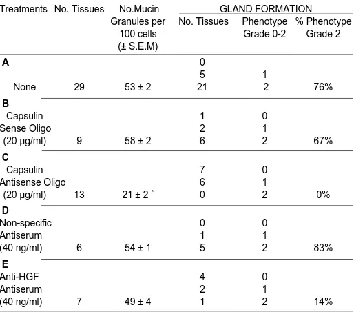
Table 1 summarizes the results of various treatments on pit/ gland formation and development of mucin granules in cultured stomach tissue: For each treatment group, the average number of mucin granules per 100 cells and the percentage of tissues displaying phenotype Grade 2 are listed. Treatment with capsulin antisense oligonucleotide #1, dramatically reduced the number of mucin granules (Table 1C), compared to control tissues treated with capsulin sense oligonucleotides (Table 1B) or tissues in standard DF-Media plus 1µM HCS, but without further treatment (Table 1A). In addition, phenotype Grade 2 was never observed in any tissue treated with capsulin antisense oligonucleotides (Table 1C) compared to the percentage of control tissues treated with capsulin sense oligonucleotides exhibiting phenotype Grade 2
Fig. 1. Comparison of in vivo fetal stomach tissue with cultured fetal stomach tissue. (A) E16.5 in vivo stomach tissue, AB/PAS (400x). (B) E19.5 in vivo stomach tissue, AB/PAS (400x). (C) E16.5 tissue cultured for 3 days in the absence of 1 µM HCS, AB/PAS (400x). (D) E16.5 tissue cultured for 3 days in the presence of 1 µM HCS, AB/ PAS (400x). (E) E19.5 in vivo stomach tissue, TFF2 (400x). (F) E16.5 tissue cultured for 3 days in the presence of 1 µM HCS, TFF2 (400x).
A
B
C
D
(Table 1B) or tissues in standard DF-Media plus 1µM HCS, but without further treatment (Table 1A). In contrast, treatment with anti-rat HGF antiserum did not affect the number of mucin granules (Table 1E) compared to standard DF-Media plus 1µM HCS, but without further treatment (Table 1A) nor treatment with non-specific antiserum (Table 1D). However, the percentage of tissues displaying Grade 2 pit/gland formation were reduced in anti-rat HGF serum treated tissues (Table 1E) compared to con-trol tissues treated with non-specific antiserum (Table 1D).
The expression of capsulin, HGF and GAPDH mRNA was examined in cultured tissues following different treatments by RT-PCR and the results are shown in Fig. 3. The levels of GAPDH expression were similar in all treatment groups. However, capsulin expression in tissue treated with capsulin antisense oligonucleotides (Fig. 3, CAP antisense) was dra-matically reduced compared to levels of expression in capsulin sense oligonucleotide treated controls (Fig. 3, CAP sense). In addition, there is a slight reduction in both level of HGF and GAPDH mRNA expression in tissue treated with capsulin antisense oligonucle-otides (Fig. 3, CAP antisense). Interestingly, HGF expression increased in tissues treated with anti-HGF antiserum (Fig. 3, anti-HGF Ab) compared to anti-HGF expression in tissues treated with non-specific anti-serum (Fig. 3, NS Ab).
lumen marking the differentiated phenotype (Familari et al., 1998). Trefoil proteins are first detectable at E.17.5, well before the columnar epithelium is evident. In comparison sparse mucus staining is not observed until E.18.5 and morphologically identi-fiable mucus granules are first detectable around E.19.5 (Yeomans, 1974). Parietal cells can be detected a day later, after the monolayer of cuboidal epithelial cells is well established, and has begun to invaginate into the underlying mesenchymal tissue (Familari et al., 1998).
In the stomach tissue cultured for three days, pit/glands and polarised epithelial cells are observed, as well as two markers of mucus cells can be detected, namely mucin granules and TFF2 expression. However, the expression of these markers is found throughout the epithelial cell layer unlike the expression found in E19.5 in vivo tissues, although sparse mucin-like granules can be detected in the basal layer of epithelial cells at E19.5 in vivo stomachs in this and other studies (Yeomans, 1974; Otani et al., 1993).
The conditions of culture in the present study appear to have contributed to over-abundant expression of mucus cell markers. Interestingly, Tsukada and colleagues reported expression of pepsinogen throughout the epithelial cell in cultured fetal stomach tissue (Tsukada et al., 1997). The conditions of culture in the present study and in the study reported by Tsukada and col-leagues (1997) required both the presence of serum (10%) and glucocorticoids but it is not clear if either or both additives to the media have contributed to the expression of mucus cell markers in the base of epithelial cells. Certainly the background staining is more intense in cultured tissues than in comparable aged in vivo tissues but may not be due to the glucocorticoids because this is true for tissues cultured with or without HCS.
Fig. 2.Effect of different treatments on cultured stomach tissue. E16.5 stomach tissues cultured for three days in the presence of: (A) 20 µg/ml sense/scrambled capsulin oligonucle-otides, AB/PAS (400x). (B) 20 µg/ml capsulin antisense oligonucleotide #1. AB/PAS (400x). (C) 20 µg/ml capsulin antisense oligonucleotide #1. TFF2 (400x). (D) 40 ng/ml anti-mouse cytokeratin 8 antiserum. AB/PAS (400x). (E) 40 ng/ml rabbit anti-rat HGF antiserum. AB/PAS (400x). (F) 40 ng/ml rabbit anti-rat HGF antiserum. TFF2 (400x).
Discussion
Mesenchyme-epithelium interactions are crucial for develop-ment of many organs including the gut. However, very little is known about the molecular mechanisms that underlie these mes-enchyme-epithelium interactions in the developing gut. Results of recent studies suggest that Sonic hedgehog, Hox and Bmp genes may be involved in epithelium-mesenchyme (ME) interactions in the earliest stages of chick and mouse gut development (reviewed in Roberts, 1998). In addition, regionalisation of the gut tube occurs early in gut development and homeobox-containing genes have been implicated in this process (Beck et al., 2000).
The details of ME interactions in the stomach are not understood and organ cultures of stomach tissue can therefore be a useful tool to help investigate the molecular mechanisms involved in this interaction. In this study we have provided evidence that capsulin, a mesenchyme-specific factor, may have an important role in pit/ gland formation and development of mucin granules.
In a previous study we examined the morphology and differen-tiation in the developing rat stomach (Familari at al., 1998). At embryonic day 16.5 (E.16.5) the stomach is lined by stratified epithelium. At this developmental stage, TFF1 and TFF2 genes are activated, well before differentiation of the epithelium (as determined by the presence of polarised columnar epithelial cells) and prior to stem cell commitment. At E.18.5, a monolayer of partially polarised, cuboidal epithelial cells is established, a few secondary lumina are evident with the beginning of granule formation in the apical region of surface cells; and at E.19.5-20.5 deep pit/glands are beginning to form in the glandular stomach lined by a single layer of mostly polarised epithelial cells facing the
A
D
B
E
However, glucocorticoids have been repeatedly reported to in-duce epithelial cell differentiation in various organs, including gas-tric epithelial cells (Henning, 1987). Previous studies have demon-strated that glucocorticoids are produced in embryonic rat adrenal tissues beginning E.13.5 (Roos, 1967). The circulating corticosteroid concentration in rat fetal plasma before birth is about 1µM, and the corticosteroid concentration in the adrenal cortex appears to be maximal at 18 days gestation, just before major structural changes occur in the gastric epithelium (Kamoun cited in Yeomans et al., 1976). Therefore the concentration of glucocorticoids used in this study appear to be within the physiological range and do not explain the expression of mucus cell markers in the base as well as apical surface of epithelial cells in cultured tissues. Interestingly, Otani and colleagues (1993) who reported using periodic acid-thiocarbohydrazide silver staining in human embryonic stomachs demonstrated that carbohydrate-like staining was also found in the basal part of the epithelium and these carbohydrate-rich sites ap-peared as vacuoles scattered throughout the epithelium. Otani and colleagues concluded that there were signifcant amounts of carbo-hydrate in the base of the epithelial cells in the stomach. Whether or not carbohydrate-like staining in human fetal stomach represents mucin granules remains unresolved, but it is worth noting that mucin granules and TFF2 expression in the base of epithelial cells in cultured rat stomach tissue in our study appear to be in vacuoles. In the present study it was necessary to count all mucin granules within an area of section containing 100 cells because at the level of light microscope there is not the resolution to distinguish between gran-ules or vacuoles achieved by studies using the electron microscope (Yeomans, 1974; Otani 1993).
Our results are consistent with the reports of Yeomans et al. (1976) and Tsukada et al., (1998), that glucocorticoids induced
cytological differentiation in the cultured fetal rat stomach tissue. Although Tsukada and colleagues (1998), not only reported the presence of pepsinogen throughout the epithelial cells in tissues after three days in culture but also that up to 80% of cells within the epithelium were pepsinogen positive.
We confirm the findings of Yeomans et al., (1976), that glucocortiocoids were found to induce pit/gland formation. In con-trast, Tsukada et al., (1998) reported an inhibitory effect of hydrocor-tisone in cultured tissue on pit/gland formation. The reason for the difference remains unclear, since essentially the same organ culture conditions and similar concentrations of glucocorticoids were used in both studies. It is worth noting that in the rat, morphologically identifiable pepsinogen-secreting chief cells are not observed prior to birth (Yeomans et al., 1976; Familari et al., 1998), although we did not test the cultured tissue for the presence of pepsinogen in the present study.
The results from this study, suggest that after three days in culture in DF-Media with 1µM HCS, pit/gland formation and mucus cells develop in stomach tissue. Although, in the conditions reported in this study, fetal development proceeded more slowly in vitro than in vivo, as has been reported by several other investigators in studies of cultured stomach tissue (Yeomans, et al., 1976; Menard et al., 1993). And this may explain why parietal cells were not detected in the cultured tissues in this and other studies (Yeomans et al., 1976). Although it does also indicate, that additional factors, missing from the present culture system, are likely to be important in normal development. Interestingly, Tømmerås et al., have demonstrated that parietal cells can be induced after 15-18 days when epithelial cells are cultured in collagen gels only in DMEM-F12 media supple-mented with 10% serum and 10µM of HCS. In the present study, stomach tissue was not cultured beyond 3 days.
The conditions outlined in this study permitted the development of pit/glands and two markers of mucus cell differentiation - mucin granules and TFF2 expression, which allowed us to examine the role of capsulin in morphogenesis and mucus cell differentiation in cultured stomach tissue. Three different antisense and sense/ scrambled oligonucleotides to rat capsulin mRNA were tested. Treatment with either #2 or #3 antisense oligonucleotides alone had little effect on gland formation. It is not surprising that two of the three antisense oligonucleotides directed against capsulin mRNA were ineffective. Others have reported differences in the effectiveness of
TABLE 1
AVERAGE NUMBER OF MUCIN GRANULES AND PERCENTAGE OF TISSUES SHOWING GLAND FORMATION
PHENOTYPE GRADE 2 PER TREATMENT GROUP
Treatments No. Tissues No.Mucin GLAND FORMATION Granules per No. Tissues Phenotype % Phenotype
100 cells Grade 0-2 Grade 2 (± S.E.M)
A 0
5 1
None 29 53 ± 2 21 2 76%
B
Capsulin 1 0
Sense Oligo 2 1
(20 µg/ml) 9 58 ± 2 6 2 67% C
Capsulin 7 0
Antisense Oligo 6 1
(20 µg/ml) 13 21 ± 2 * 0 2 0%
D
Non-specific 0 0
Antiserum 1 1
(40 ng/ml) 6 54 ± 1 5 2 83%
E
Anti-HGF 4 0
Antiserum 2 1
(40 ng/ml) 7 49 ± 4 1 2 14%
(* Significant difference between groups B & C; P < 0.001)
oligonucleotides directed against different regions of the same message and differences in effectiveness of oligonucleotides di-rected against the same region but of different length (Colman, 1990; Kronmiller, J.E., 1991).
However, when antisense oligonucleotide # 1 was added to the media by itself, pit/gland formation was inhibited and mucin granule formation was reduced. This result is consistent with the studies of Quaggin (et al., 1998), who reported that treatment with antisense oligonucleotides directed against capsulin mRNA, significantly de-creased branching morphogenesis when added to fetal kidney cultured tissues (Quaggin et al., 1998). Significantly, this involved epithelial morphogenesis even though capsulin is a mesenchymal-specific factor, suggesting that the downstream targets of capsulin are inducers of epithelial cell differentiation.
Given that capsulin is a 26kD protein localised in the nucleus (Miyagishi, et al., 2000), its effects in cultured stomach tissue are indirect and possibly mediated by a secreted factor. HGF, is an obvious candidate which could potentially mediate the effects of capsulin and our results confirm that HGF, a mesenchyme-specific factor, has a role in epithelial cell morphogenesis (Fukamachi et al., 1994b; Matsumoto and Nakamura, 1996; Kong et al., 1998). Interest-ingly, anti-rat HGF antiserum added to media inhibited branching morphogenesis in both cultured fetal tissue from kidney (Santos et
al., 1994) and lung (Ohmichi et al., 1998). In addition, we also provide
evidence that mucin granule formation is not affected when the action of secreted HGF is neutralised by anti-HGF antiserum in cultured stomach tissue. The role of HGF in stimulating epithelial cell growth and movement is well established (Masumoto and Nakamura, 1996), but the identity of the other factors required for mucus granule formation are currently unknown.
Interestingly, the level of capsulin mRNA expression was mark-edly reduced in tissues treated with capsulin antisense oligonucle-otides #1 compared to sense treated controls. This suggests that the effect of capsulin on epithelial cell morphogenesis and cytodifferen-tiation were due to a direct effect on attenuating capsulin mRNA levels but may also be due to an effect via a modest reduction in HGF mRNA levels. The level of mRNA expression does not indicate the level of protein expression for any of the genes examined in the cultured tissue, and only when capsulin protein can be detected in micro-scale Western Blots using a specific antibody will the matter be resolved. Currently the cultured tissues are too small to yield useful amounts of protein to detect differences in protein profiles between treatment groups.
Does the role of capsulin in cultured stomach, demonstrated in this study reflect a role in stomach development in vivo? Recent studies of mice with deletions in both copies of capsulin have
demonstrated that this factor is essential for lung, kidney and spleen organogensis (Quaggin et al., 1999; Lu et al., 2000). The mutant animals die within five to ten minutes after birth due to poorly developed lungs. Significantly, organ buds for the lungs, kidneys and spleen are formed but fail to develop due to defects in epithelial cell differentiation in these organs. Although capsulin is expressed in the mesenchyme of many organs including the stomach, a description of the phenotype of the gastrointestinal tract in the mutant animal has not been described.
The results of the present study, suggest a role for capsulin in pit/ gland formation in cultured fetal stomach. Although, the results cannot confirm that the role of capsulin may be mediated via HGF, it is worth noting that the 5’ regulatory region of HGF, contains five canonical bHLH transcription factor E-box consensus sequences within 700bp of the transcription start site, that HGF is a secreted factor, and that both HGF and capsulin have demonstrated roles in both fetal lung and kidney epithelial cell differentiation (Santos et al., 1994; Matsumoto and Nakamura, 1996; Quaggin et al., 1999; Lu et
al., 2000).
Our results suggest that capsulin may also be involved in morpho-genesis and epithelial cell differentiation in the stomach, however, the effect of ablating capsulin mRNA expression on epithelial cell differentiation in cultured stomach tissue was specific for mucin granule formation, since TFF2 protein expression remained unaf-fected.
Further studies are underway to detail the mechanism of action of capsulin in morphogenesis and differentiation in fetal rat stomach.
Materials and Methods
Animals
Embryos taken from pregnant Wistar rats were housed in groups of two with free access to food and water. Vaginal plug detection at 9am was defined as day 0.5 and the rat embryos were obtained at embryonic day 16.5 (E16.5). The pregnant rats were sacrificed under anesthesia followed by cervical dislocation before the embryos were removed.
Stomach dissection
The embryos were rapidly removed and rinsed in PBS (1x Dulbecco’s Phosphate Buffered Saline, ICN) then placed in 15 Media [(Leibovitz L-15 media, Life Technologies) supplemented with penicillin (100 IU/ml, ICN), streptomycin (100 µg/ml, ICN) and 0.2% (w/v) BSA (bovine serum albumin, Life Technologies)] until cultured. Tissue was dissected free of forestomach, antrum and esophagus; and only the presumptive glandular stomach was used for tissue culture. The presumptive glandular stomach was divided into four approximately equal tissues(1x1mm), placed onto nylon filters (0.45 µm, Millipore) and unfolded, mucosal side up then placed into organ culture dishes (Falcon).
Organ culture
Initially the conditions of organ culture described by Yeomans et al., (1970) and Menard et al., (1993) using Leibovitz L-15 media with or without hydrocortisone but lacking serum were tested. However the cultured tissue failed to undergo pit/gland formation nor were differentiated cell types evident. Culture conditions described by Tsukada et al. (1998) proved successful using DMEM/F12 1:1 v/v media, hydrocortisone and fetal calf serum. The tissues were cultured in 500 µl DF-Media [DMEM/F12 Media (1:1 v/v), Life Technologies, USA supplemented with penicillin (100 IU/ml) and streptomycin (100 µg/ml) and 10% fetal calf serum]. The dishes were placed in a humidified chamber and placed in an incubator at 37°C with 95% Air/5% CO2 for 3 days without changing the media. Stomach tissues were TABLE 2
SEQUENCE OF OLIGONUCLEOTIDES ADDED TO CULTURED TISSUE
grown in the presence of hydrocortisone at a final concentration of 1 µM unless otherwise stated. Hydrocortisone Sodium Succinate (Pharmacia) was dissolved in 0.9% saline to prepare a 1 M solution, then serially diluted with DF-Media to 1 µM. Several concentrations of hydrocortisone (HCS; 0.1, 1 and 10 µM) were tested and 1 µM was found sufficient to support pit/ gland formation and development of polarised epithelial cells.
To test the role of capsulin, cultured fetal stomach tissues were treated, alone or in combination, with the following as specified in Fig. 2: Sense/ scrambled or antisense oligonucleotides directed against capsulin mRNA; specific rabbit anti-rat HGF antiserum (40 ng/ml, a generous gift from Professor Nakamura, University of Osaka); a non-specific monoclonal mouse antiserum against cytokeratin 8 (40 ng/ml,BioGenex, USA);
The nucleotide sequences of sense/scrambled and antisense DNA oligonucleotides, directed to flank the translation initiation codon incorporat-ing phosphorothioate linkages (Life Technologies, USA), are shown in Table 2. Oligonucleotides were targeted to the same region in rat capsulin mRNA as that of mouse capsulin mRNA (Quaggin et al., 1998). Quaggin and colleagues demonstrated that mouse capsulin antisense oligonucleotides (at a concentration of 30 µg/ml) directed to flank the translation start codon inhibited branching morphogenesis in kidney explant cultures. Oligonucle-otides at 20 µg/ml were pre-mixed with 10 µl cationic liposomes (Liopfectamine, Life Technologies) before addition to media.
Several concentrations of anti-rat HGF antiserum (4 ng/ml, 40 ng/ml, 400 ng/ml) were tested and 40 ng/ml was found sufficient to inhibit pit/gland formation in cultured stomach tissue. These concentrations were based on the study by Santos et al. (1994), who demonstrated that (i) 4.5 µg/ml antirat HGF antiserum when added to media blocked branching morphogenesis in cultured fetal kidney tissue, and (ii) that 4.5 µg/ml of anti-rat HGF antiserum was sufficient to neutralise 5 ng/ml rat HGF in fetal kidney tissue of 5X5 mm2, i.e. tissue 5X the size of the fetal stomach tissue used in the present study. Antisera were added at appropriate concentrations directly to media.
Each treatment was tested in 2-3 experiments and each treatment group consisted of 3-6 stomach tissues per experiment.
Morphological analysis
Uncultured stomachs (E16.5 and E19.5) and 3 day cultured E16.5 tissue were fixed in 4% Paraformaldehyde/PBS for 2 hours or 30 minutes respec-tively before dehydration through an ethanol series and paraffin embedding. Light Microscopic examination was undertaken on 5 micron tissue sections for gland formation. The presence of polarised, columnar epithelial cells and the presence of mucin granules as determined by staining with AB/PAS (Alcian Blue/Periodic Acid Schiff) were taken as evidence for differentiation. The identification of mucus cells and parietal cells were also determined by TFF-2 and H,K-ATPase immunohistochemistry respectively, as previously described (Familari et al., 1998).
A system of grading the epithelial phenotype of cultured rat stomach tissue was developed by Yeomans et al. (1976). Pit/gland formation is graded from 0-2 according to: absence of pit/glands (Grade 0); presence of pits only in the epithelial layer (Grade 1); and presence of pits/glands that penetrate into the mesenchyme (Grade 2). This system was used to grade pit/gland formation in cultured stomach tissues throughout the present study.
The number of mucin granules was quantified in order to adjust for biological variability. In each treatment group, for each cultured stomach tissue, after fixing and staining, mucin granules were counted on an area of section containing approximately 100 cells. A given area of epithelial cells in each stomach tissue was counted in 3 serial sections using Image Analysis Software (Pharmacia). The average number of mucin granules was then recorded for each tissue within a treatment group. The average number of mucin granules for each treatment group was then calculated. To remove any bias, counting of mucin granules in tissue sections was undertaken by the investigators unaware of the different treatments on each section. Statistical analysis was undertaken by Student t-test and ANOVA.
Reverse Transcription –Polymerase Chain Reaction (RT-PCR)
For RT-PCR, 1-2 explants from each treatment group were pooled and total RNA was extracted using the acid-phenol-chloroform method as
developed by Chomczynski and Sacchi (1987). 20 ng of total RNA was reversed transcribed into first strand complementary DNA using 200 units of Moloney murine leukemia virus (Promega) primed with 20 ng oligo(dT) in 40 mM Tris-HCl (pH8.3), 75 mM KCl, 3 mM MgCl2, 10 mM dithiothreitol and 1 mM dNTP’s in a 20 ul volume at 37°C for 60’. 5 µl of this reaction was subsequently used as template for PCR. Reaction conditions were 10 mM Tris-HCl pH 8.3, 50 mM KCl, 2 mM MgCl 2, 200 µM dNTPs, 25 ng of each sense and antisense primer and 1.25 units of Taq polymerase (Promega) in a 50 µl volume. The sequence of the primers used were:
rat HGF sense 5’-GCA GAC ACC ACA CCG GCA-3’,
antisense 5’-TCC CTG TGT AGC ACC AAG-3’;
rat capsulin sense 5’-CTG TGA CTC CCT GAA AGT G-3’,
antisense 5’-GTC TTG AGC CTG GAG AAG-3’;
rat GAPDH sense 5’-TGT CAG CAA TGC ATC CTG CA -3’,
antisense 5’-CTG TTG AAG TCA CAG GAG AC-3’.
Parameters for each cycle were as follows: denaturation at 950C for 30s, annealing at 60°C for 30 s and extension at 72°C for 1 min for 23 cycles. Note, primer pairs for HGF, capsulin and GAPDH amplification were added to each PCR, except that GAPDH primers were added for only the last 18 cycles. Previous studies in our laboratory have demonstrated that the optimum number of cycles for each primer pair can be determined by adding fourfold serial dilutions of the RT reaction to the PCR until a linear response is obtained (Alderman et al., 2000). The expression of GAPDH is within the linear range of amplification if assessed at 18 cycles, and HGF and capsulin at 22 cycles. In order to use the expression of GAPDH as an internal control for the quantity of total RNA in each reaction for each treatment group, 18 cycles of PCR are optimum. PCR products were resolved on 1.5% agarose gels containing ethidium bromide.
Acknowledgements
The authors wish to thank Dr. Don Newgreen for critical reading of this manuscript.This work was supported by NHMRC funding to MF.
References
ALDERMAN, B.M., COOK, G.A., FAMILARI, M., YEOMANS, N.D. and GIRAUD, A.S. (2000). Resistance to apoptosis is a mechanism of adaptation of rat stomach to aspirin. Am. J. Physiol. 278:G839-G846.
BECK, F., TATA, F. and CHAWENGSAKSOPAK, K. (2000). Homeobox genes and gut development. Bioessays. 22: 431-441.
CHOMCZYNSKI, P. and SACCHI, N. (1987). Single-step method of RNA isolation by acid guanidinium thiocyanate-phenol-chloroform extraction. Anal. Biochem. 162:156-159.
COLMAN, A. (1990). Antisense strategies in cell and developmental biology. J. Cell
Sci. 97:399-409.
DUH, G., MOURI, N., WARBURTON, D. and THOMAS, D.W. (2000) EGF regulates early embryonic mouse gut development in chemically defined organ culture.
Pediat. Res. 48: 794-802
FAMILARI, M., COOK, G.A., MARRYATT, G., TAUPIN, D.R., YEOMANS, N.D and GIRAUD, A.S. (1998) Trefoil peptides are early markers of gastrointestinal maturation in the rat. Int. J. Dev. Biol. 42: 783-789.
FAMILARI, M and GIRAUD, A.S.(1998) Identification of the rat homologue of the mouse
capsulin gene by cDNA representational difference analysis. Gene 222:245-248
FUKADA, K., ISHII, Y., SAIGA, H., SHIOKAWA, K and YASUGI, S. (1994). Mesen-chymal regulation of epithelial gene expression in developing avian stomach: 5’-flanking region of pepsinogen gene can mediate mesenchymal influence on its expression. Development 120:3487-3495
FUKAMACHI, H., MIZUNO, T. and TAKAYAMA, S. (1979). Epithelial-mesenchymal interactions in differentiation of stomach epithelium in fetal mice. Anat. Embryol. 157: 151-160.
FUKAMACHI, H., ICHINOSE, M., ISHIHAMA, S., TSUKADA, S., YASUGI, S., SHIOKAWA, K., FURIHATA, C., YONEZAWA, S. and MIKI, K.(1994a). Fetal rat glandular stomach epithelial cells differentiate into surface mucous cells which express cathepsin E in the absence of mesenchymal cells in primary culture.
FUKAMACHI, H., ICHINOSE, M., TSUKADA, S., KAKEI, N., SUZUKI, T., MIKI, K., KUROKAWA, K. and MASUI, T. (1994b). Hepatocyte growth factor specifically stimulates gastro-intestinal epithelial growth in primary culture. Biochem. Biophys.
Res. Comm. 205: 1445-1451.
HENNING, S.J. (1987) Functional development of the gastrointestinal tract. In
Physiology of Gastrointestinal Tract (Ed. L.R. Johnson). Raven Press, New York,
285-300.
HIDAI, H., BARDALES, R., GOODWIN, R., QUERTERMOUS, T. and QUERTERMOUS, E.E. (1988) Cloning of capsulin, a bHLH factor expressed in progenitor cells of the pericardium and the coronary arteries. Mech. Dev 73:33-43. ICHINOSE, M., YAHAGI, N., MATSUBARA, Y., TSUKADA, S., OKA, M., SHIMIZU, Y., YONEZAWA, S., KAGEYAMA, T., MIKI, K. and FUKAMACHI, H. (1997). Substratum-dependent and region –specific control of attachment and prolifera-tion of gastrointestinal epithelial cells in primary serum-free culture. Biochem.
Biophys. Res. Comm. 230: 537-541.
ISHIZUYA-OKA, A. and MIZUNO, T. (1984). Intestinal cytodifferentiation in vitro of chick stomach endoderm induced by duodenal mesenchyme. J. Embryol. exp.
Morph. 82: 163-176.
KONG, W., LEE, L.F. and MULVIHILL, S.J (1998). Hepatocyte growth factor stimu-lates fetal gastric epithelial cell growth in vitro. J. Surg. Res. 78: 161-168 KRONMILLER, J.E., UPHOLT, W.B. and KOLLAR, E.J. (1991). EGF antisense
oligonucleotides block murine odontogenesis in Vitro. Dev. Biol. 147: 485-488. LU, J., RICHARDSON, J.A and OLSON, E.N. (1998). Capsulin: a novel bHLH
transcription factor expressed in epicardial progenitors and mesenchyme of visceral organs. Mech. Dev. 73:23-32
LU, J., CHANG, P., RICHARDSON, J.A., GAN, L., WEILER, H. and OLSON, E.N. (2000). The basic helix-loop-helix transcription factor capsulin controls spleen organogenesis. Proc. Natl. Acad. Sci. 97: 9525-9530.
MASSARI, M.E and MURRE, C. (2000). Helix-Loop-Helix Proteins: Regulators of Transcription in Eukaryotic Organisms. Mol. Cell. Biol. 20:429-440.
MATSUMOTO, K. and NAKAMURA, T. (1996) Emerging multipotent aspects of hepatocyte growth factor. J. Biochem. 119:591-600.
MENARD, D., ARSENAULT, P. and MONFILS, S. (1993). Maturation of human fetal stomach in organ culture. Gastroenterol. 104: 492-501
MIYAGISHI, M., HATTA, M., OHSHIMA, T., ISHIDA, J., FUJII, R., NAKAJIMA, T. and FUKAMIZU, A. (2000). Cell type-dependent transactivation or repression of mesoderm-restricted basic helix-loop-helix protein, POD1/Capsulin. Mol. Cell.
Biochem. 205:141-147.
OHMICHI, H., KOSHIMIZU, U., MATSUMOTO, K. and NAKAMURA, T. (1998). Hepatocyte growth factor (HGF) acts as a mesenchyme-derived morphogenic factor during fetal lung development. Develop. 125: 1315-1324
OTANI, H., YONEYAMA, T, HASHIMOTO, R., HATTA, T. and TANAKA, O. (1993) Ultrastructure of the developing rat stomach in human embryos. Anat. Embryol. 187:145-151
QUAGGIN, S.E., VANDEN HEUVEL, G.B. and IGARASHI, P. (1998) Pod-I, a mesoderm-specific bHLH protein expressed in mesenchymal and glomerular epithelial cells in the developing kidney. Mech. Dev. 71: 37-48.
QUAGGIN, S.E., SCHWARTZ, L., CUI. S., IGARASHI, P., DEIMLING, J., POST, M. and ROSSANT, J. (2000). The basic-helix-loop-helix protein Pod1 is critically important for kidney and lung organogenesis. Develop. 126: 5771-5783. ROBB, L., MIFSUD, L., HARTLEY, L., BIBEN, C., COPELAND, N.G., GILBERT, D.J.,
JENKINS, N.A. and HARVEY, R. (1998) epicardin: a novel bHLH transcription factor gene expressed in epicardium, branchial arch myoblasts, and mesenchyme of developing lung, gut, kidney and gonads. Dev. Dyn. 213: 105-113. ROBERTS, D.J. (1998) Epithelial-mesenchymal interactions in gastrointestinal
de-velopment. In Molecular Basis of Epithelial Appendage Morphogenesis (Ed. C-M Chuong). Landes Company, USA. pp.181-201.
ROOS, T. B. (1967) Steroid synthesis in embryonic and fetal rat adrenal tissue.
Endocrinol. 81: 716-728.
SAKAMOTO, N., FUKADA, K., WATANUKI, K., SAKAI, D., KOMANO, T., SCOT-TING. P.J. and YASUGI, S. (2000). Role for cGATA5 in transcriptional regulation of embryonic chicken pepsinogen gene by epithelial-mesenchymal interactions in the developing chicken stomach. Dev. Biol. 223:103-113.
SANTOS, O.F.P., BARROS, E.J.G., YANG, X-M., MATSUMOTO, K., NAKAMURA, T., PARK, M. and NIGAM, S.K. (1994). Involvement of hepatocyte growth factor in kidney development. Dev. Biol. 163: 525-529
TERANO, A., IVEY, K.J., STACHURA, J., SEKHON, S., HOSOJIMA, H., McKENZIE, W.N., KRAUSE,W.J and WYCHE, J.H. (1982). Cell Culture of rat gastric fundic mucosa. Gastroenterol. 83: 1280-91.
TØMMERÅS, K., CHEN, Y., RHEDIN, M., CABERO, J.L. and MÅRDH,S. (1997) Proliferation and differentiaiton of cells from explants of fetal rat stomach. Acta.
Physiol. Scand. 159: 155-161.
TSUKADA, S., ICHINOSE, M., YAHAGI, N., MATSUBARA, Y., YONEZAWA, S., SHIOKAWA, K., FURIHATA, C., MIKA, K. and FUKAMACHI, H. (1998) Induction of precocious pepsinogen synthesis by glucocorticoids in fetal rat gastric epithe-lium in organ culture: Importance of mesenchyme for epithelial differentiation.
Different. 62: 239-247.
YASUGI, S. (1993) Role of epithelial-mesenchymal interactions in differentiation of epithelium of vertebrate digestive organs. Develop. Growth & Differ. 35: 1-9. YEOMANS, N.D. (1974) Ultrastructural and cytochemical study of mucous granules
in surface and crypt cells of the rat gastric mucosa. Biol. Gastroenterol (Paris) 7:285-290
YEOMANS, N. D., TRIER, J. S., MOXEY, P.C. and MARKEZIN, E.T. (1976) Matura-tion and differentiaMatura-tion of cultured fetal stomach. Gastroenterol. 71: 770-777 ZGLESZEWSKI, S.E., BLEWETT, C.J., CILLEY, R.E., KRUMMEL, T.M. and CHINOY,
M.R. (1998). Esophageal/pyloric ligation enhances development of the murine fetal stomach in organ culture. J. Pediat. Surg. 33: 433-441