Developmental Expression Pattern
Temporal and spatial expression patterns of FoxN genes in
Xenopus laevis embryos
MAXIMILIAN SCHUFF, ANTJE RÖSSNER, CORNELIA DONOW and WALTER KNÖCHEL*
Abteilung Biochemie, Universität Ulm, Ulm, Germany
ABSTRACT Using RT-PCR and in situ hybridisation, we have analysed the temporal and spatial expression patterns of Xenopus Fox genes of subclass N. By screening cDNA libraries and by RT-PCR using embryonic RNA and primers derived from EST analyses, we could isolate FoxN2, FoxN4, FoxN5 and different isoforms of FoxN3. FoxN2 and FoxN3 transcripts were found during all developmental stages including early cleavage and tailbud stages. FoxN5 transcripts were only present at early cleavage stages, while FoxN4 expression began after midblastula transition. Spatial expression of FoxN2 was first detected in the early eye field and later, in the branchial arches, the vagal ganglion and in the developing retina. FoxN3 transcripts were found within the animal cap. In post-gastrula embryos, neural crest cells and the early eye field showed strong expression of FoxN3. At late tadpole stages, the branchial arches were stained. FoxN4 was expressed in the early eye field and later in the developing retina cells, the nephrostomes of the pronephric kidney and in the midbrain. A ubiquitous expression of FoxN5 was found in early cleavage stage embryos.
KEY WORDS:
X. laevis, FoxN transcription factor, eye anlage, nephrostome, branchial arch
Forkhead box (Fox) transcription factors are involved in a variety of biological processes, such as cell proliferation, maintenance of pluripotency and cellular differentiation. According to conserved amino acid positions within the DNA binding winged-helix do-main, they are divided into subclasses (FoxA - FoxS) (Kaestner et al., 2000). Subclass N gained special interest, because Foxn1 (whn) was found to be mutated in nude mice lacking immune defence (Nehls et al., 1994). Foxn1 knockout in mice results in downregulation of hair keratins, athymia and abnormal morpho-genesis of the epidermis and hair follicles. Furthermore, Foxn1 is a downstream target of the Wnt pathway (Balciunaite et al., 2002). FOXN2/HTLF (Human T-cell Leukemia Factor) was identified in search of transcription factors binding the LTR of the human T-cell leukemia virus (Li et al., 1992). FOXN3/CHES1 (checkpoint suppressor 1) suppresses the lethality, UV sensitivity and a G2 checkpoint defect of a mec1 null mutation in yeast and plays an essential role in cell cycle regulation (Pati et al., 1997; Scott et al., 2003). It was found to be downregulated in oral squamous cell carcinoma (OSCC) (Chang et al., 2005). Foxn4 is expressed in the retina (Gouge et al., 2001). A knockout analysis in mice revealed that Foxn4 is involved in the development of amacrine and horizontal cells during retinogenesis. The retinogenic factors Math3, NeuroD1 and Prox1 have been identified as putative downstream targets (Li et al., 2004). FOXN5/R1, FOXN6/R2 and
*Address correspondence to: Dr. W. Knöchel, Abteilung Biochemie, Universität Ulm, Albert-Einstein-Allee 11, D-89081 Ulm, Germany. Fax: +49-(0)-731-502-3277. e-mail: [email protected]
Abbreviations used in this paper: EST, expressed sequence tag; Fox, forkhead box transcription factor, HTLF, Human T-cell leukemia factor.
0214-6282/2006/$25.00
© UBC Press Printed in Spain www.intjdevbiol.com
their homologues in the rat and the mouse were identified in silico. It is known that human FOXN6 RNA is expressed in breast cancer cell lines and primary breast cancer (Katoh and Katoh, 2004a; 2004b).
While the information on mammalian FoxN factors is steadily increasing, so far little is known about members of this subclass in Xenopus. We therefore analysed the sequences of the Xeno-pus laevis FoxN subclass by blasting the human, mouse and zebrafish sequences against several Xenopus laevis EST librar-ies and, simultaneously, suggest their genomic organisation by blasting the genome of Xenopus tropicalis.
Results and Discussion
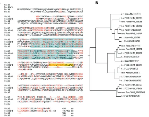
exists in Xenopus laevis, but is only expressed at later stages. FoxN2 was first identified by overlapping EST clones (BU909915, CA988030, CF27159) of Xenopus laevis which en-code the C-terminal part and two overlapping EST clones from Xenopus tropicalis (CX795231, CX800360) encoding the puta-tive N-terminal part. Blast analysis of these EST clones led to the full-length cDNA clone BC081030 (Klein et al., 2002). The open reading frame of FoxN2 encodes 430 amino acids (Fig. 1A) sharing 67% identity with the human sequence (NM_002158).
FoxN3 was identified from the 5´-EST clone (BJ046265) and the 3´-EST clone of Xenopus tropicalis (CX855631) and Xenopus laevis (BJ074333). Using primers derived from these EST clones, we succeeded in PCR amplification of the complete Xenopus sequence. Interestingly, FoxN3 shows alternative splicing, be-cause cloning of the PCR fragments yielded two ORF clones of different length, which differ in an additional 66 bp encoding exon VI (see Fig. 2). It seems, the longer isoform is also present in mice
as we identified this exon in the genomic sequence of mouse Ches1 and we found a putative Foxn3 EST clone (BG082349) containing this exon. Moreover, 5´-RACE of Xenopus laevis FoxN3 revealed two different leader exons which were also identified in the genome of Xenopus tropicalis (see Fig. 2). Sequences of the long variant containing exons I and VI (FoxN3a) and of the short variant containing exon II and lacking exon VI (FoxN3b) were deposited at EMBL (EMBL accession numbers AM114794 and AM114795, respectively).
FoxN4 was discovered in the X. tropicalis EST clone CX964167. Additional database analyses led to two other non-overlapping Xenopus laevis EST clones (BJ061286, DR724233), coding for the N-terminal part of FoxN4. A PCR fragment derived from these EST clones was used for library screening and an incomplete cDNA containing the 3´-end was isolated. The entire coding sequence was amplified from stage 32 Xenopus laevis cDNA (EMBL accession number AM114796).
Fig. 1. The FoxN subclass in Xenopus laevis.(A) Comparison of amino acids of FoxN proteins. Identical amino acids are shown in red, deletions are indicated by dashes. The fork head domains and the amino acids encoded by an additional exon VI (present in FoxN3a but not in FoxN3b) are coloured. (B) Phylogenetic tree of FoxN proteins of different species (d.r., Danio rerio; h.s., homo sapiens; m.m., mus musculus; x.l., Xenopus laevis)
created by ClustalW. The FoxN proteins can be classified into three branches (FoxN1/4, FoxN2/3 and FoxN5/6 paralogues (Katoh and Katoh, 2004b)).
FoxN5 was identified from the Xenopus laevis EST clone CA791267. A PCR fragment derived from this EST clone was used for screening a gastrula stage cDNA library. Four identical cDNA clones were isolated that all contain an open reading frame of 885 bp (deposited under EMBL accession number AM114797). A comparison with corresponding sequences of mouse, human and the X. tropicalis EST clone (CR761025), which contains a stop codon in the 5´-UTR, suggests that the open reading frame is complete. The resulting protein shares only an identity of 23% with human FOXN5 (NM_181721). The X-chromosomal-located human FOXN6 (NM_198451) encodes a protein that shares 31% identity with the Xenopus laevis protein, but FOXN6 is regarded as a retrotransposon of an ancestral FOXN5 during evolution (Katoh and Katoh, 2004b). Since no other FoxN homologues were found in Xenopus laevis, we report this sequence as FoxN5. Additional evidence for this nomenclature comes from the ge-nomic localisation and its surroundings. In the human chromo-some 11 we found Archain flanking FOXN5. The Xenopus laevis cDNA clone matched on scaffold 75 of the X. tropicalis genome, where we identified the predicted Archain gene of Xenopus.
Fig. 1A shows an alignment of the amino acid sequences of Xenopus laevis FoxN proteins. Aside from their rather conserved winged helix domains, FoxN2 and FoxN3 share additional con-served motifs within their N-terminal and C-terminal parts. In contrast, FoxN4 and FoxN5 are less conserved outside their fork head domains and the sequence of FoxN5 deviates by various substitutions even within the fork head domain. Fig. 1B shows a phylogenetic tree of all known FoxN proteins including their
early cleavage stages. The amount of transcripts is slightly decreased during gastrulation and neurulation (stages 10-20), but at stage 30, FoxN2 is upregulated again and continues to be present until stage 45. A similar pattern was observed for the two variants of FoxN3, which differ by alternative splicing of exon VI. The variant containing exon VI migrates more slowly and seems to be less abundant. Both transcripts are maternally synthesized, are present during early cleavage stages and decrease during gastrulation. An upregulation of both splice variants occurs at stage 25 and persists until stage 45. We also looked for the utilization of the two different leader exons and found that both of
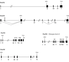
Fig. 2. Genomic organisation of FoxN genes in Xenopus tropicalis. Mapping of gene structure was analysed by Ensembl. Exons are numbered with Roman numer-als. Genes have been identified at scaffold 111 (FoxN2), scaffold 22 (FoxN3), scaffold 9 (FoxN4) and scaffold 75 (FoxN5). The FoxN4 gene has also been isolated and sequenced in Xenopus laevis.
Fig. 3. Temporal expression of FoxN genes during Xenopus devel-opment. Expression was analysed by RT-PCR using RNA isolated from embryos at the indicated developmental stages (Nieuwkoop and Faber, 1967). Primers for FoxN3 were selected to discriminate between FoxN3a
(upper band) and FoxN3b (lower band). Ornithine decarboxylase (ODC) was used as an internal control. Lane (-) shows a negative control in absence of RNA.
different isoforms from human, mouse, Xenopus laevis and zebrafish. While FoxN2, FoxN3 and FoxN4 proteins of Xenopus laevis are rather close to their orthologues from other species, Xenopus FoxN5 shows more distance to the human and mouse orthologues. Splicing of exon VI in FoxN3 leading to different protein variants was also observed for human FOXN3 (see NM_005197 and U68723 in Fig. 1B).
Fig. 2 compares the genomic organisation of the FoxN subfamily in Xenopus tropicalis. Most FoxN genes in Xenopus tropicalis contain a leader exon with the exception of FoxN5. FoxN3 has two alter-natively used leader exons separated by 1.2 kb. Moreover, exon VI is found to be alternatively spliced in embryonic RNA. The FoxN4 gene was addition-ally analysed from Xenopus laevis. Screening of a genomic library led to the isolation of two recombi-nant phages containing this gene. The structures of the orthologous genes are very similar, except that insertion or deletion of DNA sequences led to in-creased or dein-creased sizes of introns, respectively. A comparison of the genomic structures of Xenopus tropicalis FoxN5 and human FOXN5 reveals identi-cal numbers of five exons. In contrast human FOXN6 is a mono-exonal gene (data not shown).
them are used for the variants including and lacking exon VI (data not shown). Thus, at least four different sequences of FoxN3 are present during Xenopus embryogenesis. However, the temporal expression profiles are very similar. FoxN4 expression was not observed during early cleavage stages but showed an initial increase after midblastula transition. Transcripts diminish during gastrulation, but are strongly enhanced again at stage 15 and remain until stage 45. FoxN5 is expressed maternally and tran-scripts are rapidly degraded during gastrulation. At the beginning of neurulation, FoxN5 expression is already very weak and no transcripts are detected at stage 15.
The spatial expression patterns were analysed by whole mount in situ hybridisation (Harland, 1991). The earliest localized ex-pression of FoxN2 is visible at stage 23 in the eye anlagen (data not shown). At stage 26, an intense staining has been observed in the developing eye (Fig. 4A). At stage 29, FoxN2 transcripts are also localised in the branchial arches and the brain (Fig. 4C). Fig. 4B shows a magnification of the head of a stage 33/34 embryo with FoxN2 expression in the vagal ganglion. Transversal sec-tions of stained embryos (Fig. 4D-G) reveal that FoxN2 expres-sion is localised in the prospective retinal layer and a layer of cells lateral to the ventricular zone (Fig. 4E). At stage 36, when the retinal precursor cells begin to differentiate, retinal expression of FoxN2 is downregulated. Note, that FoxN2 expression is absent from the lens (Fig. 4G). Interestingly, expression in the branchial arches and the cells located lateral to the ventricular zone was also observed in the mouse embryo (Tribioli et al., 2002), suggest-ing that both genes act as functional orthologues.
Fig. 5 shows the spatial expression of the FoxN3 gene. At early cleavage stages, FoxN3 transcripts are localized within the ani-mal half (Fig. 5A). At gastrulation, the expression expands over the whole embryo, but does not include the future endodermal cells of the blastopore (data not shown). During neurulation, an intense staining in the prospective eye field is observed. Another expression domain was found in two stripes lateral to the neural tube, i.e., in the neural crest cells (Fig. 5B, stage 23 embryo). Fig. 5C shows that FoxN3 transcripts are strongly enriched in the eye vesicles (embryo stage 26, lateral view). From stage 29 onwards, FoxN3 transcripts are observed predominantly in the eye, the branchial arches and in the vagal ganglion (Fig. 5D). At stage 38, we observe diffuse staining of the whole head (Fig. 5E). However, a section reveals relatively stronger staining in the head mesen-chyme and in the eye lens (Fig. 5F).
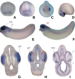
Fig. 6 shows the expression of the Xenopus FoxN4 gene. Similar to the mouse orthologue (Gouge et al., 2001), FoxN4 transcripts are predominantly localised in the developing eye. A weak staining is observed in the eye anlage at stage 18 (Fig. 6A). Between stages 21 and 26, FoxN4 expression becomes stronger in the eye field (see Figs. 6B-D). At stage 29/30, an intense staining is observed within the eye vesicle, in the midbrain and in the nephrostomes of the pronephric kidney (Fig. 6E). FoxN4 Fig. 4 (Left). Whole mount in situ hybridisation of FoxN2.(A) Stage 26, lateral view; (B) magnification of the head of a stage 33/34 embryo, lateral view; (C) lateral view of a stage 29 embryo. The red lines in (A-C) denote transversal sections shown in (D-F). (D) Stage 26; (E) stage 29;
(F) stage 33/34. (G) A transversal section through the head of an embryo at stage 36. ba, branchial arches; l, lens; prl, presumptive retinal layer; rh, rhombencephalon; r, retina; vg, vagal ganglion. The fat arrow in (E) denotes the FoxN2 stained cell layer, lateral to the ventricular zone.
Fig. 5 (Above). Whole mount in situ hybridisation of FoxN3. (A)
Transversal razor blade sectioned 8-cell stage embryo, animal cells are on top. (B) Dorsal view of a stage 23 embryo. Lateral views of (C) stage 26, (D)
stage 29 and (E) stage 38 embryos. (F) Transversal section of an embryo at stage 38. The red line in (E) indicates plane of section shown in (F). ba, branchial arches; hm, head mesenchyme; l, lens; vg, vagal ganglion.
A
B
C
D
E
F
G
A
B
C
D
expression is found in the prospective retinal layer, but is absent from the lens placode (Fig. 6G- I). After stage 30, expression of FoxN4 in the nephrostomes is downregulated. At stage 33/34, it is largely absent, while the staining in the eye is still visible (Fig. 6F). Transversal section of a stage 26 embryo shows an intense and clearly restricted staining of FoxN4 in the prospective retinal layer (Fig. 6G). After stage 33/34, expression of FoxN4 in the eye was downregulated (Fig. 6H). At stage 38, only a few cells are stained in the peripheral region of the retina (Fig. 6I). This staining co-localises with the ciliary marginal zone (CMZ), which is as-sumed to be a retinal stem cell pool in amphibians, reptiles and birds, but not in mammals.
The expression of FoxN5 was found to be ubiquititous during early cleavage and gastrula stages. In post-gastrula stage em-bryos, no staining was visible (data not shown). We suggest that maternal transcripts are evenly distributed in dividing cells, but are degraded until the onset of neurulation.
Experimental Procedures
Total RNA was extracted from Xenopus laevis embryos using TRIzol
(Gibco) reagent and RNeasy Cleanup (Qiagen). cDNA was synthesised with Transcriptor First Strand cDNA Synthesis Kit (Roche). Primers used for RT-PCR were:
Fig. 6. Whole mount in situ hybridisation of FoxN4. Anterior view of a (A) stage 18, (B) stage 21 and (C) stage 23 embryo. (D) Half-lateral view of a stage 26 embryo. Lateral view of a (E) stage 29/30 and (F) stage 33/ 34 embryo. The red line in (F) denotes the plane of the transversal sections shown in (H-I). (G) Transversal section of the head of a stage 26 embryo. (H) Section of a stage 33/34 embryo. (I) Section of a stage 38 embryo. cmz, ciliary marginal zone; l, lens; mb, midbrain; n, nephros-tomes; prl, presumptive retinal layer; r, retina.
FoxN2 forw: 5´-TCT CTG ACA TCT TCT TGC CCG TT-3´ rev: 5´-GTG CAG CCA GTT CAA GTT GGT CA-3´
FoxN3 forw: 5´-GGC AAA GGT TCT CTG TGG TGC A-3´ rev: 5´-CTG AGG CAG CCA TTG CTC CAA TA-3´
FoxN4 forw: 5´-AGG GCA CTG TGT TCT CTC CTT TG-3´ rev: 5´-CCA GGA GAG TGT ACA TTA TTT GA-3´
FoxN5 forw: 5´-TCT CCT ACA GCA CTT GAA GAC TG-3´ rev: 5´-GAT CCA CTC AGG GCA TGG AAT CA-3´
ODC forw: 5´-GGG CAA AGG AGC TTA ATG TG-3´ rev: 5´-CAT TGG CAG CAT CTT CTT CA-3´.
Sequences were amplified from cDNA obtained by RT-PCR or from Lambda ZAP gastrula and Lambda ZAP tadpole cDNA Xenopus laevis
libraries (Stratagene) with Taq Polymerase (ABgene) and cloned in pDrive cloning vector (Qiagen). For the genomic FoxN4 sequence, we
screened a genomic Lambda Fix library (Stratagene) using FoxN4 cDNA
as labelled probe. EST database analyses and comparative genomics analyses on FoxN orthologues and multiple sequence alignments were performed by using programs: http://www.ncbi.nlm.nih.gov/blast, http:// www.ensembl.org/index.html and http://www.ebi.ac.uk/clustalw/. Primers used for cloning of complete ORF sequences were:
FoxN2
forw: 5´-cgg gat ccc gAT GGG TCC AGC AAC TGG AAT GAC T-3´ rev: 5´-ccg ctc gag cgg TTG TGA GAT TGT GCC TTT GCA GT-3´
FoxN3
forw: 5´-ccg ctc gag cgg ATG GGT CCA GTC ATG CCT CCT AG-3´ rev: 5´-gcc tat gta CAT TAG AAT TCA CCT GAC GTG CTG-3´
FoxN4
forw: 5´-ccg ctc gag cgg AAT GAT AGA CAG TGA CAT CTC TGC-3´ rev: 5´-gcc tat gta ATA CAT AGA GAA GTC ATC ACA GT-3´. 5´-RACE was performed using GeneRacer Kit, Invitrogen. PCR was done with T3 or T7 primers and a gene-specific primer using a Lambda ZAP cDNA library. A second semi-nested PCR was carried out with a second gene-specific primer. The PCR products were sequenced and cloned. Whole mount in situ hybridisations were done according to standard
procedures (Harland, 1991). For vibratome sectioning the embryos were embedded in agarose (2%) or in gelatine/albumine. Serial vibratome sections (10-50 µm) were processed.
Acknowledgements
This work was supported by the Deutsche Forschungsgemeinschaft (SFB 497/A3).
References
BALCIUNAITE, G., KELLER, M.P., BALCIUNAITE, E., PIALI, L., ZUKLYS, S., MATHIEU, Y.D., GILL, J., BOYD, R., SUSSMAN, D.J. and HOLLANDER G.A. (2002). Wnt glycoproteins regulate the expression of FoxN1, the gene defective in nude mice. Nat. Immunol. 3: 1102-11088.
CHANG, J.T., WANG, H.M., CHANG, K.W., CHEN, W.H., WEN, M.C., HSU, Y.M., YUNG, B.Y., CHEN, I.H., LIAO, C.T., HSIEH, L.L. and CHENG, A.J. (2005). Identification of differentially expressed genes in oral squamous cell carcinoma (OSCC): overexpression of NPM, CDK1 and NDRG1 and underexpression of CHES1. Int. J. Cancer 114: 942-949.
GOUGE, A., HOLT, J., HARDY, A.P., SOWDEN, J.C. and SMITH, H.K. (2001). Foxn4 - a new member of the forkhead gene family is expressed in the retina. Mech. Dev. 107: 203-206.
HARLAND, R.M. (1991). In situ hybridization: an improved whole-mount method for Xenopus embryos. Methods Cell Biol. 36: 685- 695.
KAESTNER, K.H., KNÖCHEL, W. and MARTINEZ, D.E. (2000). Unified nomencla-ture for the winged helix/forkhead transcription factors. Genes Dev. 14: 142-146.
KLEIN, S.L., STRAUSBERG, R.L., WAGNER, L., PONTIUS, J., CLIFTON, S.W. and RICHARDSON, P. (2002). Genetic and genomic tools for Xenopus re-search: The NIH Xenopus initiative. Dev. Dyn. 225: 384-391.
KATOH, M. and KATOH, M. (2004a). Identification and characterization of human
A
B
C
D
E
F
FOXN5 and rat Foxn5 genes in silico. Int. J. Oncol. 24: 1339-1344.
KATOH, M. and KATOH, M. (2004b). Identification and characterization of human FOXN6, mouse Foxn6 and rat Foxn6 genes in silico. Int. J. Oncol. 25: 219-223.
LI, C., LUSIS, A.J., SPARKES, R., TRAN, S.M. and GAYNOR, R. (1992). Charac-terization and chromosomal mapping of the gene encoding the cellular DNA binding protein HTLF. Genomics 13: 658-64.
LI, S., MO, Z., YANG, X., PRICE, S.M., SHEN, M.M. and XIANG, M. (2004). Foxn4 controls the genesis of amacrine and horizontal cells by retinal progenitors. Neuron 43: 759-760.
NEHLS, M., PFEIFER, D., SCHORPP, M., HEDRICH, H. and BOEHM, T. (1994). New member of the winged-helix protein family disrupted in mouse and rat nude mutations. Nature 372: 103-107.
NIEUWKOOP, P.D. and FABER, J. (1967). Normal Table of Xenopus laevis (Daudin), 2nd ed. Elsevier/North Holland, Amsterdam.
PATI, D., KELLER, C., GROUDINE, M. and PLON, S.E. (1997). Reconstitution of a MEC1-independent checkpoint in yeast by expression of a novel human fork head cDNA. Mol. Cell Biol. 17: 3037-46.
SCOTT, K.L. and PLON, S.E. (2003). Loss of Sin3/Rpd3 histone deacetylase restores the DNA damage response in checkpoint-deficient strains of Saccha-romyces cerevisiae. Mol. Cell Biol. 23: 4522-31.
TRIBIOLI, C., ROBLEDO, R.F. and LUFKIN, T. (2002). The murine fork head gene Foxn2 is expressed in craniofacial, limb, CNS and somitic tissues during embryogenesis. Mech. Dev. 118: 161-163.
Received: November 2005
Reviewed by Referees: January 2006