Original Article
The zic1 gene is an activator of Wnt signaling
CHRISTA S. MERZDORF
#and HAZEL L. SIVE*
Whitehead Institute for Biomedical Research and Massachusetts Institute of Technology, Cambridge, USA
ABSTRACT The zic1 gene plays an important role in early patterning of the Xenopus neurectoderm. While Zic1 does not act as a neural inducer, it synergizes with the neural inducing factor Noggin to activate expression of posterior neural genes, including the midbrain/hindbrain boundary marker engrailed-2. Since the Drosophila homologue of zic1, odd-paired (opa), regulates expres-sion of the wingless and engrailed genes and since Wnt proteins posteriorize neural tissue in Xenopus, we asked whether Xenopus Zic1 acted through the Wnt pathway. Using Wnt signaling inhibitors, we demonstrate that an active Wnt pathway is required for activation of en-2 expression by zic1. Consistent with this result, Zic1 induces expression of several wnt genes, including wnt1, wnt4 and wnt8b. wnt1 gene expression activates expression of engrailed in various organisms, including Xenopus, as demonstrated here. Together, our data suggest that zic1 is an upstream regulator of several wnt genes and that the regulatory relationships between opa, wingless and engrailed seen in Drosophila are also present in vertebrates.
KEY WORDS:
zic, wingless, wnt, Xenopus, engrailed, neural
Introduction
Wnt signaling is involved in many developmental processes and despite the considerable understanding of this pathway, the upstream events that regulate wnt gene expression are not well understood (Wodarz and Nusse, 1998, Logan and Nusse, 2004, Wang and Wynshaw-Boris, 2004, Ciani and Salinas, 2005). In Drosophila, the odd-paired (opa) gene is required for activation of wingless gene expression (Benedyk et al., 1994).
Vertebrate opa homologues are members of the zic gene family of zinc finger transcription factors (Kuo et al., 1998, Mizuseki et al., 1998, Nakata et al., 1998). zic genes have been implicated in patterning the dorsal neural tube, in neural crest development and in cerebellar development (Aruga, 2004). Do zic genes regulate Wnt signaling in vertebrates? The zic1 gene (also called opl and zicr-1) (Kuo et al., 1998, Mizuseki et al., 1998, Nakata et al., 1998) may be involved in regulating wnt expression. Through-out early development in Xenopus, zic1 and several wnt genes show extensive overlap in their expression patterns in the pre-sumptive neurectoderm, in the dorsal neural tube, at the fore-brain/midbrain boundary and at the midbrain/hindbrain boundary (McGrew et al., 1992, Wolda et al., 1993, Cui et al., 1995, Chang and Hemmati-Brivanlou, 1998, Kuo et al., 1998), suggesting there may be a regulatory connection between zic1 and wnt genes. Further, wnt genes are known to posteriorize neural tissue in
*Address correspondence to: Dr. Hazel L. Sive. Whitehead Institute for Biomedical Research and Massachusetts Institute of Technology, Nine Cambridge Center, Cambridge MA 02142, USA. Fax: +1-617-258-5578. e-mail: sive@wi.mit.edu
#Present address: Department of Cell Biology and Neuroscience, Montana State University, Bozeman MT 59717, USA
Abbreviations used in this paper: C-terminus, carboxy-terminus; opa, odd-paired
gene; PCR, polymerase chain reaction; RT, reverse transcriptase.
0214-6282/2006/$25.00 © UBC Press
Printed in Spain www.intjdevbiol.com
animal cap (undifferentiated ectoderm) assays (McGrew et al., 1995, McGrew et al., 1997, Chang and Hemmati-Brivanlou, 1998, Domingos et al., 2001). This activity is shared by zic1 (Kuo et al., 1998). In particular, when the BMP inhibitor Noggin is expressed in animal caps from Xenopus, these animal caps express pan-neural genes and a subset of anterior pan-neural genes (Lamb et al., 1993), while co-expression of Wnt proteins activates more poste-riorly expressed genes (McGrew et al., 1995). Zic1 alone does not induce neural gene expression. Neither does Zic1∆C, a C-termi-nally truncated form of Zic1 that shows enhanced transcriptional activation activity compared to full length Zic1 (zic1∆C = opl∆C; Kuo et al., 1998). However, like Wnt proteins, both Zic1∆C and full length Zic1 synergize with Noggin to induce expression of poste-rior neural genes that are not activated by Noggin alone. One such gene is the midbrain/hindbrain boundary marker en-2 (Kuo et al., 1998), which is a target of Wnt signaling (Danielian and McMahon, 1996, McGrew et al., 1999).
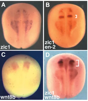
zic1 and en-2 expression domains overlap in early neurula (stage 14) embryos (Kuo et al., 1998). We began this study by showing a similar overlap in mid/late neurula (stage 17) em-bryos between the zic1 expression domain (Fig. 1A) and the expression domain of the en-2 gene (Fig. 1B).
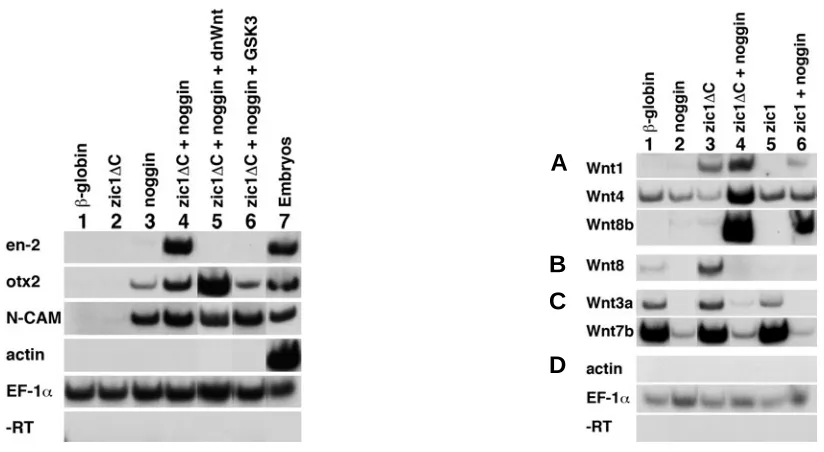
In order to investigate the regulatory relationships between zic1, en-2 and wnt genes, we utilized animal cap assays. In vitro synthesized zic1∆C and noggin RNAs were injected into 2-cell embryos, animal caps were isolated at late blastula (stage 9) and maintained in culture until control embryos reached early tailbud (stage 22), when they were harvested for RT-PCR analysis. Control experiments showed that animal caps in-jected with either β-globin or with zic1∆C alone did not show expression of either the pan-neural gene N-CAM or the anterior neural marker otx2 (Fig. 2; lanes 1 and 2). On the other hand, animal caps neuralized with Noggin showed induction of both N-CAM and otx2 expression (lane 3). In contrast, en-2 expres-sion was induced when zic1∆C and noggin were co-expressed in animal caps (lane 4). These data are consistent with previous studies showing that inhibition of BMPs is required for the induction of en-2 by zic1 in animal cap explants (Kuo et al., 1998).
We next asked whether an active Wnt signaling pathway is required for the induction of en-2 by zic1. We examined the
β
sion of GSK3 RNA and zic1∆C in neuralized animal caps also inhibited induction of en-2 expression (lane 6). The same experiments were conducted with full length zic1, where activa-tion of en-2 expression was also abolished by co-expression of full length zic1plus noggin with either of the two Wnt pathway inhibitors (not shown). Thus, induction of en-2 expression by Zic1 requires an active Wnt pathway.
Zic1 activates expression of a subset of wnt genes
One interpretation of these results is that zic1 might regulate the expression of wnt genes. To examine this possibility, we expressed zic1∆C or full length zic1 with or without noggin and assayed wnt gene expression in animal caps (Fig. 3). Animal caps were examined by RT-PCR analysis for expression of the wnt1, wnt3a, wnt4, wnt5a, wnt5c, wnt7b, wnt8, wnt8b and wnt11 genes. Animal caps taken from embryos injected with RNAs for either β-globin, noggin, zic1∆C, or full length zic1 (Fig. 3A, lanes 1-3 and 5) showed low or undetectable expression levels of wnt1, wnt4 and wnt8b. In contrast, caps removed from embryos co-injected with zic1∆C plus noggin showed strong induction of wnt1, wnt4 and wnt8b expression (lane 4) relative to control caps. Expression of full length zic1 in combination with noggin activated lower levels of wnt1 and wnt8b expres-sion than did zic1∆C and did not induce significant expression of wnt4 (lane 6). In the absence of noggin, zic1∆C, but not full length zic1, activated appreciable levels of wnt1 expression (lanes 3 and 5).
In the same assay, expression of wnt8, wnt3a and wnt7b was strong or detectable in animal caps that were not expressing Noggin (Fig. 3B, C; lanes 1, 3 and 5) and weak or absent in caps expressing Noggin (lanes 2, 4 and 6). Although Zic1∆C acti-vated expression of wnt8 (lane 3), the relevance of this is unclear. When ectopically expressed in dorsal mesoderm dur-ing gastrulation, wnt8 causes a loss of anterior structures (Christian and Moon, 1993), although the expression of en-2 is unchanged (Fredieu et al., 1997). Thus, the induction of wnt8 by zic1∆C is probably not relevant to the induction of en-2. What is the relevance of high wnt7b and wnt3a expression in control or β-globin-expressing animal caps (Fig. 3C, lane 1) and lack of expression in neuralized caps (lanes 2, 4, 6)? Since wnt7b is expressed not only in the neural plate but also in the epidermis (Chang and Hemmati-Brivanlou, 1998), Noggin may inhibit expression of the epidermal component of wnt7b expression in our assays. However, because wnt3a is expressed in the dorsal neural tube in an extensively overlapping domain with zic1 (McGrew et al., 1997, Kuo et al., 1998) and wnt3a is known to induce en-2 expression in neuralized animal caps (McGrew et al., 1995), we had not expected downregulation of wnt3a by Noggin.
We also tested induction of wnt5a, wnt5c and wnt11 expres-sion in this assay, however none of these genes showed activation of expression by the zic1∆C/zic1+noggin RNA com-Fig. 1. The zic1 expression domain overlaps with those of both
en-2 and wnt8b. In situ hybridization of stage 17 neurula embryos. (A)zic1 expression domain. (B) Double in situ hybridization with zic1 and en-2 probes shows overlap between the two expression domains. The bracket indicates en-2 expression. (C) wnt8b expression domain in the midbrain. (D) Double in situ hybridization with zic1 and wnt8b probes indicates that their expression domains overlap. The bracket indicates wnt8b expres-sion.
A
B
binations (data not shown). In sum, these assays showed selective activation of wnt gene expression by combinations of activated or full length Zic1 plus Noggin.
Expansion of the wnt8b expression domain by Zic1 in a whole embryo assay
The strong induction of wnt8b expression in animal caps co-expressing zic1∆C/zic1+noggin RNAs prompted us to investigate the ability of zic1 to induce wnt8b expression in whole embryos. First, we established that wnt8b is expressed in the midbrain during neurula stages (Fig. 1C) and overlaps with the zic1 expression domain (Fig. 1D). Subsequently, zic1∆C or full length zic1 RNAs were co-injected with lacZ tracer RNA into albino embryos. In situ hybridization showed that the region of the embryo expressing wnt8b was expanded by expression of zic1∆C (Fig. 4A) and to a lesser degree by expression of full length zic1 (Fig. 4B). Since the in situ assay is not quantitative and the
responding tissue different in the animal cap and whole embryo assays, it is not clear whether the observed expansion is equiva-lent in the whole embryo and animal cap assays.
Importantly, the region in which wnt8b expression was ex-panded was contiguous with the endogenous wnt8b expression domain. Thus, whole embryos must contain factors that regulate where wnt8b expression can be modulated and zic1 expression cannot be solely responsible for this modulation. Nonetheless, this result indicates that the animal cap assay accurately indi-cated the responsiveness of the embryo to zic1.
Regulatory relationships between wnt1 and en-2 are con-served in Xenopus
The wnt1 and en-2 expression domains overlap at the Xeno-pus midbrain/hindbrain boundary (Li et al., 2006). Although regu-latory connections between the wnt1 gene and en-2 have been shown in various organisms (McMahon et al., 1992, Sugiyama et Fig. 2 (Left). Zic1 acts through the Wnt pathway to activate en-2. Both cells of 2-cell stage embryos were injected with the in vitro synthesized RNAs listed along the top. A C-terminal truncation of the zic1 coding sequence was used (zic1∆C). Animal caps were isolated at stage 9 and cultured until sibling embryos reached stage 22. Total RNA was isolated and subjected to RT-PCR analysis with the primers shown on the left. (Lanes 1,2), β-globin or zic1∆C injected animal caps did not show expression of neural markers. (Lane 3)Noggin mRNA induced the anterior neural marker otx2 and the general neural marker N-CAM. (Lane 4) Co-injected zic1∆C plus noggin induced expression of the midbrain/hindbrain boundary marker en-2. Induction of en-2 expression is inhibited by dnWnt (lane 5) or GSK3 (lane 6), which are inhibitors of the Wnt pathway. This demonstrates that Zic1 requires an active Wnt pathway to induce en-2 expression. N-CAM and otx2 were expressed in all samples that received noggin (lanes 3-6). (Lane 7) Whole embryos at stage 22 served as positive control. Muscle actin controlled for mesodermal contamination, EF-1α served as loading control and -RT samples controlled for DNA contamination. After culture to the equivalent of stage 22, animal caps expressing β-globin and zic1 constructs were always completely round. Noggin-expressing animal caps were occasionally elongated, but the amount of noggin used was low enough that most explants were round. Co-injection of dnWnt or GSK3 RNA did not influence the shape of the animal caps beyond the effects of noggin. Injections were as follows: 200 pg β-globin, 200 pg zic1∆C, 5 pg noggin, 150 pg dnWnt8, 80 pg GSK3.
Fig. 3 (Right). Zic1 induces wnt expression. Embryos were injected into both blastomeres at the 2-cell stage with the indicated RNAs. zic1∆C was tested in addition to full length zic1. Animal caps were isolated at stage 9 and cultured until sibling embryos reached stage 22. Total RNA was isolated and subjected to RT-PCR analysis with the primers shown on the left. (Lane 1)β-globin injected animal caps. (Lane 2) Noggin-injected. (Lane 3)zic1∆C -injected. (Lane 4)zic1∆C plus noggin-injected. (Lane 5) Full length zic1-injected. (Lane 6) Full length zic1 plus noggin-injected. (A)wnt1, wnt4 and wnt8b expression was induced by zic1∆C plus noggin (lane 4). zic1∆C alone induced wnt1 expression (lane 3), while full length zic1 plus noggin did not induce wnt4 expression (lane 6). (B)zic1∆C induced wnt8 expression (lane 3). (C)wnt3a and wnt7b were expressed in β-globin, zic1∆C and zic1 -injected animal caps (lanes 1, 3 and 5), but not in samples that had been co--injected with noggin (lanes 2, 4 and 6). (D) Muscle actin controlled for mesodermal contamination, EF-1α served as loading control and -RT samples controlled for DNA contamination. Injections were as follows: 200 pg β-globin, 200 pg zic1∆C, 200 pg zic1, 5 pg noggin.
A
B
C
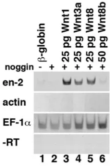
al., 1998), this has not been as fully explored in Xenopus. We therefore tested Wnt1 for its ability to induce en-2 expression and found that Wnt1 induces robust en-2 expression in neuralized animal caps (Fig. 5, lane 3). At the same time, we confirmed that Wnt3a induces en-2 expression in neuralized animal caps (lane 4) (McGrew et al., 1995) and showed that Wnt8 induces en-2 expression (lane 5). In contrast, Wnt8b did not induce en-2 expression (lane 6) and Chang and Hemmati-Brivanlou (1997) showed previously that Wnt7b does not induce en-2 in neuralized animal caps. This confirms that a regulatory pathway exists between the wnt and en-2 genes in Xenopus (McGrew et al., 1995, McGrew et al., 1999) and demonstrates that only a subset of Wnt proteins can activate en-2 expression.
expression was limited to the normal en-2 expression domain, suggesting that other factors restrict the en-2 expression domain. Expression of a dominant interfering form of zic1 (dnzic1) strongly reduced expression of en-2 (Fig. 6B) and wnt1 (Fig. 6C) in whole embryos, suggesting that Zic1 is necessary but not sufficient for the activation of wnt1 and en-2 expression.
Discussion
We show that Zic1 requires an active Wnt pathway to induce expression of the en-2 gene in neuralized Xenopus ectodermal explants. This relationship is conserved in Drosophila, where opa is required for expression of the wg and en genes (Benedyk et al., 1994).
The ability of zic1 to induce neural genes in animal cap explants is dependent on the inhibition of BMP signaling to give the explants neural character (Kuo et al., 1998). In whole em-bryos, BMP antagonists secreted from the organizer, such as Noggin and Chordin, sequester BMP proteins and therefore allow dorsal determination and formation of neural tissues (Sasai and De Robertis, 1997, Munoz-Sanjuan and Brivanlou, 2002). In-deed, zic1, which is among the first molecular indicators of neural fate determination, is expressed in direct response to interruption of BMP signaling (Tropepe et al., 2006).
In animal caps, Zic1 acts through the Wnt pathway to activate en-2 expression and we found that the expression of three wnt genes (wnt1, wnt4 and wnt8b) is activated by Zic1∆C in neuralized ectodermal explants. This suggests that Zic1 interfaces with wnt gene function by activating wnt gene expression. The expression domains of these three wnt genes overlap with that of zic1 during early embryonic stages. During gastrula stages, zic1 expression overlaps with wnt4 expression in the dorsal ectoderm (McGrew et al., 1992). During neurula stages, zic1 expression becomes restricted to the lateral edges of the neural plate and subsequently to the dorsal neural tube (Kuo et al., 1998), where its expression overlaps with that of wnt1 and wnt4 (McGrew et al., 1992, Wolda et al., 1993). Further, zic1 expression extends to the forebrain/ midbrain boundary, where the zic1 and wnt8b expression do-mains overlap in the midbrain during neurula stages (this study) and at the forebrain/midbrain boundary during tailbud stages (Cui et al., 1995). At the midbrain/hindbrain boundary, zic1 expression overlaps with expression of wnt1 and en-2 (Brivanlou and Harland, 1989, Wolda et al., 1993, Chang and Hemmati-Brivanlou, 1998, Kuo et al., 1998, Li et al., 2006).
Three consensus binding sites for LEF/TCF are present in the Xenopus en-2 promoter (McGrew et al., 1999), suggesting that the en-2 gene is a direct target of canonical Wnt signaling. In the animal cap system, wnt1, wnt3a and wnt8, but not wnt8b or wnt7b, activated en-2 expression in neuralized animal caps (this study, McGrew et al., 1995, Chang and Hemmati-Brivanlou, 1998). Based on these considerations and on spatial expression pat-terns, wnt1 is the most likely candidate through which zic1 acts to Fig. 4. Zic1 induces ectopic wnt8b expression. One cell of 2-cell albino
embryos was injected with (A) 100 pg zic1∆C RNA or with (B) 100 pg full length zic1 RNA together with 25 pg lacZ RNA as tracer. In situ hybridiza-tion with wnt8b probe at neurula stage 18 showed that zic1∆C and full length zic1 upregulate wnt8b expression on the injected (arrowheads) side.
activate expression of en-2 in Xenopus. This would be consistent with findings in mice, chick, Xenopus (this study) and Drosophila that connect wnt1 activity with activation of engrailed expression (DiNardo et al., 1988, Martinez Arias et al., 1988, Bally-Cuif et al., 1992, McMahon et al., 1992, Danielian and McMahon, 1996, Sugiyama et al., 1998).
Our data using dominant interfering constructs suggest that Zic1 is necessary for activation of wnt1 and en-2 expression. However, although Zic1 causes an increase in en-2 expression levels within its normal expression domain, ectopic Zic1 expres-sion is not sufficient to induce ectopic en-2 expresexpres-sion. Thus, other factors must be required for induction of en-2 expression and helps explain why zic1, which is broadly expressed, can activate the expression of genes that are expressed in very restricted domains. We note that expression of other zic genes overlaps temporally and spatially with that of zic1 (Nakata et al., 1997, Nakata et al., 1998, Nakata et al., 2000). Our data do not distinguish whether other genes also act or synergize with Zic1 to activate wnt and en-2 expression.
Genetic studies reveal a similar situation in Drosophila. Al-though loss of opa function gives rise to pair-rule defects in body pattern, opa is different from all other pair-rule genes in that it is expressed in a broad, unsegmented domain rather than in a segmented fashion. opa is required for the proper level and timing Fig. 6. Zic1 is required for en-2 induction. (A) One cell of 2-cell albino embryos was injected with 100 pg zic1∆C RNA, resulting in an increase of en-2 expression levels within the en-2 expression domain. Embryos injected with 100 pg of a dominant interfering zic1 construct (dnzic1) showed very significant decrease (B) in en-2 expression and (C) in wnt1 expression. All embryos were co-injected with 25 pg lacZ RNA as tracer and the injected sides are shown on the right.
of wg and en expression but not for correct positioning of the expression domains of wg and en (Ingham et al., 1988, Benedyk et al., 1994). Similarly, zic1 is expressed in a much broader domain than wnt1 or en-2 (Kuo et al., 1998). Perhaps, analogous to opa activity in Drosophila, zic1 may act to regulate the timing (and possibly the maintenance and/or level) of wnt1 and en-2 expression but not the position of their respective expression domains.
In Drosophila, the genes ftz, prd and/or eve may posi-tively regulate the position of en expression (Howard and Ingham, 1986, DiNardo and O’Farrell, 1987) and the runt gene may do so negatively in areas where ftz is not present (Kania et al., 1990, Swantek and Gergen, 2004). Expres-sion of en-2 in vertebrates may also require further regula-tory influences. For example, two pax binding sites are required for the expression of a mouse En-2 transgene (Song et al., 1996) and pax2 expression begins before
A
B
C
engrailed expression at the midbrain/hindbrain boundary in Xe-nopus (Heller and Brandli, 1997).
As reflected in our model (Fig. 7), the data indicate that Zic1 induces en-2 via activation of Wnt1 signaling. Alternatively, there is evidence that Wnt signaling may induce en-2 by an indirect mechanism that is dependent upon FGF signaling (Domingos et al., 2001). Expression of an FGF receptor in neuralized ectoderm results in the upregulation of en-2 and wnt1 expression (Umbhauer et al., 2000). Thus, Zic1 may induce en-2 by activating wnt1, which activates an FGF family member, which in turn activates en-2 expression. Consistently, Zic1 appears to be able to induce fgf8 (Li and Merzdorf, unpublished results), whose expression domain overlaps with the zic1, wnt1 and en-2 expression do-mains in Xenopus (Wolda et al., 1993, Kuo et al., 1998, Glavic et al., 2002). McGrew et al. (1997) find that Fgf induces en-2 in the presence of Wnt pathway inhibitors and Lee et al. (1997) show that wnt1 regulates en-2 expression via the Fgf pathway in mouse. Thus, there may be two pathways of en-2 induction by wnt1, one direct via LEF/TCF sites in the en-2 promoter and one indirect via FGF signaling. It will be important to determine whether the initiation and the maintenance of en-2 expression rely on different parts of the pathway, as may be the case in mouse and in Drosophila.
In conclusion, we have shown that regulatory connections described for Drosophila opa are conserved in Xenopus. As in Drosophila, Zic1 may be responsible for the level and timing of en-2 expression, rather than for its positioning. Further, the mecha-nisms underlying Zic1 activities may include activation of the expression of several wnt genes.
Materials and Methods
Growth, microinjection, dissection and culture of embryos and explants
Xenopus laevis eggs were collected, fertilized and cultured as in (Sive et al., 1989). Embryos were staged according to (Nieuwkoop and Faber,
1967). Microinjection techniques were as described (Kolm and Sive, 1995). For animal cap explant assays, both cells of 2-cell embryos were injected with a total of 100-200 pg β-globin RNA, 200 pg zic1 or zic∆C
RNA (= opl∆C ) (Kuo et al., 1998); and 3-5 pg noggin RNA in various
combinations. Further, 150 pg dnWnt8, 80 pg GSK3, 25 pg wnt1, 25 pg wnt3a, 25 pg wnt8 and 50 pg wnt8b RNAs were injected in combination
with other RNAs as detailed in the figure legends. For animal caps, late Noggin/Chordin
BMP
Zic1 + other factors
Wnt3a Wnt1 Fgf
en-2
Capped sense RNAs for microinjection were synthesized for zic1 and zic1∆C RNA in pCS2+ by SP6 transcription of a NarI/NarI fragment. The
dominant interfering zic1 (dnzic1) construct was made by PCR
amplifica-tion of the zinc finger domain and C-terminus of the zic1 coding region.
This PCR product was cloned into the Nco1 and Xba1 sites of the pCS2+ATG plasmid. The pCS2+ATG was made by inserting a Kozak sequence between the BamH1 and EcoR1 sites of the pCS2+ vector. The coding sequence of wnt1 was cloned into pCS2+ by PCR. wnt1 sense
RNA was synthesized by SP6 transcription from the NotI-digested pCS2+wnt1 plasmid. Other sense RNAs were synthesized as published: noggin (Smith and Harland, 1992), β-globin (Krieg and Melton, 1984), wnt3a (Wolda et al., 1993), wnt8 (Christian et al., 1991), wnt8b (Cui et al.,
1995), dnWnt8 (Hoppler et al., 1996), GSK3 (He et al., 1995) and lacZ
(Turner and Weintraub, 1994).
Antisense probes for in situ hybridization were transcribed as
previ-ously described: zic1 (Kuo et al., 1998), en-2 (Hemmati-Brivanlou and
Harland, 1989), wnt1 and wnt3a (Wolda et al., 1993). Since the antisense
probe synthesized from the wnt8b construct, kindly provided by Jan
Christian, does not hybridize to neurula embryos, we synthesized wnt8b
antisense RNA probe using T7 polymerase on a PCR product as tem-plate. The primers for the PCR product were:
forward: 5’-GACCTTCTTATCCCGTCTCCA-3’ and
reverse: 5’-CTAATACGACTCACTATAGGCTAAACCACAGTCACCAC AAA-3’, where the underlined bases represent the T7 RNA polymerase promoter sequence.
In situ hybridization
Whole mount in situ hybridization was performed with albino embryos
as described in (Harland, 1991). One cell of 2-cell embryos was injected with 100 pg zic1∆C RNA, 100 pg zic1 RNA, or 100 pg dnzic1 RNA
together with 25 pg lacZ RNA as tracer. β-galactosidase staining was performed as in (Kolm and Sive, 1995). The alkaline phosphate substrate NBT/BCIP (Sigma) was used and for double in situ hybridizations, BCIP
in combination with NBT/BCIP was used, although the color differences were lost during the second staining.
RT-PCR
RNA from pools of 14-25 animal cap explants or from 2 embryos was analyzed by RT-PCR as described (Kuo et al., 1998). The following
primers were used: N-CAM and en-2 primers (Hemmati-Brivanlou and
Melton, 1994), EF-1α primers (Gammill and Sive, 1997), otx2 primers
(Pannese et al., 1995), actin primers (Stutz and Spohr, 1986), wnt1 primers (forward: 5’-ATCGGGACTGTATTGCCAAG-3’
Dawid, Jan Christian and David Turner for kind gifts of plasmids. This work was supported by NIH grant 2RO1 HD33472 to H.L.S. and by NIH-NRSA postdoctoral fellowship F32-HD-8495 and NSF grant IOB-0417242 to C.S.M.
References
ARUGA, J. (2004). The role of zic genes in neural development. Mol Cell Neurosci
26: 205-21.
BALLY-CUIF, L., ALVARADO-MALLART, R.M., DARNELL, D.K. and WASSEF, M. (1992). Relationship between wnt-1 and en-2 expression domains during early development of normal and ectopic met-mesencephalon. Development 115:
999-1009.
BENEDYK, M.J., MULLEN, J.R. and DINARDO, S. (1994). Odd-paired: A zinc finger pair-rule protein required for the timely activation of engrailed and wingless in Drosophila embryos. Genes Dev 8: 105-17.
BRIVANLOU, A.H. and HARLAND, R.M. (1989). Expression of an engrailed-related protein is induced in the anterior neural ectoderm of early Xenopus embryos.
Development 106: 611-7.
CHANG, C. and HEMMATI-BRIVANLOU, A. (1998). Neural crest induction by Xwnt7b in Xenopus. Dev Biol 194: 129-34.
CHRISTIAN, J.L., MCMAHON, J.A., MCMAHON, A.P. and MOON, R.T. (1991). Xwnt-8, a Xenopus wnt-1/int-1-related gene responsive to mesoderm-inducing growth factors, may play a role in ventral mesodermal patterning during embryogenesis. Development 111: 1045-55.
CHRISTIAN, J.L. and MOON, R.T. (1993). Interactions between Xwnt-8 and Spemann organizer signaling pathways generate dorsoventral pattern in the embryonic mesoderm of Xenopus. Genes Dev 7: 13-28.
CIANI, L. and SALINAS, P.C. (2005). Wnts in the vertebrate nervous system: From patterning to neuronal connectivity. Nat Rev Neurosci 6: 351-62.
CUI, Y., BROWN, J.D., MOON, R.T. and CHRISTIAN, J.L. (1995). Xwnt-8b: A maternally expressed Xenopus wnt gene with a potential role in establishing the dorsoventral axis. Development 121: 2177-86.
DANIELIAN, P.S. and MCMAHON, A.P. (1996). Engrailed-1 as a target of the wnt-1 signalling pathway in vertebrate midbrain development. Nature 383: 332-4.
DINARDO, S. and O’FARRELL, P.H. (1987). Establishment and refinement of segmental pattern in the Drosophila embryo: Spatial control of engrailed expression by pair-rule genes. Genes Dev 1: 1212-25.
DINARDO, S., SHER, E., HEEMSKERK-JONGENS, J., KASSIS, J.A. and O’FARRELL, P.H. (1988). Two-tiered regulation of spatially patterned engrailed gene expression during Drosophila embryogenesis. Nature 332: 604-9.
DOMINGOS, P.M., ITASAKI, N., JONES, C.M., MERCURIO, S., SARGENT, M.G., SMITH, J.C. and KRUMLAUF, R. (2001). The wnt/beta-catenin pathway posteriorizes neural tissue in Xenopus by an indirect mechanism requiring fgf signalling. Dev Biol 239: 148-60.
FREDIEU, J.R., CUI, Y., MAIER, D., DANILCHIK, M.V. and CHRISTIAN, J.L. (1997). Xwnt-8 and lithium can act upon either dorsal mesodermal or neurectodermal cells to cause a loss of forebrain in Xenopus embryos. Dev Biol
186: 100-14.
GAMMILL, L.S. and SIVE, H. (1997). Identification of otx2 target genes and restrictions in ectodermal competence during Xenopus cement gland formation.
Development 124: 471-81.
GLAVIC, A., GOMEZ-SKARMETA, J.L. and MAYOR, R. (2002). The homeoprotein Xiro1 is required for midbrain-hindbrain boundary formation. Development 129:
1609-21.
HARLAND, R.M. (1991). In situ hybridization: An improved whole-mount method for Xenopus embryos. Methods Cell Biol 36: 685-95.
I.B. (1995). Glycogen synthase kinase-3 and dorsoventral patterning in Xeno-pus embryos. Nature 374: 617-22.
HELLER, N. and BRANDLI, A.W. (1997). Xenopus pax-2 displays multiple splice forms during embryogenesis and pronephric kidney development. Mech Dev
69: 83-104.
HEMMATI-BRIVANLOU, A. and MELTON, D.A. (1994). Inhibition of activin recep-tor signaling promotes neuralization in Xenopus. Cell 77: 273-81.
HEMMATI-BRIVANLOU, A.H. and HARLAND, R.M. (1989). Expression of an engrailed-related protein is induced in the anterior neural ectoderm of early Xenopus embryos. Development 106: 611-7.
HOPPLER, S., BROWN, J.D. and MOON, R.T. (1996). Expression of a dominant-negative wnt blocks induction of MyoD in Xenopus embryos. Genes Dev 10:
2805-17.
HOWARD, K. and INGHAM, P. (1986). Regulatory interactions between the segmentation genes fushi tarazu, hairy and engrailed in the Drosophila blasto-derm. Cell 44: 949-57.
INGHAM, P.W., BAKER, N.E. and MARTINEZ-ARIAS, A. (1988). Regulation of segment polarity genes in the Drosophila blastoderm by fushi tarazu and even skipped. Nature 331: 73-5.
KANIA, M.A., BONNER, A.S., DUFFY, J.B. and GERGEN, J.P. (1990). The Drosophila segmentation gene runt encodes a novel nuclear regulatory protein that is also expressed in the developing nervous system. Genes Dev 4:
1701-13.
KOLM, P.J. and SIVE, H.L. (1995). Regulation of the Xenopus labial homeodomain genes, hoxa1 and hoxd1: Activation by retinoids and peptide growth factors.
Dev Biol 167: 34-49.
KRIEG, P.A. and MELTON, D.A. (1984). Functional messenger RNAs are produced by SP6 in vitro transcription of cloned cDNAs. Nucleic Acids Res 12: 7057-70.
KUO, J.S., PATEL, M., GAMSE, J., MERZDORF, C., LIU, X., APEKIN, V. and SIVE, H. (1998). Opl: A zinc finger protein that regulates neural determination and patterning in Xenopus. Development 125: 2867-82.
LAMB, T.M., KNECHT, A.K., SMITH, W.C., STACHEL, S.E., ECONOMIDES, A.N., STAHL, N., YANCOPOLOUS, G.D. and HARLAND, R.M. (1993). Neural induc-tion by the secreted polypeptide noggin. Science 262: 713-8.
LI, S., SHIN, Y., CHO, K.W. and MERZDORF, C.S. (2006). The Xfeb gene is directly upregulated by zic1 in early neural development. Dev Dyn 235: (in press).
LOGAN, C.Y. and NUSSE, R. (2004). The wnt signaling pathway in development and disease. Annu Rev Cell Dev Biol 20: 781-810.
MARTINEZ ARIAS, A., BAKER, N.E. and INGHAM, P.W. (1988). Role of segment polarity genes in the definition and maintenance of cell states in the Drosophila embryo. Development 103: 157-70.
MCGREW, L.L., HOPPLER, S. and MOON, R.T. (1997). Wnt and fgf pathways cooperatively pattern anteroposterior neural ectoderm in Xenopus. Mech Dev
69: 105-14.
MCGREW, L.L., LAI, C.J. and MOON, R.T. (1995). Specification of the anteropos-terior neural axis through synergistic interaction of the wnt signaling cascade with noggin and follistatin. Dev Biol 172: 337-42.
MCGREW, L.L., OTTE, A.P. and MOON, R.T. (1992). Analysis of Xwnt-4 in embryos of Xenopus laevis: A wnt family member expressed in the brain and floor plate. Development 115: 463-73.
MCGREW, L.L., TAKEMARU, K., BATES, R. and MOON, R.T. (1999). Direct regulation of the Xenopus engrailed-2 promoter by the wnt signaling pathway and a molecular screen for wnt-responsive genes, confirm a role for wnt signaling during neural patterning in Xenopus. Mech Dev 87: 21-32.
MCMAHON, A.P., JOYNER, A.L., BRADLEY, A. and MCMAHON, J.A. (1992). The midbrain-hindbrain phenotype of wnt-1-/wnt-1- mice results from stepwise deletion of engrailed-expressing cells by 9.5 days postcoitum. Cell 69: 581-95.
MIZUSEKI, K., KISHI, M., MATSUI, M., NAKANISHI, S. and SASAI, Y. (1998). Xenopus zic-related-1 and sox-2, two factors induced by chordin, have distinct
activities in the initiation of neural induction. Development 125: 579-87.
MUNOZ-SANJUAN, I. and BRIVANLOU, A.H. (2002). Neural induction, the default model and embryonic stem cells. Nat Rev Neurosci 3: 271-80.
NAKATA, K., KOYABU, Y., ARUGA, J. and MIKOSHIBA, K. (2000). A novel member of the Xenopus zic family, zic5, mediates neural crest development.
Mech Dev 99: 83-91.
NAKATA, K., NAGAI, T., ARUGA, J. and MIKOSHIBA, K. (1997). Xenopus zic3, a primary regulator both in neural and neural crest development. Proc Natl Acad Sci USA 94: 11980-5.
NAKATA, K., NAGAI, T., ARUGA, J. and MIKOSHIBA, K. (1998). Xenopus zic family and its role in neural and neural crest development. Mech Dev 75: 43-51.
NIEUWKOOP, P. and FABER, J. (1967). Normal table of Xenopus laevis (daudin). North-Holland Publishing Co, Amsterdam.
PANNESE, M., POLO, C. andREAZZOLI, M., VIGNALI, R., KABLAR, B., BARSACCHI, G. and BONCINELLI, E. (1995). The Xenopus homologue of otx2 is a maternal homeobox gene that demarcates and specifies anterior body regions. Development 121: 707-20.
SASAI, Y. and DE ROBERTIS, E.M. (1997). Ectodermal patterning in vertebrate embryos. Dev Biol 182: 5-20.
SIVE, H.L., HATTORI, K. and WEINTRAUB, H. (1989). Progressive determination during formation of the anteroposterior axis in Xenopus laevis. Cell 58: 171-80.
SMITH, W.C. and HARLAND, R.M. (1992). Expression cloning of noggin, a new dorsalizing factor localized to the Spemann organizer in Xenopus embryos. Cell
70: 829-40.
SONG, D.L., CHALEPAKIS, G., GRUSS, P. and JOYNER, A.L. (1996). Two pax-binding sites are required for early embryonic brain expression of an engrailed-2 transgene. Development 122: 627-35.
STUTZ, F. and SPOHR, G. (1986). Isolation and characterization of sarcomeric actin genes expressed in Xenopus laevis embryos. J Mol Biol 187: 349-61.
SUGIYAMA, S., FUNAHASHI, J., KITAJEWSKI, J. and NAKAMURA, H. (1998). Crossregulation between en-2 and wnt-1 in chick tectal development. Dev Growth Differ 40: 157-66.
SWANTEK, D. and GERGEN, J.P. (2004). Ftz modulates runt-dependent activation and repression of segment-polarity gene transcription. Development 131:
2281-90.
TROPEPE, V., LI, S., DICKINSON, A., GAMSE, J.T. and SIVE, H.L. (2006). Identification of a BMP inhibitor-responsive promoter module required for expression of the early neural gene zic1. Dev Biol 289: 517-29.
TURNER, D.L. and WEINTRAUB, H. (1994). Expression of achaete-scute homolog 3 in Xenopus embryos converts ectodermal cells to a neural fate. Genes Dev 8: 1434-47.
UMBHAUER, M., PENZO-MENDEZ, A., CLAVILIER, L., BOUCAUT, J. and RIOU, J. (2000). Signaling specificities of fibroblast growth factor receptors in early Xenopus embryo. J Cell Sci 113 (Pt 16): 2865-75.
WANG, J. and WYNSHAW-BORIS, A. (2004). The canonical wnt pathway in early mammalian embryogenesis and stem cell maintenance/differentiation. Curr Opin Genet Dev 14: 533-9.
WODARZ, A. and NUSSE, R. (1998). Mechanisms of wnt signaling in development.
Annu Rev Cell Dev Biol 14: 59-88.
WOLDA, S.L., MOODY, C.J. and MOON, R.T. (1993). Overlapping expression of Xwnt-3a and Xwnt-1 in neural tissue of Xenopus laevis embryos. Dev Biol 155:
46-57.