The role of homeotic genes in determining the segmental
pattern of chordotonal organs in Drosophila
DARREN C.C. WONG and DAVID J. MERRITT*
School of Life Sciences, The University of Queensland, Brisbane, Queensland, Australia
ABSTRACT The homeotic genes are instrumental in establishing segment-specific characteris-tics. In Drosophila embryos there is ample evidence that the homeotic genes are involved in establishing the differences in the pattern of sense organs between segments. The chordotonal organs are compound sense organs made up of several stretch receptive sensilla. A set of serially homologous chordotonal organs, lch3 in the 1st thoracic segment, dch3 in the 2nd and 3rd thoracic segments and lch5 in abdominal segments 1 to 7, is composed of different numbers of sensilla with different positions and orientations. Here we examine this set of sense organs and a companion set, vchA/B and vch1, in the wild type and mutants for Sex combs reduced, Antennapedia, Ultrabithorax, and abdominal-A, using immunostaining. Mutant phenotypes indicate that Ultrabithorax and abdominal-A in particular influence the formation of these sense organs. Differential expression of abdominal-A and Ultrabithorax within compartments of individual parasegments can precisely modulate the types of sense organs that will arise from a segment.
KEY WORDS:
sense organs, PNS, homeosis, Drosophila, sense organs, bithorax complex, antennapedia complex
0214-6282/2002/$25.00
© UBC Press Printed in Spain
www.ijdb.ehu.es
*Address correspondence to: Dr. David J. Merritt. School of Life Sciences. The University of Queensland, Brisbane, Qld. 4072, Australia. Fax: +61-7-3365-1922. e-mail: dmerritt@zen.uq.edu.au
Abbreviations used in this paper: ch, chordotonal; dbd, dorsal bipolar dendrite.
Introduction
In Drosophila, the homeotic genes are responsible for specify-ing the diversity of segments. Most of the homeotic genes are found in two clusters, the Antennapedia Complex (ANT-C) and the Bithorax Complex (BX-C). The ANT-C contains the homeotic genes labial, proboscipedia, Deformed, Sex combs reduced, and Antennapedia (Antp). The BX-C contains the genes Ultrabithorax (Ubx), abdominal A (abd-A), and Abdominal B (Abd-B) (Lewis, 1978; Sanchez-Herrero et al., 1985). The homeotic genes are expressed in patterns that differ from segment to segment within a domain of activity covering multiple segments. The domains and patterns within domains are usually delimited by parasegmental rather than segmental boundaries (Martinez Arias and Lawrence, 1985). Parasegments are metameric units that are out of register with the conventionally defined segments of larvae and adults. The overlap of parasegments and segments defines smaller units, the anterior and posterior compartments. A feature of the homeotic genes is that they cross-regulate each other’s expres-sion (McGinnis and Krumlauf, 1992). Generally the genes with the more posterior expression domains down-regulate expression of those with more anterior expression domains. The result of these interactions is that each parasegment has a unique “homeotic code” responsible for its individual identity. Mutations affect the distribution of homeotic proteins, altering the homeotic code, and producing posterior-ward transformations of identity.
The peripheral nervous system (PNS) of Drosophila embryos provides a useful set of landmarks for investigating homeosis. Visible cuticular structures are associated with some sense organs and a number of specific antibody probes are available. The sense organ precursor cells are ectodermal in origin and thus would be expected to respond to the homeotic code in the same way as cuticular features such as denticle and spiracle patterns (Lewis, 1978). Heuer and Kaufman (1992) investigated the effects of null alleles of Scr, Antp, Ubx, abd-A and Abd-B on a range of sense organs and found that segmental transformations were approxi-mately in accord with the types of transformation seen in the epidermis. The sense organs were considered in discrete segmen-tal units by Heuer and Kaufmann (1992) rather than in compart-mental units. Here we consider a set of serially homologous chordotonal sense organs and discuss the role of homeotic genes in establishing the differences between compartments within seg-ments.
so that together with the lch/dch organs homeosis can be compre-hensively assessed in mutants.
In this study we review and reexamine the distribution of sense organs in homeotic mutant embryos, concentrating on the chordotonal organs. First we examine the phenotypes of null mutations of Scr, Antp, Ubx and abd-A. Second we examine the effects on sense organ patterns of sub-function alleles of Ubx in which the distribution of homeotic protein is altered. The sense organ phenotype of this class of mutations has not previously been examined in detail in embryos. Finally we integrate the expression patterns of homeotic genes and their role in specifying parasegmental identity to clarify their role in the specification of the chordotonal sense organs of the Drosophila larva.
Results
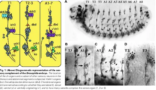
Parasegmental Pattern of Sense Organs in Wild Type Embryos The sense organs of the thoracic and abdominal segments are shown diagrammatically in Fig. 1, after 22C10 immuno-staining for sensory neurons in Fig. 2 A,B and after anti-fishhook immuno-staining for chordotonal (ch) organ scolopale cells in Fig. 3 A,B. The prothoracic (T1) and mesothoracic (T2) segments show minor differ-ences. T2 and T3 are identical. Abdominal segments A1-7 also have an identical pattern of sense organs that is markedly different to the thoracic segments. Chordotonal (ch) organs are composed of from 1
to 5 sensilla. Each sensillum is a clonal unit (i.e. the cells are derived from a common precursor) comprised of 5 cells, a bipolar neuron, an enwrapping scolopale cell, a ligament cell, a cap cell, and an ectodermal cell (Brewster and Bodmer, 1995; Orgogozo et al., 2001). One pole of the bipolar neuron gives rise to the axon that extends to the central nervous system (CNS). A single, unbranched, stretch-sensitive dendrite arises at the opposite pole.
Additional sensilla examined include the keilin’s organs, which are external sense organs composed of three cuticular hairs projecting from a common socket. Each keilin’s organ spans parasegmental borders in the thorax. Two anterior hairs sit in the anterior compartment of a parasegment and a posterior hair sits in the posterior compartment of a parasegment (Struhl, 1984). The dorsal bipolar dendrite neuron (dbd) is a multiple dendrite neuron derived from the anterior compartment of T2, T3 and A1-8.
Homeotic Gene Expression Patterns
The expression patterns of the homeotic genes during embryo-genesis have been examined in detail using antibody probes. Expression is strongest in the CNS where the pattern generally matches epidermal expression. The expression regions of the homeotic genes in the wild type are shown in diagrammatic form in Fig. 4. The altered expression patterns in the homeotic mutants are described by various authors (see Merritt and Whitington, this issue) and are taken here as a general guide only because
fine-Fig. 1. (Above) Diagrammatic representation of the sen-sory complement of the Drosophila embryo. The location of the ch organs and a subset of other sensory neurons in the thoracic and abdominal segments is depicted. Keilin’s organs (ko). Dorsal bipolar dendrite neuron (dbd). Chordotonal organs (ch) are named according to whether they are lateral (l), dorsal
(d), ventral (v) or ventrally originating (v’), and to how many sensilla comprise the sense organ (1, 3 or 5).
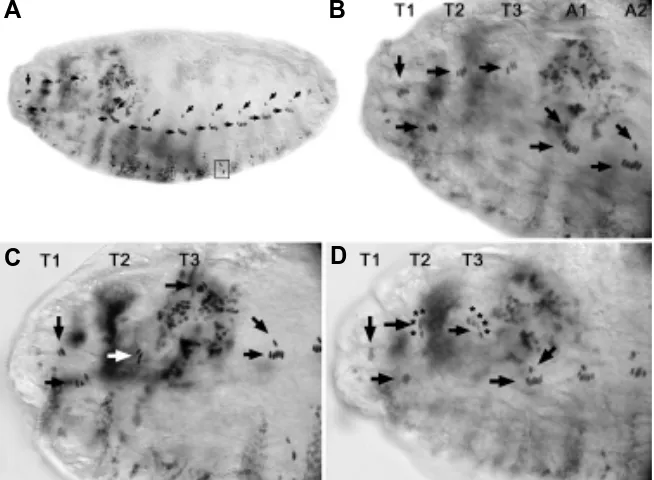
Fig. 2. (Right) Sense organs of wild type, bxd and abx embryos stained with antibody 22C10.(A) Wild type larva showing the full complement of sensory neurons on one side of the body. In this and all other figures, anterior is to the left and dorsal is up. (B) Segments T3 and A1 of a wild type larva showing the dch3 of T3 (oblique arrow) and lch5 of A1 (horizontal arrow). The dbd neurons are indicated with oblique arrowheads. (C) Segments A1 and A2 of a bxd mutant embryo. Typically in bxd embryos vchA/B (arrow)) and v’ch1 (arrowhead) are missing in A1. (D) Segments T2 and T3 of an abx mutant embryo showing an additional ch sensillum comprising dch3 in T3. T2 dch3 is normal. Individual ch sensilla are indicated with asterisks.
A
scale temporal and spatial regulation of homeotic gene expression within parasegments does occur (Duncan, 1996).
abd-A
Loss of abd-A activity transforms lch5 of seg-ments A1-7 into dch3 (Karch et al., 1990; Heuer and Kaufman, 1992). A keilin’s organ mono-hair is present in the ventral region of each abdominal segment (Sanchez-Herrero et al., 1985). V’ch1, vchA and vchB are present in the abdominal segments (Heuer and Kaufman, 1992). We have confirmed these observations using the null mu-tation, abd-AMX1 (data not shown, summarized in
Fig. 4B). The phenotype is consistent with trans-formation of the abdominal segments PS 7-12 to reiteration of T3p/A1a (PS 6) (Sanchez-Herrero et al., 1985; Heuer and Kaufmann, 1992; Kurant et al., 1998). T3p gives rise to dch3 (Hartenstein 1987) and the keilin’s organ mono-hair, thus the thorax-like dch3 is present in PS7-12.
Ubx
The Ubx null mutation affects the sense organs of segments T2, T3 and A1. The dch3 organs of T2 and T3 become transformed to the likeness of lch3 (Subramaniam et al., 1994; Heuer and Kaufman, 1992; this study). V’ch1, vchA and vchB are missing from A1, although they are present in A2-7. Vch1, usually present in the thoracic
seg-Fig. 3. Chordotonal organ phenotypes of Antp embryos.(A) Wild type embryo stained with anti-fishhook. The antibody binds to the scolopale cells of ch organs. Ch organs of the lch3/dch3/ lch5 class of serial homologs are indicated with horizontal arrow in each segment. Dch3 of T1 is indicated with a vertical arrow. V’ch1 of the abdominal segments is indicated with an oblique arrow. (B) T1-3 and A1-2 of a wild type embryo. (C,D) Antp mutant embryos showing tranformation of the T2 dch3 to an lch3 (white arrow in panel C) and transformation of dch3 of T2 and T3 to dch4 (individual sensilla indicated with asterisks).
embryos the stripe of strong Ubx expression in anterior PS5 is lost (White and Wilcox, 1985a). In the PNS a transformation of the dch3 in T2 to a T1-like lch3 was expected, however homozygous abx1 mutant embryos showed no phenotype in the PNS. When
abx1 was placed over the deficiency Df(3R)Ubx109 an additional
ch sensillum was added to the dch3 organ in either T2 or T3. 57% had an extra sensillum incorporated into the dch3 in T2 while 43% had an extra in T3 (Fig. 2D). Examination of cuticle showed no defects in the keilin’s organs. Orientation of the ch organs was normal.
bx and pbx In bx adults, PS5 is partially transformed to PS4, in particular the T3a compartment becomes transformed to T2a (Peifer and Bender, 1986), correlating with a reduction of Ubx expression in T3a. In adults, pbx mutations are reported to transform T3p to T2p (Bender et al., 1983). No change is expected in the sensory comple-ment of either bx or pbx embryos because the sense organs of the posterior compartments of T2 and T3 are identical. Both 22C10 staining and observations under DIC optics showed no changes in the PNS of embryos.
Antp
Antp is normally expressed in PS4 to 12 and is most strongly expressed in PS4 (Carroll et al., 1986). Investigations of epider-mal phenotypes indicate that loss of function of Antp produces a transformation of T2/T3 to a T1 identity however the transforma-tion is not clear-cut (Wakimoto and Kaufman, 1981). The PNS of embryos indicated a possible transformation of T2 and T3 toward a more anterior head segment identity, based on a general ments only, is seen in A1. Keilin’s organ twin hairs are present in
A1. The sensory complement of A2-7 is normal (data not shown, summarized in Fig. 4C).
The sense organ phenotype is in accord with a transformation of PS5 and 6 to PS4 identity. On a compartmental basis, the phenotype corresponds with transformation of T2 and T3 to T2a/ T1p identity, and A1 to a T2a/A1p identity.
Ubx Subfunction Alleles
The subfunction alleles of Ubx have been shown by White and Wilcox (1985a,b) to alter the distribution of the Ubx gene product in subregions of its normal expression. The four subfunction alleles that cover the Ubx region are bx, abx, bxd, and pbx. Mutants are viable and their segmental transformations have been recorded in adults. The effect of the mutations on the embryonic PNS has not been investigated in detail.
bxd55i In adults bxd causes transformation of PS6 toward PS5
(Duncan, 1987). In larvae the mutation is reported to affect the sense organ complement of A1 (Lewis, 1978). Homozygous bxd embryos stained with 22C10 show a loss of vchA, vchB and v’ch1 from A1a (Fig. 2C). An ectopic vch1, usually characteristic of T1a, T2a and T3a, is present in A1a (Fig. 4D). There is no keilin’s organ twin-hair in A1 of the mutant. The sensory complement of T3p is normal. The sense organ phenotype is consistent with a transfor-mation of PS6 (T3p/A1a) to a PS5 identity.
abx 1 abx homozygotes have been shown in adult flies to cause
a transformation of PS 5 to a PS 4 identity (Duncan, 1987). In
A
B
reduction in the number of sense organs and presence of ectopic ch organs in T2 and T3 (Heuer and Kaufman, 1992).
A re-examination of Antp embryos using 22C10 immuno-staining for neurons and dendrites showed that the dch3 organs in T1 and T2 were frequently misoriented or malformed, in T2, T3 or both (67% of embryos). Anti-fishhook immuno-staining for scolopale cell nuclei (Ma et al., 1998; D. Merritt, personal observations) showed that the dch3 of T2 is transformed to an lch3 in 54% of embryos (Fig. 3C). In 8% of embryos the dch3 in both T2 and T3
had an additional sensillum, becoming trans-formed to dch4 (Fig. 3D). In 78% of embryos the axonal projection of lch3 in T1 is abnormal, running into the T1 segmental nerve rather than T2, also shown by Heuer and Kaufman (1992). The pattern of the remaining sense organs, especially in the lateral and dorsal clusters of T2 and T3 is abnormal, frequently showing loss of many sense organs. The dbd sense organ, normally present in T2, T3 and A1-7 (Fig. 5A), was missing from T2 in 50% of embryos (Fig. 5B).
Scr
Scr is normally expressed in PS 2 and 3. Investigations of Scr mutants indicate that T1 is transformed to a more posterior thoracic iden-tity (Wakimoto and Kaufman, 1981; Pattatucci et al., 1991). Heuer and Kaufman (1992) inves-tigated the effects of Scr loss of function on the sense organs and found that lch3 of T1 (derived from the PS 4 component of the segment) was transformed to dch3. No effect upon the pre-existing dch3 of T1 (derived from the anterior compartment) was mentioned. We reexamined the defects in Scr embryos using anti-fishhook staining and found that dch3 is frequently in an abnormal position with fewer than three compo-nent sensilla (50%) and that lch3 (derived from the posterior compartment of T1) is sometimes missing (14%) (Fig. 5D). The dbd neurons show their normal distribution in T2, T3 and the ab-dominal segments (Fig. 5C).
Discussion
The subset of ch organs, lch3 (T1), dch3 (T2/ 3), lch5 (A1-7) and lch3/lch1 (A8) show a num-ber of features indicating that they are serially homologous. First, they are among the few sense organs that arise in posterior compart-ments (Hartenstein, 1987). Second, the precur-sor cells for this subset of ch organs, C1-3, originate in a similar location in each segment (Salzberg et al., 1994; Younossi-Hartenstein and Hartenstein, 1997; Rusten et al., 2001). The genetic interactions that give rise to the different numbers and orientation of sensilla comprising lch3 and lch5 are also relevant. In the thorax, C1-3 each divide to give rise to a neuron, a cap cell, ligament cell, sheath cell and an accessory cell (Brewster and Bodmer, 1996). In the abdominal segments, C3 recruits two secondary precursors from the adjacent ectoderm via epidermal growth factor receptor (EGFR) signaling (Elstob et al., 2001). All five precursors divide to form sensilla. In the abdominal segments only, precursor C1 also recruits a cluster of cells from the ectoderm that will differentiate to become oenocytes, a type of secretory cell (Elstob et al., 2001; Gould et al., 2001). A suite of genes is involved in the EGFR pathway, including argos, pointed,
A
B
C
D
E
F
G
spitz, rhomboid and the proneural gene, atonal (reviewed in Gould et al., 2001).
The different position and orientation of the lch5 and dch3 organs is attributed to migration and rotation of the developing sense organs, most likely due to ventral-ward migration of the basal attachment point, the ligament cells (Hartenstein, 1987). The apical cap cells of the lch3, dch3 and lch5 remain attached to the epidermis where they originate at similar dorso-ventral positions in the abdomen and thorax (Matthews et al., 1990). A number of mutations affecting the ventral migration of lch5 have been de-scribed (Salzberg et al., 1994; Kurant et al., 1998).
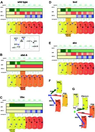
The homeotic genes are the obvious candidates for regulation of the segmental differences in the orientation, position, and numbers of chordotonal organs. First, considering lch3 (T1), dch3 (T2/3) and lch5 (A1-7) (Fig. 4), the working model is: (1) that the ground state for this class of sense organs (lch3, normally found in PS4) occurs in the absence of both Ubx and abd-A expression, (2) that Ubx expression results in the formation of dch3 (Heuer and Kaufman, 1992), normally found in PS5 and 6, and (3) that abd-A expression results in the formation of lch5 (Karch et al., 1990), normally found in PS7-12. The model, summarized in Fig. 4F, is supported by the phenotypes of homeotic mutants. In Ubx mutants, Antp expression is up-regulated throughout PS5 and 6 resulting in the formation of lch3 (Fig. 4C). Loss of abd-A function results in up-regulation of Ubx in PS7-12 resulting in the formation of dch3 organs in the posterior compartments of these segments (Fig. 4B). The abx mutation frequently produces dch4 in T2 and T3. This transformation is not consistent with a simple homeotic transformation and could be interpreted as a partial posteriorward transformation.
While Antp is the predominant homeotic gene expressed in the ground state PS4, it does not appear to play a major role in the formation of this class of sense organ. Loss of Antp function affects the sense organs in PS4 and 5 however there is no highly penetrant homeotic transformation, rather in some cases a sensillum is added to the dch3 so that it is comprised of 4 sensilla, or in some cases there is partial rotation of the dch3 to take on some features of lch3, both features indicative of a posteriorward transformation. Antp is not required for formation of the lch5 because these organs are normal in Antp mutants. Scr mutants show various degrees of disruption of the normal segmental pattern in PS4 and 5. As pointed out by Heuer and Kaufman (1992) Scr and Antp may together play a role in the formation of the ground state lch3. A precise determi-nation requires an examidetermi-nation of double mutants for Scr and Antp. Oenocytes form only in the abdominal segments (Gould et al., 2001) and their formation is linked with the formation of the dch3/ lch3/lch5 class of sense organs because oenocytes are induced via signaling from C1 (Gould et al., 2001). EGFR-mediated recruitment of oenocytes or additional ch precursors does not appear to take place in the thoracic segments (zur Lage et al., 1997) so it would not be surprising if homeotic genes played a role in EGFR-mediated recruitment of oenocytes as well as recruit-ment of secondary ch precursors. Gould et al. (2001) cite prelimi-nary evidence that the bithorax complex is involved in oenocyte formation because the cells are absent in mutants lacking Ubx, abd-A and Abd-B. We propose that, more specifically, oenocyte formation would be under control of abd-A because abd-A is required for formation of lch5. However recruitment of vchA from C5 (see below) occurs in the absence of abd-A expression so Ubx may play a role as well (Fig. 4G).
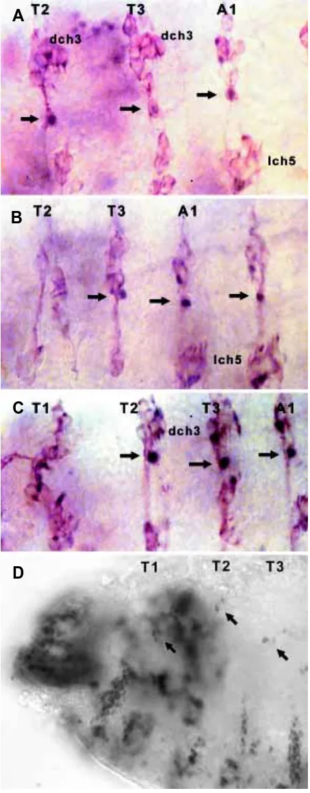
Fig. 5. Location of the dbd sense organ in wild type, Scr and Antp embryos. (A) Wild type embryo stained with 22C10 (brown) and with pdm1 (blue/black) to highlight the dbd neuron. The dbd neuron (arrow) is normally present in T2-3 and A1-8. (B) In this Antp embryo dbd is missing from T2. (C) An Scr embryo shows dbd is present in T2, T3 and A1. (D) Scr mutant embryo stained with anti fishhook has a dch3 in each of T1, T2 and T3. The lch3 is missing from T1.
A
B
C
The remaining sets of ch organs arise from the anterior compart-ments. The vch1 organ found in each thoracic segment is likely to be the serial homolog of vchB. As mentioned above, the three precursors from the posterior compartment (C1-3) give rise to lch3, dch3 and lch5. Two additional precursors (C4, 5) arise in the anterior compartment and give rise to v’ch1 (C4) and vchB (C5) in the abdomen (Okabe and Okano, 1997; zur Lage et al., 1997). The vchA precursor is then recruited from adjacent ectoderm by EGFR signaling from C5, in the same way that C3 recruits additional precursors. While the thoracic segments have not been investi-gated in detail it appears that recruitment does not operate in PS3-5 (zur Lage et al., 1997). The CPS3-5 precursor most likely gives rise to vch1 in T1-3 and does not recruit a secondary precursor, hence the thorax has no serial homolog of vchA. The homology, if any, between lch3 of T1 and the anterior compartment-derived ch of the abdomen remains unknown.
VchA and vchB form in parasegments where either Ubx or abd-A is expressed and vch1 forms where abd-Antp or Scr is expressed (Fig. 4G). Once again the ground state appears to occur in PS4. Ubx and abd-A are able to substitute for each other because vchA and vchB form normally in PS6-12 of the abd-A mutant and PS7-12 of the Ubx mutant. Abdominal segment 1 of Ubx mutants contains a vch1 instead of vchA/B due to the absence of expression of both Ubx and abd-A in this compartment. The bxd mutation shows the same phenotype as Ubx in the anterior compartment of PS6.
Transformations in the PNS of homeotic mutants of Drosophila are consistent with homeotic regulation of sense organ type. Multiple genetic interactions including proneural gene expression, precursor selection, precursor recruitment, lineage determination, and migration are involved in specifying the differences in the ch organs between segments. Precisely which processes come un-der homeotic control is not yet certain, although a regulation of the recruitment process could explain a number of the differences between the abdomen and thorax (Gould et al., 2001).
The relatively few studies of ch organ distribution outside of Drosophila indicate that sense organs considered here are strikingly conserved across distantly related insects suggesting that the chordotonal organs may be fundamental elements of a ground plan sensory system common to all insects. The grasshopper embryo has a set of ch organs in T2 and T3 that are highly likely to be homologs of Drosophila dch3, while the sternal and pleural ch organs of the grasshopper abdomen are likely homologs of the lch5 and vchA/B of Drosophila (Meier et al., 1991). In the late stage embryo of the moth Manduca sexta the distribution of ch organs in the abdominal segments shows a striking similarity to Drosophila, with a pair of ventral ch, a single lch and a parallel cluster of 4 ch in the lateral region (Grueber and Truman, 1999) which positionally correspond to Droso-phila vchA and B, v’ch1 and lch5, respectively. It is interesting that the lch is composed of four sensilla in M. sexta compared with the five of Drosophila. Adult moths preserve a three sensillum dch in the thoracic segments, except noctuoid moths, which have evolved a hearing organ around the sense organ and reduced the number of associated sensilla (Hasenfuss, 1997). As in M. sexta larvae, the lch5 homolog in the first abdominal segment is composed of four sensilla (Hasenfuss, 1997). The wealth of knowledge concerning the devel-opment of the Drosophila sensory nervous system and the role of homeotic genes in the regulation of segmental identity holds promise that a parallel investigation of sense organ development and homeotic genes in other arthropods could contribute greatly to a phylogenetic perspective on nervous system development.
Materials and Methods
Fly Stocks and Mutant Lines
Two classes of mutant embryos were analyzed, the null mutants and the sub-function allele mutants. Mutant lines include the following: Antp 25 red1 e1/ TM3, Sb1 (Antp), Scr4 red1 e1/ TM6B, Tb1 (Scr), Ubx bxd-55i /TM1 (bxd 55i ), T(2;3) abd-A MX1, mwh 1, jv 1, st 1, red 1, Sbsbd-2, abd-A MX1 (abd-A MX1), Df(3R) Ubx 109, Sb1/ Dp (3;3) P5 (Df(3R) Ubx 109), Ubx abx-1 /TM1 (abx 1) and Sb 1/ 1n (3LR) Ubx 101 (Ubx 101 ). Lines Df(3R) Ubx 109 and abx 1 were crossed to obtain hemizygous abx 1 embryos.
Immunostaining
Embryos were dechorionated in 50% household bleach (sodium hy-pochlorite), rinsed with double distilled water, and fixed in a 1:1 heptane:fixative (3.7% formaldehyde) solution. Embryos were devitellinized in a 1:1 solution of heptane and methanol. The MAb22C10 (Developmental Studies Hybridoma Bank, Iowa City, USA) antibody was used at a concen-tration of 1:7. The antibody, anti-pdm-1, used at a concenconcen-tration of 1:100 and anti-fishhook at a concentration of 1:100 (Ma et al., 1998). For a brown staining antibody reaction, a 1:1 solution of 100 µl 3 mg/ml diaminobenzidine (DAB) in 100 µl 1% PBS was used in the final step. For a blue staining antibody reaction 0.2% nickel chloride was added to the DAB solution. The staining reaction was developed by the addition of 1 µl of 30% hydrogen peroxide. A Zeiss Axioskop microscope with Nomarski optics equipped with a Dage DC330 3 CCD camera (Dage-MTI, Michigan City, USA) was used to image embryos and larvae. A series of focal planes were projected onto a single plane using the program Adobe Photoshop (Adobe Systems Inc., San Jose, USA).
Acknowledgements
We thank the Bloomington Stock Centre and E. Lewis for flies. The work was supported by grants from the Human Frontier Science Program Organization and the Australian Research Council.
References
BENDER, W., AKAM, M.E., KARCH, F., BEACHY, P.A., PEIFER, M., SPIERER, P., LEWIS, E.B. and HOGNESS, D.S. (1983). Molecular genetics of the Bithorax complex in Drosophila melanogaster. Science 221: 23-29.
BREWSTER, R. and BODMER, R. (1995). Origin and specification of type II sensory neurons in Drosophila. Development 121: 2923-2936.
BREWSTER, R. and BODMER, R. (1996). Cell lineage analysis of the Drosophila peripheral nervous system. Dev. Genet. 18: 50-63.
CARROLL, S.B., LAYMAN, R.A., MCCUTCHEON, M.A., RILEY, P.D. and SCOTT, M.P. (1986). The Localization and regulation of Antennapedia protein expression in Drosophila embryos. Cell 47: 113-122.
DUNCAN, I. (1987). The Bithorax complex. A. Rev. Genet. 21: 285-319.
DUNCAN, I. (1996). How do single homeotic genes control multiple segment identi-ties? Bioessays 18(2): 91-94.
ELSTOB, P.R., BRODU, V. and GOULD, A.P. (2001). spalt-dependent switching between two cell fates that are induced by the Drosophila EGF receptor. Devel-opment 128: 723-32.
GOULD A.P., ELSTOB, P.R. and BRODU, V. (2001). Insect oenocytes: a model system for studying cell-fate specification by Hox genes. J. Anat. 199: 25-33.
GRUEBER, W.B. and TRUMAN, J. W. (1999). Development and organization of a nitric-oxide-sensitive peripheral neural plexus in larvae of the moth, Manduca sexta. J. Comp. Neurol. 404: 127-141.
HARTENSTEIN, V. (1987). The influence of segmental compartmentalisation on the development of the larval peripheral nervous system in Drosophila melanogaster. Roux’s Arch. Dev. Biol. 196: 101-112.
HASENFUSS, I. (1997). Precursor structures and evolution of tympanal organs in Lepidoptera (Insecta, Pterygota). Zoomorphology 117(3): 155-164.
KARCH, F., BENDER, W. and WEIFFENBACH, B. (1990). abdA expression in Drosophila embryos. Gene Develop 4: 1573-1587.
KURANT, E., PAI, C.Y., SHARF, R., HALACHMI, N., SUN, Y.H. and SALZBERG, A. (1998). Dorsotonals/homothorax, the Drosophila homologue of meis1, interacts with extradenticle in patterning of the embryonic PNS. Development 125: 1037-48.
LEWIS, E.B. (1978). A gene complex controlling segmentation in Drosophila. Nature 276: 565-570.
MA, Y., NIEMITZ, E.L., NAMBU, P.A., SHAN, X.L., SACKERSON, C., FUJIOKA, M., GOTO, T. and NAMBU, J.R. (1998). Gene regulatory functions of Drosophila fish-hook, a high mobility group domain sox protein. Mech. Dev. 73: 169-182.
MCGINNIS, W. and KRUMLAUF, R. (1992). Homeobox genes and axial patterning. Cell 68: 283-302.
MARTINEZ ARIAS, A. and LAWRENCE, P.A. (1985). Parasegments and compart-ments in the Drosophila embryo. Nature 313:639-642.
MATTHEWS, K.A., MILLER, D.F.B. and KAUFMAN, T.C. (1990). Functional implica-tions of the unusual spatial distribution of a minor α-tubulin isotype in Drosophila: a common thread among chordotonal ligaments, developing muscle, and testis cyst cells. Dev. Biol.137: 171-183.
MEIER, T., CHABAUD, F. and REICHERT, H. (1991). Homologous patterns in the embryonic development of the peripheral nervous system in the grasshopper Schistocerca gregaria and the fly Drosophila melanogaster. Development 112: 241-253.
OKABE, M. and OKANO, H. (1997). Two-step induction of chordotonal organ precur-sors in Drosophila embryogenesis. Development 124: 1045-1053.
ORGOGOZO, V., SCHWEISGUTH, F. and BELLAICHE, Y. (2001). Lineage, cell polarity and inscuteable function in the peripheral nervous system of the Drosophila embryo. Development 128(5): 631-43.
PATTATUCCI, A.M., OTTESON, D.C. and KAUFMAN, T.C. (1991). A functional and structural analysis of the Sex combs reduced locus of Drosophila melanogaster. Genetics 129: 423-41.
PEIFER, M. and BENDER, W. (1986). The anterobithorax and bithorax mutations of the Bithorax complex. EMBO J. 5: 2293-2303.
RUSTEN, T.E., CANTERA, R., URBAN, J., TECHNAU, G., KAFATOS, F.C. and BARRIO, R. (2001). Spalt modifies EGFR-mediated induction of chordotonal precursors in the embryonic PNS of Drosophila promoting the development of oenocytes. Development 128(5): 711-22.
SALZBERG, A., DEVELYN, D., SCHULZE, K.L., LEE, J.K., STRUMPF, D., TSAI, L. and BELLEN, H.J. (1994). Mutations affecting the pattern of the PNS in Drosophila reveal novel aspects of neuronal development. Neuron 13: 269-287.
SANCHEZ-HERRERO, E., VERNOS, I., MARCO, R. and MORATA, G. (1985). Genetic organization of Drosophila bithorax complex. Nature 313: 108-113.
STRUHL, G. (1984). Splitting the bithorax complex of Drosophila. Nature 308: 454-457.
SUBRAMANIAM, V., BOMZE, H.M. and LOPEZ, A.J. (1994). Functional differ-ences between Ultrabithorax protein isoforms in Drosophila melanogaster: evidence from elimination, substitution and ectopic expression of specific isoforms. Genetics 136: 979-991.
WAKIMOTO, B.T. and KAUFMAN, T.C. (1981). Analysis of larval segmentation in lethal genotypes associated with the Antennapedia gene complex in Drosophila melanogaster. Dev. Biol. 81: 51-64.
WHITE, R.A.H. and WILCOX, M. (1985a). Distribution of Ultrabithorax proteins in Drosophila. EMBO J. 4(8): 2035-2044.
WHITE, R.A. H. and M. WILCOX (1985b). Regulation of the distribution of Ultrabithorax proteins in Drosophila. Nature 318: 563-567.
YOUNOSSI-HARTENSTEIN, A. and HARTENSTEIN, V. (1997). Pattern, time of birth, and morphogenesis of sensillum progenitors in Drosophila. Microsc. Res. Tech. 39:479-491.