OncoTargets and Therapy
Dove
press
O r i g i n a l r e s e a r c h
open access to scientific and medical research
Open access Full Text article
alteration of asic1 expression in clear cell renal
cell carcinoma
Yan li1
guoxiong Xu2 Kai huang1 Jun Wang3 Jihong Zhang2 Jikai liu1 Zhanyu Wang1 gang chen1
1Department of Urology, 2central
laboratory, Jinshan hospital, Fudan University, 3Department of Urology,
shanghai First People’s hospital, Medical college of shanghai Jiao Tong University, shanghai, People’s republic of china
Background: Acidic extracellular pH is a major feature of tumor tissue. Acid-sensing ion channels (ASICs) represent an H+-gated subgroup of the degenerin/epithelial Na+ channel
family and are activated by acidic microenvironment. Little is known about the expression and clinical significance of ASICs in solid tumors. The purpose of this study was to examine the expression of ASIC1 in human clear cell renal cell carcinoma (CCRCC) and to determine if the expression of ASIC1 is associated with clinicopathological features.
Methods: The expression of ASIC1 in CCRCC tissues at the mRNA and protein levels was determined by real-time quantitative polymerase chain reaction and Western blot analysis, respectively. A tissue microarray was used to assess the expression of ASIC1 protein in tumor tissue and matched adjacent normal tissues from 75 patients with CCRCC.
Results: ASIC1 expression was detected in normal renal and CCRCC samples. The expressions of ASIC1 protein and mRNA were significantly decreased in the CCRCC tissues compared with matched normal renal tissues (P,0.05). The staining density measurement showed that the expression of ASIC1 was significantly decreased in stage I (P=0.037), stage II (P=0.026), and stage III (P=0.026), grades I–II CCRCC (P=0.004), and CCRCC from male patients (P=0.00002). However, no significant difference was observed for ASIC1 expression between CCRCC and normal tissue in patients with stage IV CCRCC (P=0.236), patients with grades III–IV CCRCC (P=0.314), and female patients (P=0.095). Spearman correlations demonstrated that ASIC1 expression did not correlate to tumor stage (correlation coefficient [CC =0.168], P=0.149) and the age of patients (CC −0.147, P=0.688) but showed a positive correlation to higher tumor grades (CC =0.270, P=0.018).
Conclusion: ASIC1 is downregulated in CCRCC. ASIC1 expression may be potentially used as a novel biomarker and even a CCRCC therapeutic target. Further efforts will be made to clarify the mechanism of ASIC1 in occurrence, progression, and metastasis of CCRCC.
Keywords: ASIC1, biomarkers, clear cell renal cell carcinoma
Introduction
Renal cell carcinoma (RCC) constitutes approximately 5% of newly diagnosed male patients with cancer and 3% of newly diagnosed female patients with cancer.1 It was
estimated that 61,560 people would be diagnosed with RCC and 14,080 would die of RCC in the United States in 2015.1 Despite early diagnosis, more than 30% of patients
are diagnosed with metastatic disease. Patients with locally advanced renal tumors have a high risk of relapse with 5-year disease-free survival of 60%, a median time to relapse of 6.8 years, and a probability of developing metastases of 20%–30%.2 Clear
cell renal cell carcinoma (CCRCC) represents the major proportion in RCC. Over the past decade, significant progress has been made in the development of vascular endothelial growth factor and mammalian target of rapamycin (mTOR) signaling pathway-targeted drugs for patients with RCC. However, despite the advancement
correspondence: gang chen
Department of Urology, Jinshan hospital, Fudan University, 1508 longhang road, shanghai 201500, People’s republic of china
Tel +86 18 9308 19999 email chgan305@163.com
Journal name: OncoTargets and Therapy Article Designation: Original Research Year: 2015
Volume: 8
Running head verso: Li et al
Running head recto: Alteration of ASIC1 expression DOI: http://dx.doi.org/10.2147/OTT.S86927
OncoTargets and Therapy downloaded from https://www.dovepress.com/ by 118.70.13.36 on 26-Aug-2020
For personal use only.
Number of times this article has been viewed
This article was published in the following Dove Press journal: OncoTargets and Therapy
Dovepress
li et al
of these new agents, most patients with advanced RCC progress on therapy, but the prognosis is still poor.3 Many
genetic alterations and many kinds of biomarkers have been observed in recent years.4,5 However, there is still a lack of
efficient biomarkers for RCC. Thus, identifying early-stage diagnostic biomarkers and further understanding the under-lying mechanisms of RCC progression, recurrence, and metastasis is warranted.
Acid-sensing ion channels (ASICs) represent an H+
-gated subgroup of the degenerin/epithelial Na+ channel
family and are activated by extracellular protons.6 There
are four ASIC genes in mammals (ASIC1–ASIC4), which encode at least six distinct ASIC subunits, namely 1a, 1b, 2a, 2b, 3, and 4. ASICs are ubiquitous in the mammalian nervous system and are activated in response to a drop in pH to below 7.0.7 Activation of these channels by changes
in the extracellular pH is associated with a variety of physi-ological and pathphysi-ological processes, such as nociception, mechanosensation, and acidosis-mediated neuronal injury.8
Recent studies reported that ASICs are also expressed in non-neuronal cells and play an important role in their physi-ological and pathphysi-ological functions, including pH homeo-stasis, inflammation, and cellular migration.9–12 Acidic
extracellular pH is a major feature of tumor tissue. It has been proposed that the fast growth of solid tumors can lead to the acidification of the microenvironment and that acid pH can promote local invasive growth and metastasis.13,14
Tumor cells may develop an enhanced acid resistance to survive in the microenvironment, where normal cells will die, and ASICs are thought to be involved in this process.15
Evidence suggests that ASIC1 may play a role in tumor formation and metastasis.16 Knockdown of ASIC1 inhibits
glioblastoma cell migration.17 However, the expression and
function of ASIC1 in solid tumors, particularly in RCC, remains undefined. In this study, we examined the expres-sion of ASIC1 in human CCRCC tissues compared with their normal counterparts and the association of ASIC1 expression with clinicopathological features.
Materials and methods
Patients and tumor samples
The clinical and pathological data of patients who were diagnosed with CCRCC and underwent surgery at the Department of Urology of Jinshan Hospital, Fudan Uni-versity, People’s Republic of China, from 2013 to 2014 were collected. The study group consisted of eight patients (female: five patients; male: three patients; Stage I: three patients; Stage II: four patients; Stage III: one patient)
whose original pathological specimens were available for Western blot and quantitative polymerase chain reaction (qPCR). Paired CCRCC and adjacent non-tumor tissues were obtained, snap-frozen in liquid nitrogen, and kept at −80°C until used. The tissue microarrays were obtained from Shanghai Outdo Biotech, Shanghai, People’s Republic of China (HKid-CRCC150CS-02). Patients were staged according to the tumor-node-metastasis criteria proposed by the American Joint Committee on Cancer (stage I: T1 N0 M0; stage II: T2 N0 M0; stage III: T3 or N1 with M0; stage IV: T4 or M1). Tumor grading was performed using the Fuhrman grading scale (1–4).18 Patients were selected on
the basis of the following inclusion and exclusion criteria: (1) definite pathological diagnosis of CCRCC according to the World Health Organization criteria; (2) suitable formalin-fixed, paraffin-embedded tissue; (3) complete clinicopathological data for the patients. The Ethics Com-mittee of Jinshan Hospital, Fudan University, approved the use of tumor specimens.
Western blot analysis
Frozen tumor tissue samples with corresponding adjacent normal tissue were homogenized in SDS lysis buffer with 1% phenylmethanesulfonyl fluoride (PMSF, Beyotime, Haimen, Jiangsu, People’s Republic of China). The protein concentra-tion was assessed using bicinchoninic acid assay (GenBio). A total of 30 μg of protein from the tissues was separated on 10% SDS–PAGE gel and then transferred to polyvinylidene difluoride membrane (Millipore, Billerica, MA, USA). The membrane was blocked with 5% nonfat dry milk in Tris-buffered saline solution with 0.1% Tween-20 for 2 hours at room temperature. Membranes were then incubated with rabbit polyclonal anti-ASIC1 (ASC-014; Alomone Company; 1:200 dilutions) primary antibody in blocking solution at 4°C overnight. After washing with Tris-buffered saline solution with 0.1% Tween-20 for three times, the membrane was incubated with a horseradish peroxidase-conjugated goat anti-rabbit IgG second antibody (1:3,000 dilutions, Boster Bio, Wuhan, Hubei, People’s Republic of China) in blocking solution for 1 hour at room temperature. Loading equivalency was determined using a rabbit anti-human glyceraldehyde 3-phosphate dehydrogenase (GAPDH) antibody (1:2,000 dilution, Weiao Bio, Shanghai, People’s Republic of China). Immunoreactive bands were visualized by Immobilon Western Chemiluminescent HRP Substrate (Millipore) and semiquan-tified using Tanon-4500 Gel Imaging System with GIS ID Analysis software v4.1.5 (Tanon Science and Technology Co., Ltd., Shanghai, People’s Republic of China).
OncoTargets and Therapy downloaded from https://www.dovepress.com/ by 118.70.13.36 on 26-Aug-2020
Dovepress alteration of asic1 expression
rna extraction and real-time qPcr
RNA isolation and real-time qPCR were performed as previously described.19 Total RNA from tissues was extracted using TRIzol
reagent (Invitrogen, Carlsbad, CA, USA) according to the manu-facturer’s protocol and reversely transcribed into cDNA using the reverse transcription kit (Takara Biotechnology Co., Ltd., Dalian, Liaoning, People’s Republic of China). The primer sequences of
human ASIC1 were 5′-GACTACGCCTACGAGGTCATT-3′
(forward) and 5′-CTTGTCCGCACTGCTCCTT-3′
(reverse). The primer sequences of human GAPDH were
5′-AGAAGGCTGGGGCTCATTTG-3′ (forward) and
5′-AGGGGCCATCCACAGTCTTC-3′ (reverse). The PCR
amplification was performed at 95°C for 15 seconds and 60°C for 1 minute for 40 cycles in a 20 μL reaction system with SYBR Select Master Mix (Invitrogen) using the ABI PRISM 7300 Real-Time PCR system (Applied Biosystems, Foster City, CA, USA). All reactions were done in triplicate and repeated three times. The identity was confirmed by sequencing. The amount of target (ASIC1) normalized to an endogenous control (GAPDH) given by 2ΔΔCt.
immunohistochemistry staining
Immunohistochemistry was performed as reported previously.20
Arrays were deparaffinized in xylene and hydrated in an alco-hol series. After deparaffinization, the arrays were treated with boiling citrate buffer (10 mmol/L; pH 6.0) for 2 minutes for antigen retrieval. The treated slides were rinsed with phosphate buffered saline (PBS) three times for 5 minutes. The endog-enous peroxidase activity was blocked during 5–10 minutes incubation in 3% hydrogen peroxide at room temperature. The slides were then incubated overnight at 4°C with primary antibody against ASIC1 (ASC-014) at a 1:100 dilution in 5% bovine serum albumin solution. After three sequential washes in 0.1% Tween 20 PBS, the slides were then treated with polymer accentuator for 20 minutes. The slides were thoroughly rinsed with 0.1% Tween 20 PBS for 5 minutes followed by horserad-ish peroxidase-labeled goat anti-rabbit IgG (1:150 dilutions, Gene Tech Co., Ltd., Shanghai, People’s Republic of China) in 5% bovine serum albumin solution for 30 minutes. The arrays were then processed with a chromogen using freshly prepared diaminobenzidine solution. Finally, the slides were washed, counterstained with hematoxylin, dehydrated, and evaluated with light microscopy. Negative control slides were incubated with a rabbit IgG isotype antibody. Image analysis was performed using Image pro-plus 6.0. Each slide was carefully examined. The mean staining density was calculated for each sample with the same condition. The staining intensity was scored as: 1, weak; 2, medium; and 3, intense staining. Immunohistochemical results
were independently assessed by two pathologists without the knowledge of patient characteristics.
statistical analysis
Data were expressed as the mean ± standard error of the mean. The difference of ASIC1 expression between RCC tissue and normal tissue was measured by paired t-test. The association between ASIC1 expression and pathological parameters of CCRCC was performed with Spearman’s test. P-values ,0.05 were considered to indicate statistical significance.
Results
asic1 was downregulated in human
ccrcc
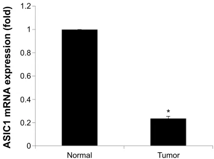
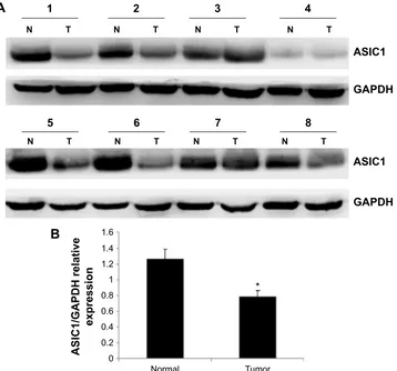
ASIC1 expression in tumor tissue was compared with ASIC1 expression in matched normal tissue samples and normal-ized to the expression of the housekeeping gene GAPDH. The results of qPCR showed that the expression of ASIC1 mRNA was significantly decreased in CCRCC compared with matched normal tissues (Figure 1, n=8, P,0.05). Sub-sequently, we examined the expression of ASIC1 protein in these tissues using Western blot analysis. The results showed a band migrating at 55 kDa for ASIC1. Densitometric analy-sis showed that ASIC1 protein expression was significantly decreased in the CCRCC tissues compared with matched normal tissue (Figure 2, n=8, P,0.05).
Pathological parameters and asic1
expression on ccrcc tissue
To analyze ASIC1 as a valuable diagnostic tissue marker for CCRCC, we performed tissue-microarray based on immu-nohistochemical analysis and performed a statistical analysis
Figure 1 asic1 mrna expression in matched tumor and normal kidney samples.
Notes: The expression level of asic1 mrna was sixfold higher in the normal tissue than that in the tumor tissue. *represents P,0.05 and n=8.
Abbreviation: asic1, acid-sensing ion channel 1.
1RUPDO 7XPRU
$6,&P51$
H[SUHVVLRQIROG
OncoTargets and Therapy downloaded from https://www.dovepress.com/ by 118.70.13.36 on 26-Aug-2020
Dovepress
li et al
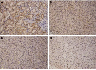
of the immunohistochemical ASIC1 protein expression. CCRCC tissues and matched adjacent normal tissues from 75 patients were included in this study. The clinicopatho-logical characteristics of the study population and the ASIC1 staining intensity in different groups are depicted in Table 1. The mean age of patients was 57.5 (range 34–82). Figure 3 shows representative examples of the expression of ASIC1 in human CCRCC and normal renal tissues. Of the 75 renal carcinomas, 30 cases (40.0%) were weakly positive, 14 cases (18.7%) showed a moderate ASIC1 expression, and 31 cases (41.3%) were strongly positive. Of adjacent normal tissues, 20 cases (26.7%) were weakly positive, 15 cases (20%) showed a moderate ASIC1 expression, and 40 cases (53.3%) were strongly positive. CCRCC tissues showed a downregu-lation of ASIC1 in 68% of the cases. The staining density measurement showed that the expression of ASIC1 was sig-nificantly decreased in stage I (P=0.037), stage II (P=0.026), and stage III (P=0.026), grades I–II CCRCC (P=0.004) and CCRCC from male patients (P=0.0004). However, no
significant difference was observed for ASIC1 expression between CCRCC and normal tissue in patients with stage IV CCRCC (P=0.236), patients with grades III–IV CCRCC (P=0.314), and female patients (P=0.095). In Spearman cor-relations, ASIC1 expression did not correlate to tumor stage (CC =0.168, P=0.149) and age of patients (CC =−0.147, P=0.688) but showed a positive correlation to higher tumor grades (CC =0.270, P=0.018).
Discussion
The metabolic pattern of cancer cells associates with an elevated production of lactate, proton accumulation, and a reversed intraextracellular pH gradient, causing a drop in extracellular pH.21 Low pH values have been shown to be
involved in cancer development, migration, invasion, and cancer progression.21,22 ASICs are H+-gated Na+ channel
subfamilies that are mainly expressed in peripheral sensory and central nervous system neurons. They are acid respon-sive and permeable to cations. Recent studies indicated that
Figure 2 (A) asic1 protein expression in matched tumor and normal kidney samples. (B) histogram shows the semiquantitative analyses of the gels from Western blots.
Notes: The expression level of asic1 protein markedly decreased in rcc compared with corresponding adjacent normal tissue. *Denotes P,0.05 and n=8. T represents tumor sample; n represents matched normal sample.
Abbreviations: asic1, acid-sensing ion channel 1; rcc, renal cell carcinoma; gaPDh, glyceraldehyde 3-phosphate dehydrogenase.
1RUPDO 7XPRU
$6,&*$3'+UHODWLYH
H[SUHVVLRQ
%
1 7
1 7
1 7
1 7
1 7
1 7
1 7
1 7
$6,&
*$3'+
$6,&
*$3'+
$
OncoTargets and Therapy downloaded from https://www.dovepress.com/ by 118.70.13.36 on 26-Aug-2020
Dovepress alteration of asic1 expression
ASICs are also expressed in non-neuronal cells. Their role of sensing pH change makes it a potential target in tumor growth and metabolism. Previous study demonstrated that the co-expression of ASIC1 and ASIC2 controls the cation current in grade IV gliomas, and inhibition of this conduc-tance decreases the growth of glioma and cell migration.23
Kapoor et al also showed that knockdown of ASIC1 and ENaC inhibits glioblastoma cell migration.17 Although these
data suggest a functional relevance of regulation of ASICs in the process of cancer cell growth and migration, little is known about the expression and clinical significance of ASICs in solid tumors. In the present study, we report for the
Table 1 Pathological characteristics of patients and the asic1 staining intensity in tissue-microarray
Patient characteristics Number of patients Mean staining intensity P-value CCRCC tissue Normal tissue
Overall 75 0.038±0.002* 0.046±0.003 0.0003
sex
Male 48 0.033±0.003* 0.043±0.003 0.0004
Female 27 0.047±0.003 0.052±0.003 0.095
Fuhrman grade
i–ii 52 (39 M, 13 F) 0.033±0.002* 0.044±0.003 0.00002
ii–iii 23 (11 M, 12 F) 0.050±0.005 0.052±0.005 0.314
Tumor stage
i 29 (18 M, 11 F) 0.031±0.003* 0.037±0.002 0.037
ii 23 (11 M, 12 F) 0.047±0.005* 0.055±0.005 0.026
iii 17 (14 M, 3 F) 0.034±0.005* 0.046±0.005 0.026
iV 6 (5 M, 1 F) 0.052±0.01 0.058±0.01 0.236
Notes: *Significant difference of mean ASIC1 staining intensity between CCRCC tissues and matched normal tissue, P,0.05. F represents female patients; M represents male patients.
Abbreviations: asic1, acid-sensing ion channel 1; ccrcc, clear cell renal cell carcinoma.
Figure 3 asic1 immunohistochemistry.
Notes: (A) normal renal tissue with prominent asic1 expression. (B–D) asic1 expression in ccrcc with high- (B), intermediate- (C), and low- (D) staining density, respectively.
Abbreviations: asic1, acid-sensing ion channel 1; ccrcc, clear cell renal cell carcinoma.
OncoTargets and Therapy downloaded from https://www.dovepress.com/ by 118.70.13.36 on 26-Aug-2020
Dovepress
li et al
first time the expression of ASIC1 in CCRCC at the mRNA and protein levels. Our results also indicated that the expres-sion of ASIC1 is associated with the clinicopathological features of human CCRCC.
Ion channels are major signaling molecules expressed in a wide range of tissues where they have significant involve-ment in determining a variety of cellular functions includ-ing proliferation, solute transport, volume control, enzyme activity, secretion, invasion, gene expression, excitation-contraction coupling, and intercellular communication.24,25
The exact role of ASIC1 in cellular function and behavior is still unclear. Several studies showed that ASIC1 was involved in the acid-induced cell death.26–28 Blockade of ASIC1 by
the blocker amiloride significantly decreased the cell death and inhibited cell apoptosis by regulating Bcl-2 family gene expression and activity of caspase 3/9.29 Silencing ASIC1
by short hairpin RNA not only protected the cell from death but also protected the cell from apoptosis.30 These studies
suggested that ASIC1 may act as a cancer suppressor protein in the acid tumor microenvironment.
In the present study, the molecular weight of 55 kDa does not match the about 70 kDa of ASIC1 expressed in nervous system, suggesting ASIC1 channel responsible for the proton-induced currents are homomeric or heteromeric. ASICs may have a variable composition, and hence, tissue-specific differences in biophysical parameters may result from different channel compositions in different tissues.16
The results of our present study also suggested that ASICs may play different roles in different tissues.
The immunohistochemistry demonstrated lower ASIC1 expression in CCRCC tissues compared with matched nor-mal adjacent renal tissues in nor-male patients, but not in fenor-male patients. Previous studies also found that the expression and function of ASICs showed significant sex differences.31,32
ASIC1 might contribute to the sex difference of incidence and mortality in patients with RCC.
Most types of cancer, including RCC, go undetected and are non-painful in early stage. Chronic or breakthrough pain usually indicates cancer progression and metastasis. Studies have demonstrated that ASICs modulation may contribute to the generation and maintenance of pain.33 Tissue acidosis is an
important feature of the tumor and a direct cause of pain and hyperalgesia. It has been shown that ASICs can detect moder-ate acidifications, mediating acute pain in acidic solution.34
In the present study, we found significant decrease of ASIC1 in early stage and low-grade CCRCC tissues. However, ASIC1 did not decrease in stage IV and grades III–IV CCRCC tissues. The result suggests that ASIC1 may decrease in the
early stage of cancer and make cancer a non-painful disease even in the acidic extracellular microenvironment. However, this hypothesis is new and obviously requires validation.
Conclusion
The current study indicates that ASIC1 is downregulated in CCRCC. These findings suggest that ASIC1 may be potentially used as a novel biomarker and even as a CCRCC therapeutic target. Further efforts will be made to clarify the mechanism of ASIC1 in occurrence, progression, and metastasis of cancers.
Disclosure
The authors report no conflicts of interest in this work.
References
1. Siegel RL, Miller KD, Jemal A. Cancer statistics, 2015. CA Cancer J
Clin. 2015;65(1):5–29.
2. Puente Vázquez J, Alonso Gordoa T, Moreno J, et al. New challenges in kidney cancer management: integration of surgery and novel therapies.
Curr Treat Options Oncol. 2015;16(3):337.
3. Srinivasan R, Ricketts CJ, Sourbier C, Linehan WM. New strategies in renal cell carcinoma: targeting the genetic and metabolic basis of disease. Clin Cancer Res. 2015;21(1):10–17.
4. Sato Y, Yoshizato T, Shiraishi Y, et al. Integrated molecular analysis of clear-cell renal cell carcinoma. Nat Genet. 2013;45(8):860–867. 5. Tun HW, Marlow LA, von Roemeling CA, et al. Pathway signature
and cellular differentiation in clear cell renal cell carcinoma. PLoS One. 2010;5(5):e10696.
6. Wemmie JA, Price MP, Welsh MJ. Acid-sensing ion channels: advances, questions and therapeutic opportunities. Trends Neurosci. 2006;29(10): 578–586.
7. Krishtal O. The ASICs: signaling molecules? Modulators? Trends
Neurosci. 2003;26(9):477–483.
8. Wemmie JA, Taugher RJ, Kreple CJ. Acid-sensing ion channels in pain and disease. Nat Rev Neurosci. 2013;14(7):461–471.
9. Nitta CH, Osmond DA, Herbert LM, et al. Role of ASIC1 in the development of chronic hypoxia-induced pulmonary hypertension.
Am J Physiol Heart Circ Physiol. 2014;306(1):H41–H52.
10. Tong J, Wu WN, Kong X, et al. Acid-sensing ion channels contribute to the effect of acidosis on the function of dendritic cells. J Immunol. 2011;186(6):3686–3692.
11. Uchiyama Y, Cheng CC, Danielson KG, et al. Expression of acid-sensing ion channel 3 (ASIC3) in nucleus pulposus cells of the intervertebral disc is regulated by p75NTR and ERK signaling. J Bone
Miner Res. 2007;22(12):1996–2006.
12. Holzer P. Acid-sensing ion channels in gastrointestinal function.
Neu-ropharmacology. 2015;94:72–79.
13. Parks SK, Chiche J, Pouyssegur J. pH control mechanisms of tumor survival and growth. J Cell Physiol. 2011;226(2):299–308.
14. Estrella V, Chen T, Lloyd M, et al. Acidity generated by the tumor microenvironment drives local invasion. Cancer Res. 2013;73(5): 1524–1535.
15. Damaghi M, Wojtkowiak JW, Gillies RJ. pH sensing and regulation in cancer. Front Physiol. 2013;4:370.
16. Ye JH, Gao J, Wu YN, Hu YJ, Zhang CP, Xu TL. Identification of acid-sensing ion channels in adenoid cystic carcinomas. Biochem Biophys
Res Commun. 2007;355(4):986–992.
17. Kapoor N, Bartoszewski R, Qadri YJ, et al. Knockdown of ASIC1 and epithelial sodium channel subunits inhibits glioblastoma whole cell current and cell migration. J Biol Chem. 2009;284(36):24526–24541.
OncoTargets and Therapy downloaded from https://www.dovepress.com/ by 118.70.13.36 on 26-Aug-2020
OncoTargets and Therapy
Publish your work in this journal
Submit your manuscript here: http://www.dovepress.com/oncotargets-and-therapy-journal
OncoTargets and Therapy is an international, peer-reviewed, open access journal focusing on the pathological basis of all cancers, potential targets for therapy and treatment protocols employed to improve the management of cancer patients. The journal also focuses on the impact of management programs and new therapeutic agents and protocols on
patient perspectives such as quality of life, adherence and satisfaction. The manuscript management system is completely online and includes a very quick and fair peer-review system, which is all easy to use. Visit http://www.dovepress.com/testimonials.php to read real quotes from published authors.
Dovepress
Dove
press
alteration of asic1 expression
18. Fuhrman SA, Lasky LC, Limas C. Prognostic significance of mor-phologic parameters in renal cell carcinoma. Am J Surg Pathol. 1982; 6(7):655–663.
19. Huang K, Chen G, Luo J, Zhang Y, Xu G. Clinicopathological and cellular signature of PAK1 in human bladder cancer. Tumour Biol. 2015;36(4):2359–2368.
20. Li W, Wang X, Li B, Lu J, Chen G. Diagnostic significance of over-expression of Golgi membrane protein 1 in prostate cancer. Urology. 2012;80(4):e951–e957.
21. Cairns RA, Harris IS, Mak TW. Regulation of cancer cell metabolism.
Nat Rev Cancer. 2011;11(2):85–95.
22. Kato Y, Ozawa S, Miyamoto C, et al. Acidic extracellular microenvi-ronment and cancer. Cancer Cell Int. 2013;13(1):89.
23. Berdiev BK, Xia J, McLean LA, et al. Acid-sensing ion channels in malignant gliomas. J Biol Chem. 2003;278(17):15023–15034. 24. Becchetti A. Ion channels and transporters in cancer. 1. Ion channels
and cell proliferation in cancer. Am J Physiol Cell Physiol. 2011;301(2): C255–C265.
25. Fraser SP, Diss JK, Chioni AM, et al. Voltage-gated sodium channel expression and potentiation of human breast cancer metastasis. Clinical
Cancer Res. 2005;11(15):5381–5389.
26. Jetti SK, Swain SM, Majumder S, Chatterjee S, Poornima V, Bera AK. Evaluation of the role of nitric oxide in acid sensing ion channel medi-ated cell death. Nitric Oxide. 2010;22(3):213–219.
27. Hu W, Chen FH, Yuan FL, et al. Blockade of acid-sensing ion channels protects articular chondrocytes from acid-induced apoptotic injury.
Inflamm Res. 2012;61(4):327–335.
28. Yuan FL, Chen FH, Lu WG, et al. Inhibition of acid-sensing ion chan-nels in articular chondrocytes by amiloride attenuates articular cartilage destruction in rats with adjuvant arthritis. Inflamm Res. 2010;59(11): 939–947.
29. Rong C, Chen FH, Jiang S, et al. Inhibition of acid-sensing ion channels by amiloride protects rat articular chondrocytes from acid-induced apoptosis via a mitochondrial-mediated pathway. Cell Biol
Int. 2012;36(7):635–641.
30. Weng XC, Zheng JQ, Jin QE, Ma XY. Inhibition of acid-induced apoptosis by targeting ASIC1a mRNA with short hairpin RNA. Acta
Pharmacol Sin. 2007;28(10):1621–1627.
31. Kobayashi H, Yoshiyama M, Zakoji H, Takeda M, Araki I. Sex differ-ences in the expression profile of acid-sensing ion channels in the mouse urinary bladder: a possible involvement in irritative bladder symptoms.
BJU Int. 2009;104(11):1746–1751.
32. Burnes LA, Kolker SJ, Danielson JF, Walder RY, Sluka KA. Enhanced muscle fatigue occurs in male but not female ASIC3-/-mice. Am J
Physiol Regul Integr Comp Physiol. 2008;294(4):R1347–R1355.
33. Duan B, Wu LJ, Yu YQ, et al. Upregulation of acid-sensing ion channel ASIC1a in spinal dorsal horn neurons contributes to inflammatory pain hypersensitivity. J Neurosci. 2007;27(41):11139–11148.
34. Callejo G, Castellanos A, Castany M, et al. Acid-sensing ion channels detect moderate acidifications to induce ocular pain. Pain. 2015;156(3): 483–495.
OncoTargets and Therapy downloaded from https://www.dovepress.com/ by 118.70.13.36 on 26-Aug-2020