Tissue tectonics and the multi-scale regulation of
developmental timing
Lara Busby* and Benjamin Steventon *#
* Department of Genetics, University of Cambridge,#Corresponding author: [email protected]
Abstract
Development encompasses processes that occur at multiple length-scales, includ-ing gene regulatory interactions, cell movements and reorganisation, cell signallinclud-ing and growth. It is essential that the timing of events in all of these different processes are coordinated to generate well patterned tissues and organs. However, how the timing of intrinsic cell state changes is coordinated with events at the multi-tissue and whole organism level is unknown. Here, we argue that an important mechanism which accounts for integration of timing across levels of organisation is provided by tissue tectonics: i.e. how morphogenetic events driving tissue shape change result in the relative displacement of signalling and responding tissues and coordinate devel-opmental timing across scales. In doing so, tissue tectonics provides a mechanism by which the cell specification events intrinsic to cells can be modulated by the temporal exposure to extracellular signals. This exposure is in turn regulated by higher-order properties of the embryo such as their physical properties, rates of growth and the combination of dynamic cell behaviours impacting tissue morphogenesis. Tissue tec-tonics creates a downward flow of information from higher to lower levels of biological organisation, providing an instance of downward causation in development.
1
Introduction
Time is central to biological phenomena: biological processes are inherently dynamic and
this is true across all biological fields. Developmental biology provides a strong context
to study biological time, as it allows for the study of developmental timing at many
different levels of biological organisation - opening the possibility for the identification
of mechanisms which coordinate these different length scales. Developmental timing can
be thought of in terms of the absolute timing of a given event, the ordering of events
relative to one another, the directionality of developmental processes, and the more general
tempo (speed) at which development proceeds (Duboule, 2003; Ebisuya and Briscoe, 2018;
Johnson and Day, 2000). We will focus specifically in this review on how theabsolute
timingof a given event in development is controlled, and propose a mechanism by which
timing may be coordinated across different levels of organisation in the embryo.
As a cell moves through developmental time, it undergoes a series of cell state
tran-sitions that ultimately define its fate. In considering the mechanisms that regulate the
timing of cell state transitions, a distinction has been made between intrinsic and extrinsic
timing mechanisms: intrinsictimers function within the cell, whilstextrinsicmechanisms
implicate the importance of the external cellular environment in providing inputs to the
timer (Figure 1). Whether a timer is controlled through intrinsic or extrinsic mechanisms
has primarily been investigated using classical experimental embryological methods. For
example, physically grafting cell populations between embryos of different ages
(hete-rochronic grafting) allows any influence of external factors on a timer to be identified. If
the timer of interest progresses as expected from the age of the donor tissue once placed
in this novel environment, it suggests that the functioning of the timer is intrinsic to the
cell population. Conversely, if the timer is accelerated, decelerated, reset or advanced in
the host context, it suggests that external factors which the cell population is exposed
to in this context are important for the normal functioning of the timer and that the
mechanistic basis for the functioning of the timer is extrinsic.
To determine the contribution of extrinsic or intrinsic components to timing an event,
an additional experimental embryological approach involves removing groups of cells from
the embryonic environment and culturing them in a neutral environment (explant culture).
If the timer is able to progress outside of the embryo, it may provide evidence that its
underlying mechanisms act cell-intrinsically. Note that these experiments are very similar
to those used to investigate cell specification and determination. A cell may be defined as
specified to form a particular structure if, when isolated from the embryo and placed in a
(Slack, 1991). A cell is determined when it gives rise to this structure in any context,
including any embryonic context (Slack, 1991). This differs somewhat to the employment
of these assays in the field of timing, but the assays are nonetheless fundamentally the
same and highlight the importance of understanding the mechanisms by which intrinsic
timers are modulated by the local signalling environment that they encounter during
development.
Figure 1: Intrinsic and extrinsic timers. Schematic summarising the distinction between intrinsic and extrinsic
timers. Each of the ‘blobs’ represents either a cell population or cell, dependent upon context. In an intrinsic timer mechanism, each population (cell) has its own internal timer which is not affected by external information.
In contrast, in an extrinsic timer mechanism, information from the surroundings is integrated by the population (cell) to infer the state of the timer.
The specification of distinct cell types during development is inherently linked to the
timing at which cells receive either the inhibition or activation of extracellular signals.
One example of this is in the patterning of the early ectoderm into epidermis, neural plate
border or neural cell states, and the subsequent patterning of these embryonic territories.
Neural specification requires a continued modulation of FGF, Wnt and BMP pathway
activity from pre-gastrulation stages onwards (Linker and Stern, 2004; Streit et al., 2000;
Tuazon and Mullins, 2015; Tucker et al., 2008), and neural plate border specification and
regionalisation requires a distinct series of temporal exposure to these same pathways
(Streit and Stern, 1999; Steventon et al., 2009; Steventon and Mayor, 2012; Patthey et al.,
2009; Britton et al., 2019). Hence, an important unanswered question in developmental
biology is how the temporal exposure to extracellular signals is regulated during early
development, and how is this linked to alterations in the morphogenetic properties of
tissues as they undergoes shape change and growth. This understanding is essential, as it
likely holds the key to understanding the regulative and self-organising properties of the
early embryo.
The timing at which cells receive external signals to modulate intrinsic timers of cell
and responding populations) become apposed to one another in the embryo, or become
shifted relative to the position of cells releasing secreted modulators of the signalling
pathway activity. Therefore, a key regulator of developmental timing acts at the
multi-tissue level, and is based on the progressive spatial re-positioning of multi-tissues as they alter
in size and shape through morphogenesis. To emphasize the importance of this
higher-level regulation of developmental timing through the spatial displacement of signalling
and responding tissues, we re-introduce the term tissue tectonics. This term has been
introduced elsewhere in relation to the tension and stress forces acting within tissues to
drive morphogenesis (Blanchard et al., 2009). Here, we extend the concept to consider how
it can act as a important regulator of timing in development, and provides a mechanism
by which intrinsic developmental timers can be regulated by morphogenetic events. While
the direct regulation of cellular signalling and gene expression states by mechanochemical
coupling has received increasing attention (Hannezo and Heisenberg, 2019; Naganathan
and Oates, 2017), this review highlights tissue tectonics as an additional mechanism by
which morphogenesis and patterning can be coordinated in development. We propose
that changes to tissue tectonics in evolution, and more generally alterations to the timing
of developmental events (heterochrony), are important for producing new forms.
As a complete coverage of the literature on developmental timing would be beyond the
scope of a single review, we instead provide a series of case studies in which the concept of
developmental timing has been approached at different levels of biological organisation.
We will first present a series of studies in which the relative contributions of intrinsic and
extrinsic timers to temporal control have been elucidated. Timers act at different rates
(tempos) in different species, so we will follow this with a discussion of recent studies
that have investigated the molecular basis for species-specific developmental tempos. We
will then briefly discuss how alterations in the timing of one developmental process over
another can act as a mechanism for evolutionary change in reference to a fundamental
concept in evolutionary developmental biology: heterochrony. Finally, we will review
some recent work that demonstrates the importance of tissue tectonics in coordinating
multi-tissue morphogenetic events with the timing of cell fate decisions and the emergence
2
Intrinsic and extrinsic timers in development
2.1 Single cell intrinsic timers
As as starting point to consider how developmental timing is regulated during embryonic
development, we will first consider some examples where the intrinsic capability of
indi-vidual cells has been demonstrated experimentally. The concept of cell intrinsic timers
is inherently linked to the concept of competence in development, i.e. how cells change
in their ability to respond to a given inductive cue over developmental time. Induction
may be defined as the process in which an inducing tissue releases a signal that results
in a change in the direction of differentiation of a responding tissue (Gurdon, 1987). In
Xenopus animal cap explants, there is a clear delineation in time in the transition
be-tween a competent and a non-competent state for mesoderm induction: the competence
of animal cap tissue to respond to contact with vegetal tissue is lost at the early gastrula
stage (Gurdon et al., 1985). To investigate what determines the timing of competence in
development, experiments have been performed which isolated animal cap ectoderm from
the embryo and showed that even outside of the embryonic environment, competence is
still lost at the same point in time (Grainger and Gurdon, 1989), indicating that
compe-tence loss is intrinsically timed. Further, even when cells are dissociated from the animal
cap, single cells maintain the expected timing of competence: this suggests that timing
acts in this case cell-autonomously (Grainger and Gurdon, 1989). By placing dissociated
single cells in a solid gelatin matrix, the authors inhibited cell division and showed that
loss of competence in this context is independent of cell division (Grainger and Gurdon,
1989). Together, these results implicate a cell-intrinsic timer in animal cap ectoderm cells
that modulates the ability of the cell to respond to induction by vegetal signals.
An additional example of single-cell intrinsically timed developmental events comes
from the Drosophila nervous system. Here, neuroblasts give rise to the precursors of
the nervous system, ganglion mother cells (GMCs). GMCs are produced by
asymmet-ric divisions of a neuroblast that produce a daughter neuroblast and a daughter GMC.
Through the sequential expression by the neuroblast of ‘temporal identity genes’ (
hunch-back, kruppel, pdm1, castor), each of the sequentially generated GMCs has a specific
identity (Isshiki et al., 2001). How are the transitions between each of these gene
ex-pression profiles controlled over time? Experiments that cultured isolated neuroblastsin
vitro showed that gene expression transitions occur in isolated cells outside of the
embry-onic environment, suggesting that this gene expression timer is controlled by cell-intrinsic
mechanisms (Grosskortenhaus et al., 2005). In G2-arrested embryos, the timing and order
transitions is regulated by a mechanism independent of the cell cycle (Grosskortenhaus
et al., 2005). This is also not a simple linear positive transcriptional cascade; mutations
in thehb and Kr genes have little effect on later gene expression of the other temporal
identity genes. More recently, mathematical modelling work has provided insights into
the mechanistic basis for theDrosophila neuroblast temporal identity timer.
Experimen-tal data is consistent with a repressor-decay timer, where the decay of a previous timer
component (e.g. hb) times the onset of expression of a later component (e.g. pdm1),
through the relief of repressive interactions (Averbukh et al., 2018).
A final example of an intrinsic timer that functions in individual cells is given by the
oscillatory component of the segmentation clock. During the production of the embryonic
anteroposterior (head to tail) body axis, paraxial mesoderm on either side of the
mid-line is sequentially segmented into blocks termed somites (somitogenesis). The primary
model for somitogenesis is the Clock and Wavefront Model, in which an anteroposterior
gradient of FGF and Wnt signalling (the wavefront) is combined with a cell-intrinsic
os-cillator based on Notch signalling to segment blocks of tissue (Cooke and Zeeman, 1976;
Pourqui´e, 2011). A set of genes including those that encode transcription factors of the
Hes/ Her family are expressed with oscillatory dynamics in the presomitic mesoderm
(PSM) (Aulehla and Johnson, 1999; Lewis, 2003). Strikingly, when pre-somitic cells are
removed from zebrafish embryos and culturedin vitro, oscillations in Her1 expression are
still observed (Webb et al., 2016). Mathematical modelling was used to demonstrate that
the observed patterns of gene expression are consistent with all of the cells essentially
hav-ing the same oscillatory behaviour captured at different points in their dynamics (Webb et
al., 2016). Thus, in development, individual cells have the capacity to generate oscillatory
gene expression, which must be coordinated across the PSM population through extrinsic
signals (recently reviewed by Oates, 2020). This cell-intrinsic oscillator is important,
to-gether with extrinsic signals (the wavefront) in segmenting blocks of paraxial mesoderm
along the anteroposterior axis.
Together, these three examples provide good evidence for the existence of intrinsic
timers that are able to operate in isolated single cells. Furthermore, they provide examples
of the utility of experimental assays in investigating the intrinsic or extrinsic control of
developmental timers. Together with methods for the improved imaging and analysis
of real-time changes in single cell gene expression, experimental assays such as those
described above are key in enabling questions to be asked relating to the degree to which
single cell intrinsic timers can be coordinated across groups of cells in a given tissue.
We will next consider three case studies in which the balance of intrinsic vs. extrinsic
regulation of developmental timing has been investigated, before considering how these
2.2 Balancing intrinsic and extrinsic timing in avian limb development
Heterochronic grafting has provided good evidence for a number of population-level
in-trinsic timing mechanisms. A first example comes from avian limb development. Here,
cells of the polarising region (or zone of polarizing activity, ZPA) expressSonic Hedgehog
(Shh) for a defined duration between HH20–27 (approximately embryonic days 3.5–5.5)
(Riddle et al., 1993). Sonic Hedgehog (Shh) protein functions in the specification of
an-teroposterior positional values and proliferation of limb bud cells (Towers et al., 2008;
Yang et al., 1997). If the polarising region of a HH20 embryonic wing is grafted in place
of the endogenous polarising region in a HH24 embryo, the donor polarising region
con-tinues to expressShh 32 hours post-graft. At this time point, the host polarising region
in the contralateral wing has downregulated Shh expression and transcripts are not
de-tectable byin situ hybridisation (Chinnaiya et al., 2014). This result suggests that the
mechanisms controlling timing ofShh transcription in this tissue are not dependent upon
extrinsic signalling. Population-level analyses of cell cycle parameters showed that there
are distinct stereotyped changes to the progression of the cell cycle between HH20-30,
including a marked increase in the proportion of cells in G1 (Chinnaiya et al., 2014).
Inhibiting cell division in the limb bud using the drug colchicine leads to an extension in
the period for which Shh is expressed (Chinnaiya et al., 2014). Together, these
exper-iments point to an intrinsic timer for Shh expression in the polarising region, with cell
cycle progression as an important input.
A second cell-population intrinsic timer acting in avian limb development controls the
termination of limb bud outgrowth (Pickering et al., 2018). The limb field is initially
specified as a portion of the lateral plate mesoderm, before outgrowth to form a bud
beginning at HH18. The cells of the limb bud proliferate and drive outgrowth. The
pro-cesses controlling the termination of outgrowth are important for regulating the size and
shape of the limb. The classical model for limb outgrowth termination implicated the
breakdown of a feedback loop between FGF signalling from the apical ectodermal ridge
(AER) and BMP signalling in the underlying mesenchyme (Verheyden and Sun, 2008). A
recent study has produced evidence that a cell intrinsic timer is key in controlling limb bud
outgrowth termination (Pickering et al., 2018). When donor HH29 distal mesenchyme
cells were grafted into a younger (HH20) host limb, grafted tissue had a cell cycle profile
more similar to HH29 control (ungrafted) tissue than to HH24 contralateral tissue
(Pick-ering et al., 2018). Furthermore, expression of genes involved in the TGFß pathway, as
well as functional BMP signalling, was maintained in grafted tissue with a transcriptional
profile similar to HH29 ungrafted tissue (Pickering et al., 2018). The authors argue that
for the termination of limb bud outgrowth.
An example of an extrinsically controlled developmental timer is also found in avian
limb development. The proximodistal axis of the limb comprises three segments : the
stylopod, zeugopod and autopod (from proximal to distal). The most proximal
seg-ments of the wing are specified early in its development (HH18-19) through an extrinsic
mechanism (Saiz-Lopez et al., 2015). This has been demonstrated by grafting of HH20
distal mesenchyme into HH24 limb buds. The tissue-level contribution of the donor cells
in these experiments was indistinguishable from HH24-HH24 homochronic grafts
(Saiz-Lopez et al., 2015). That is, behaviour of the HH20 donor cells is reset by grafting into an
older host environment, causing cell contribution only to structures distal to the autopod,
rather than their normal contribution to the zeugopod. Interestingly, in this system, the
extrinsic timer controlling proximal fate specification is combined with an intrinsic timer
controlling the specification of more distal proximodistal values (Saiz-Lopez et al., 2015).
Taken together, these examples from avian limb development clearly demonstrate the
utility of experimental embryological techniques in investigating the contribution of
cell-intrinsic and extrinsic timers to the regulation of developmental events. Heterochronic
grafting methods allow the external ”time” experienced by a cell population to be
ma-nipulated, and these methods allow insights which would not be possible otherwise. The
avian limb provides a rich example of how cell-population level intrinsic and extrinsic
timers can work together to allow the patterning and morphogenesis of a higher-level
structure in development.
2.3 Balancing intrinsic and extrinsic timing in mammalian neurogenesis
Population-level intrinsic timers have also been described in the development of the
ner-vous system. The mammalian brain is complex and comprises a large repertoire of diverse
cell types. In many cases, neural progenitor cells undergo temporal changes in the types
of daughter cells that they produce, allowing for the production of a diverse set of
daugh-ter cells over time. For example, progenitor cells of the mouse cortex may be maintained
in a culture system, where they express molecular markers with a similar timing to that
observed in the embryo (Gaspard et al., 2008). As differential daughter cell identities are
observed over time in the absence of any external signals, this suggests that the
transcrip-tional progressions observed in normal development are mediated by an intrinsic timer.
These cultured cells are functional: when grafted back into the embryo, their repertoire of
projections closely resembles those found in normal development (Gaspard et al., 2008).
reveal-ing an important role in diverse species for cell-intrinsic timers in the generation of the
nervous system.
The timing of differentiation of cells that make up the nervous system may also
be determined through cell-intrinsic mechanisms. Experiments that removed rat optic
nerve cells from the embryonic environment and cultured them in PDGF demonstrated
that oligodendrocyte precursor cells (OPCs) divide and differentiate with a timing which
closely replicates that observed within the embryo (Temple and Raff, 1986). If single
OPCs are plated in individual microwells, they divide (with a maximum of eight divisions
observed) before differentiating. If separated into individual wells, the daughters of the
same OPC divide the same number of times before differentiating (Shen et al., 2006).
What determines the timing of differentiation in these cells? It appears that the
un-derlying mechanism is cell-intrinsic, given that cells, even in isolation, differentiate with
replicable timing. In contrast to the aforementioned examples from limb development,
however, this mechanism does not appear to be cell-cycle dependent. When OPC cultures
are held at a reduced temperature (33oC as opposed to the standard 37oC), they divide
more slowly. However, the timing of differentiation occurs earlier in these cultures (Gao
et al., 1997). This suggests it is not the number of cell divisions which is important, so
much as the absolute passage of time. Further investigation has led to a model for OPC
differentiation that places gradual changes in the level of several proteins, including p27,
p18 and Id4, at the centre of the intrinsic timer (reviewed by Raff, 2006).
Gradual changes in transcription over time have been implicated in another study
focusing on mammalian cortex development. Here, apical progenitor cells (APCs), which
give rise to cells of the dorsal cortex, were subjected to transcriptional analysis at several
time points in development (Okamoto et al., 2016). Through computational manipulation
of the resulting dataset, the authors were able to distinguish two orthogonal axes in
the clustered dataset – a temporal axis and a differentiation axis. That is, a subset
of genes was identified whose expression changed over time in a manner distinct from
the phenomenon of differentiation. These genes show gradual changes in expression over
developmental time (Okamoto et al., 2016). Manipulations which overexpressed a Cyclin
dependent kinase (Cdk) inhibitor through electroporation did not disturb the temporal
change in the temporal axis genes (Okamoto et al., 2016). This is evidence for another
cell-intrinsic timer in mammalian cortex development, that is not dependent upon cell cycle
progression for its function. These results, together with those of the studies described
above, implicate an important role for cell intrinsic timers in generating the highly diverse
cell type repertoire of mammalian brains.
mechanisms, heterochronic grafting has provided evidence for a number of
population-level extrinsically controlled timer mechanisms in development. Progenitor cells of the
mammalian neocortex produce different daughter cell fates sequentially during
develop-ment. Radioactive labelling of progenitor cells in ferrets has shown that the deepest layers
of the cerebral cortex are generated before the more superficial ones (Angevine and
Sid-man, 1961; Jackson et al., 1989; Luskin and Shatz, 1985; Rakic, 1974). In experiments
which grafted dissociated cells from E29 embryos (which would normally contribute to
layer 6 (L6) of the cortex) to embryos at a later stage (in the process of generating L2/3)
it was found that dependent upon the cell cycle status of the donor cells, a different
out-come was observed (McConnell and Kaznowski, 1991). If donor cells were transplanted
into the older host embryo prior to cell cycle completion (during S-phase), daughter cells
contributed to L2/3 of the cortex, like host progenitor cells at this stage. However, if cells
were allowed to complete their cell cycle before transplantation (the graft was performed
24 hours later), daughter cells were found in L6 (McConnell and Kaznowski, 1991). These
experiments suggest that there are extrinsic inputs to the timer mechanism that controls
progressive generation of different neuronal fates by these progenitor cells. Further, it was
found that the donor cells for the pre-cell cycle experiment had doubled DNA content
(McConnell and Kaznowski, 1991). This was taken as evidence that the daughter cell
contribution (status of the timer) is determined within the progenitor cell, not within the
daughter cells themselves.
This case study of mammalian neurogenesis shows the importance of intrinsic timer
mechanisms in generating a huge repertoire of neural cell types from a common progenitor
population. The above studies have demonstrated that intrinsic mechanisms may regulate
the transition between generation of different neural cell types, as well as the timing of
differentiation of individual progenitor cells. In many cases, these transitions appear to
be independent of cell division.
2.4 Balancing intrinsic and extrinsic timing during body axis elongation
An additional case study which demonstrates the importance of both intrinsic and
extrin-sic timer mechanisms in normal development is body axis elongation. During development
of the vertebrate anteroposterior (AP, head to tail) body axis, progenitor cells found in
the tailbud of the embryo contribute to various axial and paraxial tissues. These
pro-genitor cells sequentially express Hox genes over time, with a timing corresponding to
the organisation of theseHox genes on the chromosome – this is a phenomenon termed
temporal collinearity (reviewed by Deschamps and Duboule, 2017). VertebrateHox genes
is organised both in space and in time in a manner that closely mirrors the organisation of
genes on the chromosome (Izpis´ua-Belmonte et al., 1991; Mallo et al., 2010). Genes at the
3’ end of the cluster are expressed earlier in development and with more anterior domain
boundaries than those found toward the 5’ end of the cluster. Of particular interest to
this review is the temporal aspect of colinearity (summarised inFigure 2). This schematic
shows the generalised expression domains of various combinations ofHox genes in chicken
embryos at three stages in development: HH4, HH8 and HH18. The more 3’Hox genes
are expressed early in development (Figure 2b) whilst those which are found toward the
5’ end of the cluster are not expressed until much later in development (Figure 2d). This
expression pattern is particularly interesting given results which suggest a possible
func-tional role forHox gene expression in cell behaviours. Experiments which overexpressed
various posteriorly-expressedHox genes in chicken primitive streak cells suggest that this
overexpression is sufficient to delay the ingression of cells that will contribute to
meso-derm through the streak (Iimura and Pourquie, 2006). What are the mechanisms which
account for the timing ofHox gene expression during body axis elongation?
To investigate the balance of intrinsic and extrinsic regulation in Hox gene expression
during body axis elongation, heterochronic grafts were performed in a study conducted by
McGrewet al. 2008. Chordoneural hinge (CNH) donor cells taken from a HH15 embryo
were grafted to the location they would have been found in at HH8, in the posterior node
region. Donor cells were assayed for expression of theHoxa10 gene, which is not expressed
at HH8-10, but is expressed at HH15. It was found that within 8 hours post-graft,Hoxa10
transcripts were no longer present in the donor tissue (nor the host) (McGrew et al.,
2008). This result suggests that the mechanisms governing the progression ofHox gene
expression by axial progenitor cells are under extrinsic control, because placing these
cells in an ‘earlier’ host environment was able to ‘reset’ the expression of Hox genes to
match host tissue time. However, the degree to which this is a common feature of Hox
gene expression across the processes of embryo elongation and patterning has yet to be
determined. In order to gain a more complete understanding of the mechanistic basis of
temporal Hox colinearity, we will briefly review what is known about the regulation of
Hox gene expression within the cell.
Analyses ofHox gene cluster chromatin structure have revealed changes that advance
along the cluster over time, comprising a loss of H3K27 trimethylation coincident with
a gain of H3K4 trimethylation (Soshnikova and Duboule, 2009). These histone
modifi-cations are widely associated with transcriptionally silent and active promoters
respec-tively (Bannister and Kouzarides, 2011). In embryonic stem (ES) cells, silent Hox loci
have both of these modifications, which are resolved during differentiation to one or the
Figure 2: Temporally colinear Hox gene expression in the chicken embryo. (A) Schematic of a generalisedHox cluster. The direction of transcription is indicated. The schematics in (B)-(D) summarise the expression ofHox
genes at three stages of chicken embryonic development: (B) HH4, (C) HH8 and (D) HH18. Each colour code represents a combination of differentHox genes. The axial progenitor domain is highlighted at each stage by the
bracket labelled with an asterisk. As development proceeds, axial progenitor cells express more 5’Hox genes. This pattern of gene expression has been termed the ’Hox clock’. This schematic was inspired by a similar one in
structuring of chromatin in mice has found the segregation of active and silent genes to
separate chromatin compartments during development (Nordmeer et al., 2011). In cells
of the presomitic mesoderm (PSM), as transcriptional activity progresses along theHox
cluster toward the 5’ end, genes are transferred into the active chromatin compartment
(Nordmeer et al., 2011). This process has been suggested to be important for the timing
of Hox gene regulation, through ‘protection’ of the most 5’ (posteriorly-expressed) Hox
genes from potential activating factors.
An important set of experiments in mouse used genetic modifications to insert a
reporter gene at different locations within the HoxD cluster, in order to test the role
of centromeric and telomeric influences on expression dynamics (Tschopp et al., 2009).
They found evidence for a timer of Hox gene expression where the location of a gene
within the cluster is sufficient to determine the timing with which it is expressed. If the
gene was inserted closer to the 5’ end of theHoxD cluster, it was expressed with a later
timing (Tschopp et al., 2009). Note that the location within the cluster was not sufficient
to predict the ultimate tissue-specific spatial gene expression pattern – it appears that
spatial regulation is more complex and tissue-specific.
Studies have also shown that an important upstream factor for initial Hox gene
ex-pression is Wnt signalling. Addition of Chiron (a Wnt agonist) to cells in culture results
in the sequential induction of expression ofHox genes (Neijts et al., 2016). Further, the
exposure of pre-gastrulation mouse embryos to Chiron results in precocious expression
of Hox genes (Neijts et al., 2016). A number of Beta catenin-binding regions have been
identified in a region 3’ to the HoxA cluster, which are in close contact with the most
3’ genes of the cluster before their activation. These Wnt responsive enhancer regions
are functionally required for the expression of HoxA genes in development (Neijts et al.,
2016).
Together, these results support a chromatin organisation-based mechanism that
gov-erns the timing of Hox gene expression activation in development and is initially
acti-vated through Wnt signalling. As transcription of a given gene is actiacti-vated, the locus is
transferred from an inactive chromatin compartment to an active one. There are clearly
extrinsic inputs into this timer, as demonstrated by heterochronic grafting experiments.
It is not yet clear what the driving force for progressive chromatin opening is, or how the
pace of gene activation is set. As developmental tempo varies widely between different
vertebrates, it is likely that the rate of transfer of genes between the inactive and active
3
Developmental tempo
The pace (tempo) and duration of embryonic development are highly variable amongst
different animal species. For example, even amongst mammals, variation in gestation
period is vast. Mouse embryonic development typically spans 20-30 days (dependent on
the species), whilst human development takes nine months. Many of the developmental
processes encompassed within embryogenesis differ in their pace between species, and
so an active field of research focuses on asking how different tempos of development are
achieved in different organisms. A study which differentiated mouse EpiS cells and human
ES cells to neural fates in culture demonstrated that species-specific developmental timing
is maintained in culture, with neural differentiation markers expressed with accelerated
timing in the mouse cells (Barry et al., 2017).
A recent study used mouse and human cells in culture to investigate the basis for
species-specific differences in the tempo of the segmentation clock (Matsuda et al., 2019).
Matsuda and colleagues set up a cell culture system to differentiate pluripotent stem cells
to a PSM-like state. They utilised a luciferase reporter gene under the control of the
HES7 promoter in order to image oscillations in gene expression over time, finding that
the period of oscillations differs between human and mouse cells (in culture) - the period
of oscillations is 2-3 times longer in human cells than mouse cells. This cell culture assay
allowed the authors to ask how different species’ cells, cultured in identical conditions,
exhibit different oscillatory periods of gene expression. Experiments that swapped the
HES7 loci between cells of each species (inserting the mouseHES7 locus into the human
cell genome, and vice versa) demonstrated that the difference in period is not a result
of sequence differences at this locus. Measurement of biochemical reaction parameters
revealed differences in the rate of degradation of HES7 protein in human cells relative
to mouse cells, regardless of whether this protein was encoded by the mouse or human
gene sequence. Further, the delay in transcription and translation of HES7 was greater
in human cells than mouse cells. Using mathematical modelling, the authors showed that
altering biochemical reaction parameters in line with these observations was sufficient to
account for the different periods of oscillation in human and mouse cells (Matsuda et al.,
2019). These results suggest that some fundamental difference in the status of biochemical
reaction parameters may be responsible for differences in tempo of the segmentation clock.
Similar experiments which differentiated mouse and human spinal cord progenitors
to form motor neurons in culture revealed global differences in protein stability between
mouse and human cells (Rayon et al., 2019). Motor neuron differentiation in the embryo
2014). The pace of gene expression progression in progenitor cells in culture closely
resembles the differences observed in embryos during this process. For example, expression
of motor neuron marker genes including Isl1 occurs after 2-3 days of culture in mouse cells
but not until approximately 6 days in human cells (Rayon et al., 2019). These differences
do not result from genomic sequence differences, as introduction of the human Olig2
gene into mouse cells reveals that timing of expression of this gene is determined by the
cellular context: the humanOlig2 gene in mouse cells is expressed with the same timing
as the mouseOlig2 gene in mouse cells. Given that the observed differences in timing of
motor neuron differentiation do not result from sequence differences in the known GRN
components, the authors assayed kinetic parameters of gene expression in order to look
for differences between mouse and human cells. They found that whilst mRNA
half-life was similar in the two cell types, protein half-half-life was significantly shorter in mouse
progenitors than in human progenitors. The authors argue that this is a general pattern,
because introduction of an exogenous reporter protein showed that this protein also has
a species-specific half-life dependent on its cellular context (Rayon et al., 2019).
These studies investigating the basis for species-specific developmental tempos are
intriguing. A species-specific complement of transcription rate, translation rate, mRNA,
and protein stability may determine the tempo of embryonic development. Interestingly,
a study which compared eight different rodent species showed that protein degradation
rates are negatively correlated with species lifespan (Swovick et al., 2018). An important
open question remains: what is responsible for these differences in kinetics? Are genomic
sequence differences in the machinery of transcription/ translation/ protein degradation
responsible? Could there be inputs from metabolism? Metabolism and nutrition are key
factors that vary dramatically between species: many species that have a prolonged period
of embryogenesis also access a large nutrient supply. Studies in wild red deer have shown
a modest extension of gestation time in mothers who had lower access to food, suggesting
input from metabolism and nutritive factors to development (Asher et al., 2005; Verme,
1965). Further research effort is required in this area in order to understand the factors
that cause differences in biochemical kinetics and give rise to species-specific tempos.
In summary, development at the organismic level occurs with a conserved timing and
tempo in a given species. Our understanding of developmental tempo, which varies wildly
between species, has been advanced by recent studies which have compared mouse and
human cells in culture (Matsuda et al., 2019; Rayon et al., 2019). These studies have
suggested an important role for global differences in biochemical reaction parameters in
coordinating different tempos of developmental processes, though how these differences
in reaction parameters are achieved in the evolution of species remains elusive. While
evolution-ary change, there are also multiple examples of where the rate of development of one
particular developmental process has been altered with respect to another to generate
novel biological forms and functions.
4
Time in the evolution of development: heterochrony
Heterochrony, defined as a shift in the developmental timing of events, has long been
recognised as an important concept in the field of evolutionary developmental biology
(evo-devo). Both De Beer and Gould published influential texts arguing that changes in
developmental timing of events relative to an ancestral form are an important force in the
evolution of morphology (Gould, 1977; De Beer 1951). Dramatic changes to morphology
may be achieved through changes in the timing of the development of specific organs
relative to one another, as demonstrated by the following examples.
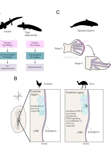
The classic example of developmental heterochrony is given by the Mexican axolotl,
Ambystoma mexicanum. Most salamanders have a life history that involves progression
via a metamorphosis event from an aquatic larval morph to a terrestrial adult morph. This
metamorphosis involves considerable changes to body form and physiology, such that the
adult form is specialised for life primarily on land. However, in the axolotl, reproducing
adults closely resemble the larval form – this species does not undergo metamorphosis,
but does have functional reproductive organs (Gould, 1977). This is an example of
pae-domorphosis, defined by Gould (1977) as the permanent retention of ancestral juvenile
traits in the adult stage of a derived species. Many comparisons have focused on
differ-ences betweenA. mexicanum and its sister taxon,A. tigrinum (the tiger salamander). In
axolotls, the production of functional gonads in an otherwise morphologically larval state
is understood to occur through neoteny – a retardation in somatic development (Gould,
1977). Thus, a dramatically different morphological form is achieved through changes in
the timing of some organs relative to others. In this case, the development of all organs
but the gonads are temporally slowed.
Metamorphosis in salamander species closely related to the axolotl occurs through
secretion of thyroid hormone (TH) from the thyroid gland (Gudernatsch, 1912).
Exper-iments conducted by Laufberger that bathed axolotls in TH showed that exogenous TH
is able to induce metamorphosis in the axolotl – an event that would never be observed
during normal development (reviewed by Huxley and Hogben, 1922). Further, the axolotl
has a functional thyroid gland, which has been observed to synthesise and release T4
if stimulated with thyroid stimulating hormone (TSH) (Taurog, 1974). However,
Figure 3: Heterochrony between organs can produce new forms in evolution. (A) Axolotl paedomorphosis results
from a loss of the metamorphosis event. In a close relative of the axolotl, the tiger salamander, metamorphosis is triggered by the pituitary-thyroid axis. However, TSH release by the pituitary gland does not occur in the
axolotl, causing a loss of the downstream events and associated metamorphosis. (B) Schematic of the chicken and emu lateral plate mesoderm (LPM) at HH18. In most avian species, including the chicken, reciprocal signalling
between the forelimb region of the LPM and the overlying ectoderm (purple) promotes proliferation of LPM cells and limb bud outgrowth. However, the emu forelimb region of the LPM produces the signalling ligand FGF10 at an insufficient level to induce FGF8 production in the ectoderm (purple). Consequently, cells of the LPM forelimb
region do not proliferate and the limb bud does not grow out from the body at HH17. (C) Analysis of spotted dolphin developing limb buds has shown that the apical ectodermal ridge (AER) persists over digits II and III for
longer than over the other digits. These digits exhibit hyperphalangy (many finger bones) and it has been suggested that the local persistence of the AER may be related to this trait. Silhouette images of animals were taken from
stimulate TH release (reviewed by De Groef et al., 2018) (Figure 3a). Experimentally
transplanting the pituitary gland of the axolotl into a host tiger salamander results in
the inhibition of metamorphosis in the host (Blount, 1950). The reciprocal graft has
also been performed, and induced metamorphosis in the axolotl (Blount, 1950). These
results indicate that in the axolotl, the thyroid axis is disrupted and TSH is not released
from the pituitary gland to stimulate TH release and metamorphosis. Genomic analyses
have implicated the met1, met2 and met3 genes in these species differences. After
ax-olotl treatment with exogenous T4, the effects of non-paedomorphic salamander alleles
for these genes are additive in reducing time to metamorphosis (Voss and Smith, 2005).
It is still not clear mechanistically how mutations in these genes in the axolotl result in a
loss of TSH release. Nonetheless, this is a striking example of how a simple change to an
endocrine pathway can shift the timing of development of one organ (the gonads) relative
to the rest of the body, with striking morphological effects.
A second example in which developmental heterochrony has been implicated in
mor-phological novelty is given by the emu, Dromaius novaehollandiae. The emu is one of
many species of flightless bird within the ratites (a group within the Palaeognathae) and
has very small wings that have a single digit. The timing of forelimb development in the
emu is significantly delayed relative to that of other birds and other amniotes. The limb
bud does not grow out from the flank of the emu embryo until Hamburger Hamilton stage
20 (HH20) – whilst in the vast majority of birds, outgrowth begins at HH17 (Hamburger
and Hamilton, 1951). A recent study has revealed the underlying developmental basis
for the delayed outgrowth of the emu wing bud (Young et al., 2019). Prior to limb bud
outgrowth, limb precursor cells reside in the lateral plate mesoderm (LPM). In both the
emu and the chicken, the epithelial to mesenchymal transition (EMT) of mesenchymal
precursors from the somatopleure and their movement to the LPM is intact (Young et
al., 2019). However, in the forelimb field of the emu LPM these precursors do not
pro-mote outgrowth of the limb bud at HH17. Analyses of proliferation in chicken and emu
fore- and hindlimb regions of the LPM show that at stages at which chicken fore- and
hindlimb regions and emu hindlimb regions are proliferating, the cells of the emu forelimb
LPM do not proliferate (Young et al., 2019). This difference results from a disruption of
reciprocal signalling between the LPM and overlying ectoderm. In chicken development,
the production of FGF10 by the LPM induces reciprocal signalling via FGF8 from the
ectoderm to the LPM (Ohuchi et al., 1997). FGF8 signalling of the ectoderm to the
LPM is required to promote proliferation in the limb bud field (Ohuchi et al., 1997).
However, in the emu, the ectoderm overlying the LPM does not express Fgf8 at HH18
(Young et al., 2019). Grafting of donor chicken LPM into the emu limb field (under the
difference in limb bud outgrowth timing results from changes to the LPM in the emu
(Young et al., 2019). ThoughFgf10 is expressed by the emu LPM in the forelimb region,
the authors suggest that the quantitative level of expression of this signalling ligand is
insufficient to induce the expression of Fgf8 in the ectoderm (Figure 3b). In support of
this hypothesis, overexpression ofFgf10 in the emu LPM results inFgf8 induction in the
overlying ectoderm and precocious limb bud outgrowth (Young et al., 2019). An enhancer
mutation responsible for the observed differences betweenFgf10 expression in the chicken
and emu embryo was also identified. Together, these results reveal that subtle changes to
the timing of expression of a signalling ligand in development are able to alter the timing
of development of the emu forelimb, contributing to changes to the gross morphology of
this structure.
A third example of heterochrony in morphological evolution is provided by the
dol-phin flipper. Dolphins are aquatic mammals possessing many adaptations for life in
water, including the modification of the forelimb to form a flipper. The flippers act
dur-ing swimmdur-ing as rudders, and in many species the digits exhibit hyperphalangy: relative
to the ancestral state, they possess numerous finger bones (phalanges) (Kukenthal, 1893).
Careful study of spotted dolphin (Stenella frontalis) embryos over the period of limb
development revealed that hyperphalangy is localised to digits II and III of the dolphin
forelimb (Richardson and Oelschl¨ager, 2002). This character correlates closely with the
prolonged maintenance of an apical ectodermal ridge (AER) over digits II and III,
sug-gesting that changes to the dynamics of AER development may be responsible for this
morphological change (Richardson and Oelschl¨ager, 2002) (Figure 3c). Thus, localised
persistence of the AER over digits II and III may promote the formation of additional
phalanges in these digits.
In summary, it is clear that alterations in phylogeny to the timing of development
of specific organs relative to the rest of the body can allow for pronounced changes in
form. This pattern was recognised by De Beer (1951) and Gould (1977), and the examples
discussed demonstrate that organ-level heterochrony may occur in diverse contexts. The
second and third examples in this section also provide good support for the importance of
multi-tissue inductive interations in the timing of developmental events. In each of these
cases (the emu forelimb and the spotted dolphin flipper), changes in signalling tissue
dynamics cause a change to the timing of a developmental event. For example, in the
emu forelimb, reduced FGF signalling from the LPM to the ectoderm results in substantial
delay in time of a signalling event, and the outgrowth of the limb bud. It is clear that the
apposition of signalling and responding tissues, as well as underlying signalling dynamics,
are important points of control in timing of events. These signalling events can have effects
population-level changes (e.g. the outgrowth of limb buds) and organ-population-level changes (e.g. the timing
of development and final morphology of digits of the limb).
5
Tissue tectonics as a mechanism to coordinate
develop-mental timing across scales
5.1 Pattern emergence in development: how intrinsic and extrinsic
tim-ing act together to generate spatial patterns of gene expression.
In the experiments enumerated above, we have seen multiple examples where cells display
an intrinsic ability to move through successive gene expression states in the absence of
extrinsic signals or cues. This demonstrates that cells are not passive entities that await
exposure to extracellular signals, but set their own developmental pace of differentiation
through a combination of mechanisms. Such mechanisms include the metabolic rate of
the cell and the associated tempo of a cell’s mRNA and protein turnover and also
alter-ations in the accessibility of transcription factors to bind and regulate gene expression at
the chromatin level. Ultimately, these biochemical alterations in a cell’s physiology and
nuclear architecture will impact the rates of transcription factor production and
degra-dation, as well as the efficiency to regulate either activation or repression of target genes.
The impact of these parameters can be modelled together with the higher level
regula-tive structure of gene regulatory networks to generate predictions on the dynamics of cell
state transitions through the use of sets of ordinary differential equations (Jaeger and
Monk, 2014; Strogatz, 2014). When used to simulate the temporal changes in gene
ex-pression across a field of cells, this dynamical systems approach has been highly effective
in determining how transcription factor networks operate as a function of these dynamic
modulators to give rise to changes in gene expression states over time. Two well studied
examples of how dynamical systems approaches have been used to investigate the
func-tion of gene regulatory networks are the gap gene system in dipteran insects, and the
dorsal-ventral patterning of the vertebrate neural tube (Jaeger, 2018; Verd et al., 2019;
Sagner and Briscoe, 2019; Kicheva et al., 2012). Locally, autocrine and paracrine signals
pass between cells undergoing cell state transitions, allowing for non-cell autonomous
reg-ulation of gene expression dynamics across cell popreg-ulations. When viewed at the tissue
level through a series of snapshots of gene expression analyses, coupling cell intrinsic and
extrinsic gene expresssion regulation in such a way results in the formation of gene
ex-pression patterns that are highly striking to the experimental observer. However, it is
point in time, or through an instantaneous response of gene regulation to external signals.
Rather they are an emergent property of cell intrinsic regulatory interactions coupled to
cell extrinsic control of their their inherent dynamics.
Broadly speaking, extrinsic timers offer a mechanism to provide tissue and
multi-organ coordination of developmental processes. Extrinsic signals are inherently linked to
the concept of induction in development, and ultimately to the role that morphogens
play in patterning tissues as they develop. Historically, the study of morphogens has
focused on their ability to generate gene expression patterns at a given fixed point in
time, and ignores the dynamics of their exposure to cells and that of the emerging gene
expression pattern in space. While much can be learned from asking how morphogens
can provide sufficient precision in the spatial domain to generate a given pattern, the
eventual fate of a cell is not determined by its expression state at any given point in
time but is rather an output of the sum of all state transitions it undergoes during
development (Verd and Jaeger, 2020). Caution against viewing pattern formation as a
mere ’snapshot’ of a continuous developmental process has been conceptualised as part
of the “general relativistic positional information framework” (Jaeger et al. 2008). This
framework places emphasis on our understanding of how biological systems may generate
the full dynamic profile of a given set of gene expression states within a tissue of interest,
and highlights cells as dynamic entities that integrate multiple sources of information
through time (rather than receiving positional information at one critical timepoint).
This consideration re-focuses the question of how gene expression patterns are established
away from the generation and interpretation of concentration gradients and towards the
regulation of the temporal exposure to morphogens during development.
5.2 Tissue tectonics as a higher-order regulator of morphogen exposure
during multi-tissue morphogenesis.
The timing at which cells and cell populations receive signals is again a highly distributed
phenomenon. Signal timing depends not only on when a signal reaches a cell (which itself
is a composite of multiple mechanisms of both extracellular and intracellular transport,
reviewed by Rogers and Schier, 2011; Sagner and Briscoe, 2017), but also on the rate of
production and transport of signal inhibitors. Across longer time-scales, cells will also
move relative to sources of these signals and inhibitors, creating a patterning mechanism
that is acting at the multi-tissue level. The relative rate of tissue movement is an output
of the state of the cells in question, but also of the mechanical properties of the
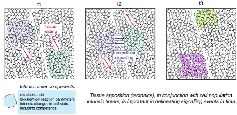
environ-ment in which it is moving. Here, we term the relative displaceenviron-ment of signalling and
in the lithosphere (Figure 4). The rate at which these multi-tissue interactions occur is
inherently linked to alterations in the mechanical properties of tissues as they are formed
during development. To fully illustrate how alterations in tissue tectonics can impact the
spatial and temporal regulation of patterning in development, we will briefly review two
examples.
Figure 4:Tissue Tectonics. Three snapshots in time are shown in this schematic, at timepoints t1, t2 and t3. Two
populations of cells are shown in purple and green, each of which is found in different tissue sheets. As the tissues slide relative to one another, the populations of interest come into close apposition, allowing intercellular signalling
to occur. At t3, the inductive signalling event has occurred and both populations’ cells have experienced a change in cell state. Clearly, tissue tectonics is an important determinant of the timing of signalling events in development.
5.2.1 Vertebrate gastrulation
During gastrulation, multiple tissue interactions act together to both specify and pattern
the three principle germ layers along three principle axes of orientation: anterior-posterior,
dorsal-ventral and left-right. In addition to establishing these essential coordinate systems
of the body plan, a series of cell movements act in a well orchestrated manner to
pro-gressively separate layers of tissues, and to begin the process of embryo elongation along
the anterior-posterior axis. A particularly well studied aspect of patterning during this
process is the initial specification of neural tissue with the ectoderm, and its subsequent
patterning along the anterior-posterior axis. While signals from the early gastrula-stage
organiser are important for ’activating’ the initial anterior character of neural tissue,
sub-sequent ’transforming signals’ then act to convert this character to more posterior neural
tissue as gastrulation proceeds (for a recent review see Martinez Arias and Steventon,
2018). During both the initial specification of neural tissue and its subsequent
modulation of FGF, BMP, Wnt and retinoic acid signalling pathways (Streit et al., 2000;
Linker and Stern, 2004; Stern et al., 2006). A conserved element of these interactions is a
requirement for the down-regulation of BMP signalling during neural plate specification,
and a subsequent posteriorisation by the Wnt signalling pathway (Niehrs, 2010). The
temporal exposure of ectodermal cells to these pathways has been shown to be a key
component of the patterning mechanism (Tuazon and Mullins, 2015; Tucker et al., 2008),
highlighting the question of what regulates the temporal exposure of cells to patterning
signals during gastrulation.
We propose that tissue tectonics is a key aspect of the temporal regulation of signal
exposure, and it is essential for ensuring appropriate coordination between the
morpho-genetic and patterning aspects of gastrulation. One aspect of this coordination is well
studied, and requires information to flow from gene-regulatory network activity through
to the control of cell movements, tissue morphogenesis and embryo elongation. Critically
however, it also requires information flow in the opposite direction, i.e. cells must be
able to determine the state of embryo elongation and tissue morphogenesis to coordinate
these processes with cell specification and patterning. A recent study has approached this
question using explants of zebrafish embryonic cells that were cultured away from the yolk
and yolk syncytial layer (Trivedi et al., 2019). Such aggregates go on to break
morpho-logical symmetry and generate multiple germ layers in an organised manner (Trivedi et
al., 2019; Schauer et al., 2020). As the explants continue to elongate, progressive bands
of the hindbrain marker Krox20 appear concomitantly with the movement of a pole of
Wnt/beta-catenin away from a source of BMP4/7 expression at the opposite end,
suggest-ing that the elongation itself may be an important upstream regulator to determine the
timing of exposure to both BMP and Wnt signal activity. Indeed, blocking convergence
and extension of the explants results in an alteration in the spatial-temporal exposure to
these signalling pathways and the specification of hindbrain (Trivedi et al., 2019).
To-gether, these results provide an initial insight into the role that tissue tectonics plays
in providing a causal link between the mechanisms of global embryo elongation and the
patterning of the nervous system during gastrulation.
5.2.2 Cavefish eyefield specification and the evolution of gastrulation.
Alterations in the morphogenesis of gastrulation are common and require a mechanism
for such alterations to impact patterning in a manner that a conserved body plan can be
generated at later developmental stages. A recent study examined differences between
two different morphs of the characid fish during gastrulation: a wildtype river-dwelling
a number of morphological differences relative to surface fish, including a complete loss of
eyes. This opens the question of how alterations in the morphogenesis aspect of
gastru-lation might impact the patterning aspect of gastrugastru-lation in the adaption of popugastru-lations
to new ecological environments. An examination of the expression of the homologs of
various genes expressed by the organizer in the embryos of characid fish revealed
sub-stantial differences in the expression of these genes during gastrulation (Torres-Paz et al.,
2019). For example, dickkopf1b (dkk1b) is expressed in two distinct populations at 50%
epiboly in the river-dwelling morph, but in one continuous domain at the same stage in
the cavefish morph. This difference in expression domain is associated with advanced
internalisation of these cells (which contribute to the anterior prechordal plate) in the
cavefish morph relative to the river-dwelling morph. Notably, the expression ofdkk1b is
also downregulated earlier in the cavefish morph than the river-dwelling morph. As a
consequence of shifted timing of AP axis formation (heterochrony) in the cave morph,
the eyefield which forms within the overlying neurectoderm is reduced in size. Through
functional experiments which mimicked the impact of advanced Wnt signalling
activa-tion in the eyefield (through treatment with LiCl, becausedkk1b is an antagonist of Wnt
signalling), the authors showed that increasing Wnt signalling in early surface-dwelling
embryos results in a reduced eyefield and later a misshaped retina. Together, these results
give an example of a developmental signalling event which is altered in timing through
changes to the timing of apposition of tissues (here, the anterior neurectoderm and the
anterior prechordal plate). In this example, changes to the timing of these events have a
marked morphological effect, accounting for the loss of eyes in the cavefish morph. This
study opens a set of fascinating questions over the limits of developmental constraint and
robustness in the evolution of gastrulation morphogenesis, and the causal role that tissue
tectonics might play in linking these two aspects of body plan development and evolution.
5.3 Tissue tectonics as a mediator of downward causation in
develop-ment.
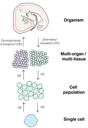
Up until now, developmental biologists have focused on the emergent properties of
devel-opment: how processes at a lower level of a complex system (i.e. at the level of a cell
and the mechanisms driving its cell state transitions) can impact observable features at
higher levels (i.e. the patterned expression of genes across a field of cells when observed
at a fixed time-point). However, one of the salient properties of developmental systems
is their ability to regulate pattern upon the loss or experimental removal of certain parts
of the embryo. Some striking examples include, but are in no way limited to, the ability
of the chicken embryo to develop normally after surgical removal of a large portion of
the primitive streak, and the formation of monozygotic twins (Dreisch 1892; Psychoyos
and Stern, 1996). This regulative (or self-organising) ability of developmental systems
fascinates experimental embryologists to this day and has recently come back into focus
through observations regarding the ability of embryonic cells to break symmetry and
gen-erate patterns when aggregated and cultured as multi-cellular aggregates (Beccari et al.,
2018; van den Brink et al., 2020; Veenvliet et al., 2020; Warmflash et al., 2014).
Regulative development requires a mechanism that enables the sensing of changes to
the properties of a system at higher levels (including alterations in the size and/ or shape
of an embryo or primordium), and to convey changes in the state of the system at lower
levels (i.e. alterations in the intrinsic state of cells and cell population) (Figure 5). To
achieve this, there must be an element of downward causation in the system. Essentially,
a mechanism by which information can be passed downwards to confer alterations in a
cell’s gene expression state in response to multi-tissue level perturbation. This downward
causation runs in the opposite direction to the emergence of gene expression patterns
(Figure 5), and similarly requires an understanding of how alterations in the timing of
exposure to signals and their inhibitors is regulated through time. We propose that
tissue tectonics is also an essential consideration in understanding the mechanisms of
downward causation in development, because downward causation is intimately associated
with signalling between cells. It is clear to see how manipulations to the embryo (for
example, removal of the anterior primitive streak) would impact signalling events between
cells, changing the apposition of different tissues. A full understanding of the mechanical
properties of tissues will allow us to follow how tissues respond to injury, how this impacts
the timing of exposure to extrinsic timers, and how this in turn regulates the operation
of intrinsic timers and the emergence of patterns during regulative development and
self-organisation.
5.4 Conclusions
In this review, we have given an overview of studies in developmental biology that have
asked how developmental events are timed. These studies have focused at a variety of
levels of organisation: from the level of chromatin modifications within single cells to
the coordination of multi-tissue or multi-organ events. Cell-intrinsic and extrinsic timer
mechanisms both contribute to the overall timing of events during development, and we
have described the utility of experimental embryology (in particular, heterochronic
graft-ing) in distinguishing between these modes of developmental timer control. Through
out of close apposition, allowing for the controlled timing of developmental signalling
events. Signalling events provide an important level of control for developmental timing,
being converged on by both low-level events including gene expression as well as dramatic
changes in the morphology of the embryo. We propose that tissue tectonics is a key
mech-anism that integrates timing information across scales of organisation within the embryo
during development. As we have seen, changes to the timing of signalling events can have
a dramatic effect on morphology; for example, in the development of the emu wing, in
the development of the cavefish eyefield and in the development of the dolphin flipper.
It is conceivable that the diversity of gastrulation-stage embryonic forms in vertebrates
are associated with changes to the timing of developmental events, as tissue tectonics will
be markedly different (Martinez Arias and Steventon, 2018). In summary, timing is a
highly distributed phenomenon in developmental biology that is coordinated over diverse
levels of organisation. A huge number of timer mechanisms, some intrinsic and others
extrinsic, work together to reproducibly time developmental events in the embryos of a
given species. We have shown the importance of inter-cell signalling events as a point
of control and coordination across these levels of organisation, and have argued for the
importance of tissue tectonics (the movement of signalling and responding tissues relative
Figure 5: Summary. This schematic summarises the different levels of organisation where time has been studied in developmental biology. Interaction between the various levels of organisation occurs bidirectionally. Information
passes from higher to lower levels throughdownward causation, exemplified by pattern regulation in the embryo (see text for examples). Information also passes from the lower levels to higher levels, throughdevelopmental emergence.
For example, changes in cell state can lead to changes to inter-population signalling and ultimately direct higher-level changes to the embryo in morphogenesis. We argue that a link between these higher-levels of organisation is provided by the concept of ‘tissue tectonics’. The ways in which signalling and responding tissues are displaced relative to
6
Acknowledgements
The authors would like to thank the following for helpful comments on the review: Chloe
Bash, Toby Andrews, Aleksandra Marconi, Tim Fulton, Dillan Saunders, Matthew
Tow-ers, Alfonso Martinez Arias and Berta Verd. We apologise to any whose work may have
been excluded from the text; this is by no means a comprehensive review of all research
that focuses on developmental timing. The silhouette images inFigure 3 were taken from
www.phylopic.org and are attributed to Darren Naish (emu), Chris Huh (dolphin), Sarah
Werning (Xenopus) and Soledad Miranda-Rottmann (mouse).
7
References
• Angevine, J.B., and Sidman, R.L. (1961). Autoradiographic Study of Cell Migration
during Histogenesis of Cerebral Cortex in the Mouse. Nature 192, 766–768.
• Asher, G.W., Mulley, R.C., O’Neill, K.T., Scott, I.C., Jopson, N.B., and Littlejohn,
R.P. (2005). Influence of level of nutrition during late pregnancy on reproductive
productivity of red deer: I. Adult and primiparous hinds gestating red deer calves.
Anim. Reprod. Sci. 86, 261–283.
• Aulehla, A., and Johnson, R.L. (1999). Dynamic expression of lunatic fringe
sug-gests a link between notch signaling and an autonomous cellular oscillator driving
somite segmentation. Dev. Biol. 207, 49–61.
• Averbukh, I., Lai, S.L., Doe, C.Q., and Barkai, N. (2018). A repressor-decay timer
for robust temporal patterning in embryonic drosophila neuroblast lineages. Elife
7, 1–19.
• Azuara, V., Perry, P., Sauer, S., Spivakov, M., Jørgensen, H.F., John, R.M., Gouti,
M., Casanova, M., Warnes, G., Merkenschlager, M., et al. (2006). Chromatin
signatures of pluripotent cell lines. Nat. Cell Biol. 8, 532–538.
• Bannister, A.J., and Kouzarides, T. (2011). Regulation of chromatin by histone
modifications. Cell Res. 21, 381–395.
• Barry, C., Schmitz, M.T., Jiang, P., Schwartz, M.P., Duffin, B.M., Swanson, S.,
Bacher, R., Bolin, J.M., Elwell, A.L., McIntosh, B.E., et al. (2017). Species-specific
developmental timing is maintained by pluripotent stem cells ex utero. Dev. Biol.