Electronic Thesis and Dissertation Repository
8-20-2013 12:00 AM
Complex Shoulder Instability: The Role of the Latarjet Coracoid
Complex Shoulder Instability: The Role of the Latarjet Coracoid
Transfer
Transfer
Ryan DegenThe University of Western Ontario Supervisor
Dr. Jim Johnson
The University of Western Ontario Joint Supervisor Dr. George Athwal
The University of Western Ontario
Graduate Program in Medical Biophysics
A thesis submitted in partial fulfillment of the requirements for the degree in Master of Science © Ryan Degen 2013
Follow this and additional works at: https://ir.lib.uwo.ca/etd
Part of the Orthopedics Commons, and the Surgical Procedures, Operative Commons
Recommended Citation Recommended Citation
Degen, Ryan, "Complex Shoulder Instability: The Role of the Latarjet Coracoid Transfer" (2013). Electronic Thesis and Dissertation Repository. 1454.
https://ir.lib.uwo.ca/etd/1454
This Dissertation/Thesis is brought to you for free and open access by Scholarship@Western. It has been accepted for inclusion in Electronic Thesis and Dissertation Repository by an authorized administrator of
Integrated Article Format
by
Ryan Michael Degen
Graduate Program in Medical Biophysics
A thesis submitted in partial fulfillment of the requirements for the degree of
Master of Science
The School of Graduate and Postdoctoral Studies The University of Western Ontario
London, Ontario, Canada
ii
Abstract
Recurrent anterior shoulder instability is a common clinical entity that is debilitating for
patients, and often requires surgical stabilization. Recurrence rates following soft-tissue
stabilization procedures are moderately high and have been attributed to associated bony
defects of either the glenoid or humeral head. Complex shoulder instability, which is defined
as instability associated with bony defects around the shoulder, is a challenging clinical
problem. The Latarjet coracoid transfer has been proposed as a treatment option and its
resultant stabilizing effects have been explored in this biomechanical cadaver-based study
and compared to alternative procedures.
For both glenoid and humeral head defects, the Latarjet coracoid transfer adequately
stabilized the shoulder, outperforming other procedures often clinically utilized for these
scenarios. It did, however, result in increased superior translation of the shoulder compared
to other procedures, the clinical significance of which is presently unknown.
The Latarjet coracoid transfer is a useful procedure for complex shoulder instability. Further
study should assess for any potential deleterious clinical effects following this procedure.
Keywords
iii
Co-Authorship Statement
Chapter 1 Ryan Degen – Sole Author
Chapter 2 Ryan Degen – Study Design, Specimen Preparation, Data Collection,
Manuscript Review
Josh Giles – Study Design, Specimen Preparation, Data Collection, Data
Analysis, Manuscript Preparation
Jim Johnson – Study Design, Manuscript Review
George Athwal – Study Design, Specimen Preparation, Manuscript Review
Chapter 3 Ryan Degen – Study Design, Specimen Preparation, Data Collection, Data
Analysis, Manuscript Preparation
Josh Giles – Study Design, Specimen Preparation, Data Collection, Data
Analysis, Manuscript Review
Jim Johnson – Study Design, Manuscript Review
George Athwal – Study Design, Specimen Preparation, Manuscript Review
Chapter 4 Ryan Degen – Data Collection, Data Analysis, Manuscript Preparation
Josh Giles – Study Design, Specimen Preparation, Data Collection, Data
Analysis, Manuscript Review
Harm Boons – Study Design, Specimen Preparation, Manuscript Review
Jim Johnson – Study Design, Manuscript Review
George Athwal – Study Design, Specimen Preparation, Manuscript Review
iv
Chapter 2 A version of this manuscript has been submitted for publication in the Journal
of Bone and Joint Surgery, American Volume and is awaiting review.
Chapter 3 A version of this manuscript has been submitted for publication in Clinical
Orthopaedics and Related Research and is awaiting review.
Chapter 4 A version of this manuscript has been accepted for publication in the
International Journal of Shoulder Surgery. Permission to reprint this version
of the manuscript has been granted by the Publisher.
v
Acknowledgments
I would like to thank a few people who were instrumental in the completion of this thesis. None of
this work would have been possible without my supervisors Dr. George Athwal and Dr. Jim Johnson,
as well as my co-investigator Josh Giles. I would like to thank all of you for your efforts in
completing this research.
Dr. Athwal and Johnson – I appreciate your help not only organizing and overseeing this research, but
in helping prepare me for a career in orthopedic research. From advisory committee meetings, to
rehearsal presentations, and constructive criticisms on manuscript preparation, the advice and
assistance you have both provided will go a long way towards helping me start my own academic
research practice.
Josh – thank you for your tremendous contributions to this work. The simulator that you have
designed and continually improved on, your mastery of any and all software programs for data
collection and analysis and your patience during our marathon testing sessions were all very much
appreciated. I have no doubt that your future is bright and wish you well in all future endeavours.
To Dr. King, Dr. Canham and Dr. Johnson – thank you for your attentiveness and feedback during
our communication course sessions. These sessions have certainly improved my understanding of
some fundamental principles in biomechanical research, but have undoubtedly improved my personal
communication and presentation skills. All presentations I have given since these sessions have been
met with praise and I attribute this largely to your advice, so thank you.
I would also like to thank the Department of Medical Biophysics for being understanding of the
challenges faced with being a graduate student and medical resident, particularly for being
accommodating with course selections and alternate low-level comprehensive examinations.
Lastly, I would like to thank my wife, Lindsay. Thank you for being so helpful and patient with me
while I took on yet another task that added stress to our lives and ate away at weekends and vacations
over the past two years. This master’s degree will be an integral part of my career, and our life,
vi
Abstract ... ii
Co-Authorship Statement... iii
Acknowledgments... v
Table of Contents ... vi
List of Tables ... x
List of Figures ... xi
Chapter 1... 1
1 Introduction ... 1
1.1 The Shoulder... 1
1.1.1 Osteology ... 1
Labrum, Capsule and Ligaments ... 7
1.2 Shoulder Kinematics ... 14
1.2.1 Static Stabilizers... 14
1.2.2 Dynamic Stabilizers ... 16
1.3 Shoulder Instability... 20
1.3.1 Pathophysiology... 20
1.4 Treatment Options ... 28
1.5 Study Rationale... 39
1.6 Objectives ... 40
1.7 Hypotheses ... 40
1.8 Thesis Overview ... 41
1.9 References... 42
vii
2 Bristow versus Latarjet Coracoid Transfer for Recurrent Shoulder Instability with
Glenoid Deficiency ... 52
2.1 Introduction... 52
2.2 Materials & Methods ... 55
2.2.1 Specimen Preparation and Shoulder Simulator ... 55
2.2.2 Experimental Testing Protocol ... 58
2.2.3 Stability and Range of Motion... 60
2.2.4 Outcome Variables & Statistical Analyses ... 61
2.3 Results... 62
2.3.1 Joint Stiffness and Stability ... 62
2.3.2 Range of Motion ... 67
2.4 Discussion ... 69
2.5 Conclusion ... 72
2.6 References... 73
Chapter 3... 76
3 Remplissage versus Latarjet Coracoid Transfer for Hill-Sachs defects: A Biomechanical Comparison ... 76
3.1 Introduction... 76
3.2 Materials & Methods ... 77
3.2.1 Specimen Preparation ... 77
3.2.2 Shoulder Simulator ... 78
3.2.3 Surgical Protocol... 79
3.2.4 Experimental Protocol & Outcome Variables ... 80
3.2.5 Data and Statistical Analysis ... 81
3.3 Results... 81
3.3.1 Joint Stiffness... 81
viii
3.4 Discussion ... 90
3.5 Conclusions... 93
3.6 References... 94
Chapter 4... 97
4 A Biomechanical Assessment of Superior Shoulder Translation after Reconstruction for Anterior Glenoid Bone Loss: The Latarjet Procedure versus Allograft Reconstruction... 97
4.1 Introduction... 97
4.2 Materials and Methods... 100
4.2.1 Specimen Preparation ... 100
4.2.2 Shoulder Simulator ... 102
4.2.3 Testing Protocol ... 103
4.2.4 Stability Testing ... 106
4.2.5 Statistical Analysis... 106
4.3 Results... 107
4.3.1 Humeral Head Translation: Neutral Rotation ... 107
4.3.2 Humeral Head Translation: Internal Rotation... 109
4.3.3 Humeral Head Translation: External Rotation ... 111
4.3.4 Humeral Head Translation: Load Effect... 113
4.4 Discussion ... 113
4.5 Conclusion ... 117
4.6 References... 118
Chapter 5... 122
5 General Discussion & Conclusions... 122
5.1 Bristow versus Latarjet Coracoid Transfer (Chapter 2)... 123
ix
5.3 Superior Shoulder Instability following Latarjet Coracoid Transfer (Chapter 4)125
5.4 Cadaveric Testing ... 126
5.5 Conclusion ... 127
5.6 References... 127
Appendix A: Glossary... 130
Appendix B: Abbreviations List ... 132
Appendix C: Copyright Permissions ... 133
x
Table 2-1. Incidents of Glenohumeral Joint Dislocation during Two Stability Tests. ... 66
xi
List of Figures
Figure 1-1. Osteology of the Shoulder... 2
Figure 1-2. Glenoid Retroversion and Inclination ... 4
Figure 1-3. Humeral Head Retroversion... 6
Figure 1-4. Glenoid with Soft-tissue Restraints... 9
Figure 1-5 Anterior view of Shoulder with Rotator Cuff Muscles ... 11
Figure 1-6 – Posterior view of Shoulder with Rotator Cuff Muscles ... 11
Figure 1-7. Glenohumeral Joint Concavity-Compression Effect. ... 18
Figure 1-8. Bankart Lesion and Hill-Sachs Defect ... 23
Figure 1-9. Hill-Sachs Defect Engagement in External Rotation... 25
Figure 1-10. Bony Bankart Lesion... 27
Figure 1-11. Bankart Stabilization... 30
Figure 1-12. Bristow Coracoid Transfer ... 33
Figure 1-13. Latarjet Coracoid Transfer ... 35
Figure 1-14. Remplissage Procedure ... 37
Figure 2-1 – The Bristow and Latarjet Coracoid Transfers ... 54
Figure 2-2– In-vitro Shoulder Simulator... 57
xii
Rotation... 64
Figure 2-5. Axial Rotation in Adduction and Abduction, and Horizontal Extension in Abduction... 68
Figure 3-1. Joint Stiffness in Adduction and Neutral Rotation ... 83
Figure 3-2. Joint Stiffness in Adduction and External Rotation... 83
Figure 3-3. Joint Stiffness in Abduction and Neutral Rotation ... 84
Figure 3-4. Joint Stiffness in Abduction and External Rotation... 84
Figure 3-5. Internal/External Range of Motion in Adduction ... 86
Figure 3-6. Internal/External Range of Motion in Abduction ... 86
Figure 3-7. Horizontal Extension Range of Motion in Abduction and External Rotation (60°) ... 87
Figure 4-1. Mounted Shoulder Specimen ... 101
Figure 4-2. Coracoid Reconstructions of a 30% Glenoid Defect ... 105
Figure 4-3. Superior Translation in Adduction and Neutral Rotation. ... 108
Figure 4-4. Superior Translation in Adduction and Internal Rotation... 110
Chapter 1
1
Introduction
Overview: The purpose of this thesis will be to introduce the concept of anterior
shoulder instability focusing on complex shoulder instability, which involves osseous
defects of the shoulder that predispose to further instability. In this chapter, a brief
overview of anatomy of the shoulder will be provided. Shoulder kinematics will also be
reviewed, paying particular attention to the structures that serve as primary restraints to
shoulder instability. Treatment options for primary and complex shoulder instability will
briefly be reviewed. The objective, hypothesis, rationale and outline of the thesis will
also be reviewed.
1.1 The Shoulder
The shoulder, primarily thought of as the glenohumeral joint, is a ball-and-socket
joint that allows a wide range of multiaxial joint movement in all three planes (sagittal,
coronal, axial or transverse) through a complex interaction of bones, supporting
ligaments and surrounding musculature (1). It is the most mobile joint in the body and,
as a result of being less constrained, is also the most frequently dislocated joint. There
are a number of factors that contribute to its stability, which will be introduced here.
1.1.1
Osteology
The primary articulation of the shoulder, as stated above, is between the humeral
head and the glenoid concavity of the scapula, representing the glenohumeral joint
(Figure 1-1). However, the shoulder complex also includes the clavicle, or collar bone.
Together, these three osseous structures define the shoulder, which has four major
articulations: Sternoclavicular (SC) joint – between the sternum and clavicle;
Acromioclavicular (AC) joint – between the scapula and clavicle; Glenohumeral joint –
between the humeral head and glenoid; and the Scapulothoracic articulation – between
Figure 1-1. Osteology of the Shoulder
1.1.1.1
The Clavicle
The clavicle, an “S-shaped” bone, articulates medially with the sternum to make
up the SC joint, and laterally with the acromion, an extension of the scapular spine, to
form the AC joint. The clavicle serves as a site of muscular attachment and also acts as a
strut to support the glenohumeral joint. The SC joint is the only true point of attachment
of the shoulder girdle (or appendicular skeleton) to the trunk (or axial skeleton). Inferior
migration of the shoulder complex is prevented through strong ligamentous attachments
between the clavicle and coracoid (an extension of the scapula) (4).
1.1.1.2
The Scapula
The scapula is a broad, triangular bone that comprises the posterior aspect of the
shoulder, overlying the 2nd through 7th posterior ribs (4). It serves as an attachment site
for several muscles. Its anterior concave surface articulates with the posterior convexity
of the ribs via muscular attachments, making up the scapulothoracic articulation, which
stabilizes the scapula and provides support to the glenohumeral articulation.
Laterally, the scapula forms a flat projection, known as the glenoid fossa, which
articulates with the humeral head, forming the glenohumeral joint. The glenoid fossa is
relatively small, only 1/4 to 1/3 the size of the humeral head, and thus provides only a
small contribution to glenohumeral joint stability on its own, relying heavily on the
complex interaction of the static and dynamic stabilizers, which will be reviewed later in
this chapter (5). The lack of constraint provided by the glenoid allows for such mobility
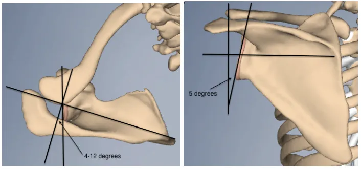
of the glenohumeral joint. Relative to the axis of the scapula, the glenoid is retroverted
approximately 4-12º (average of 7º) and is superiorly inclined approximately 5º (Figure
Figure 1-2. Glenoid Retroversion and Inclination
The glenoid has a variable orientation relative to the scapula in both axial/transverse and coronal planes. Looking on an axial view (left), the glenoid face is retroverted (averaging 4-12º)
The superior process of the scapula, known as the scapular spine, separates two of
the rotator cuff muscles, and also acts as a site of muscular attachment. This process
continues laterally and anteriorly and becomes the acromion, which articulates with the
clavicle (AC joint).
Anterior and medial to the glenoid, the scapula has an additional bony extension,
known as the coracoid process which projects anteriorly and laterally. Often referred to
as the “lighthouse” of the shoulder, this is an important anatomic reference point during
surgery and serves as an attachment site for several ligaments and muscles that confer
stability to the shoulder complex. Particularly important are the coracoclavicular
ligaments, strong ligaments running between the coracoid and the clavicle that prevent
inferior displacement of the shoulder girdle (coracoclavicular ligaments), and the
coracohumeral ligament, running from the coracoid to the greater tuberosity which also
prevents inferior humeral head displacement (4).
1.1.1.3
The Humerus
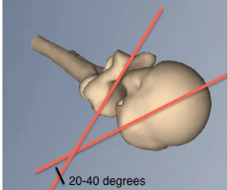
The humerus is the largest bone in the upper extremity. The proximal end, or
humeral head, articulates with the glenoid. The head is retroverted relative to the
humeral shaft (compared to trans-epicondylar axis of the distal humerus) by
approximately 30º (Figure 1-3). The head has three distinct areas – the greater tuberosity
(GT), lesser tuberosity (LT) and the bicipital groove located between them. The
tuberosities represent insertion sites for the rotator cuff muscles, which dynamically
stabilize the glenohumeral joint. As its name implies, the bicipital groove is the location
where the long head of the biceps tendon runs, as it continues proximally to its insertion
above the glenoid fossa. Slightly more distal along the humeral shaft, there is a region
Figure 1-3. Humeral Head Retroversion
Labrum, Capsule and Ligaments
1.1.1.4
Labrum
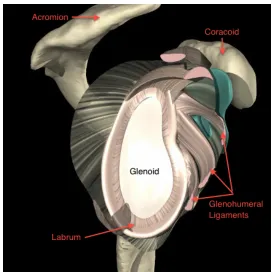
The glenoid labrum is a fibrocartilaginous complex attached circumferentially to
the edge of the glenoid cavity (Figure 1-4). It serves to deepen the concavity of the
glenoid by approximately 50% and contributes to shoulder stability by resisting
translatory forces acting on the humeral head. Additionally, it serves as an attachment
site for the glenohumeral ligaments and long head of the biceps (1,2). Disruptions of this
structure are common following dislocation and are frequently associated with recurrent
shoulder instability.
1.1.1.5
Joint Capsule
The articular capsule is a fairly loose, redundant structure that attaches around the
scapular neck and inferior aspect of the neck of the humerus, near the lesser tuberosity.
There are three focal areas of thickening of the capsule, known as the glenohumeral
ligaments (GHL), that act as “check-reins” to excessive rotation or translation of the
humerus. Running from the inferior aspect of the humeral head, or the humeral neck,
these structures insert or coalesce with the glenoid labrum (4,6,7).
1.1.1.6
Superior Glenohumeral Ligament
The superior GHL, running from the supraglenoid tubercle above the glenoid face
to the lesser tuberosity of the humerus, has a parallel course to the coracohumeral
ligament (Figure 1-4). The two are felt to act together as a restraint to inferior translation
and external rotation of the humeral head with the arm resting at one’s side (position of
adduction) (4).
1.1.1.7
Middle Glenohumeral Ligament
The middle glenohumeral ligament is the most variable, with some patients
having a so-called “cord-like” middle GHL, known as a “Buford complex”, and up to
30% of patients being deficient of this ligament altogether (4,8). It also runs from the
supraglenoid tubercle to the lesser tuberosity, although some fibers coalesce with the
abduction, the middle GHL becomes taut, limiting further external rotation of the
humerus in this position. Maximal tension in the middle GHL is reached at
approximately 45º of abduction, at which point it is also able to resist anterior translation
of the humeral head in this position (10).
1.1.1.8
Inferior Glenohumeral Ligament
The inferior GHL is a hammock-like structure, with origins from both the
anteroinferior and posteroinferior aspects of the glenoid (9). This ligament has two
separate bands, an anterior and posterior band with an intervening segment of capsule.
The anterior band inserts at the inferior margin of the articular surface of the humeral
head, just below the lesser tuberosity. In abduction with the arm externally rotated, the
so-called ‘position of apprehension’, the anterior band of the inferior GHL moves to the
front of the shoulder where it is maximally taut and serves to resist anterior translation of
Figure 1-4. Glenoid with Soft-tissue Restraints
1.1.1.9
Coracohumeral Ligament
The coracohumeral ligament (CHL) is a broad ligament originating from the
superior portion of the joint capsule at the base of the coracoid process and inserting on
the greater tuberosity. This acts in conjunction with the superior GHL, as described
above, along with the anterior joint capsule to make up the “rotator interval”, which
functions to resist inferior translation of the humeral head in adduction (1,12). The
rotator interval structures also resist anterior translation with the arm in adduction.
1.1.1.10 Coracoacromial Ligament
The coracoacromial ligament (CAL) runs from the coracoid process to the
anterior margin of the acromion. This structure provides a restraint against superior
translation of the humeral head, largely in response to a superiorly directed force exerted
axially on the humerus (13). Additionally, this structure, occasionally referred to as the
more expansile coracoacromial “veil”, interacts with other structures of the rotator
interval and prevents inferior translation of the humeral head (14).
1.1.1.11 Coracoclavicular Ligaments
Comprised of two separate bands, the trapezoid (lateral) and the conoid (medial)
ligaments make up the complex commonly referred to as the coracoclavicular ligaments.
As previously mentioned, these run from the superior edge of the coracoid process to the
undersurface of the clavicle, and serve primarily as a restraint to inferior translation of the
scapula, and subsequently the glenoid (4).
1.1.1.12 Muscles
The muscles surrounding the shoulder provide a component of dynamic stability,
helping stabilize the joint while permitting motion. The rotator cuff is a muscular
complex that surrounds the joint capsule and is comprised of the supraspinatus
(superiorly), subscapularis (anteriorly), infraspinatus and teres minor muscles
(posteriorly), which serves to provide a compressive load to stabilize the joint and
Figure 1-5 Anterior view of Shoulder with Rotator Cuff Muscles
Anterior view of the shoulder with surrounding rotator cuff musculature. Here, the subscapularis, responsible for internal rotation of the shoulder, can be seen anterior to the glenohumeral joint inserting on the lesser tuberosity of the humeral shaft (3)
Figure 1-6 – Posterior view of Shoulder with Rotator Cuff Muscles
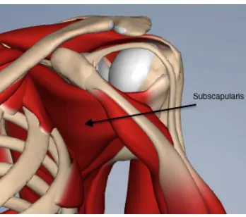
The supraspinatus originates in the supraspinatus fossa on the cranial side of the
scapula, above the scapular spine and inserts on the greater tuberosity. It is innervated by
the suprascapular nerve and is responsible for shoulder abduction.
The subscapularis muscle originates in the subscapularis fossa on the anterior
aspect of the scapula and inserts on the lesser tuberosity. It is innervated by the upper
and lower subscapular nerves and is responsible for internal rotation of the shoulder.
The infraspinatus and teres minor muscles originate on the posterior surface of the
scapula and insert on the posterior aspect of the greater tuberosity. The infraspinatus is
innervated by the suprascapular nerve, while the teres minor is innervated by the axillary
nerve; together, these muscles are responsible for external rotation of the shoulder.
The long head of the biceps originates from the glenoid labrum at the
supraglenoid tubercle, just above the glenoid articular surface, and runs in the bicipital
groove between the greater and lesser tuberosities. It joins the short head of the biceps,
which originates from the coracoid process, and runs down the humerus with a common
muscle belly, inserting on the bicipital tuberosity of the radius. Together, the short and
long heads are innervated by the musculocutaneous nerve and are responsible for elbow
flexion and forearm supination.
In addition to the short head of the biceps, the coracobrachialis muscle also
originates from the coracoid process. Together, the two are referred to as the conjoined
tendon. The coracobrachialis muscle inserts distally on the humerus. It is also innervated
by the musculocutaneous nerve and is responsible for forward elevation and adduction of
the shoulder.
Overlying all of these muscles is the deltoid, separated from the rotator cuff by
the subdeltoid bursa (fluid-filled sac). It has three separate heads: the anterior, arising
from the lateral clavicle; the middle, arising from the acromion; and the posterior, arising
from the scapular spine. The three heads have a common insertion, onto the deltoid
responsible for forward flexion (anterior head), abduction (lateral head) and extension
1.2
Shoulder Kinematics
As stated earlier, the shoulder is one of the most mobile joints in the body.
Motion of the shoulder includes:
1. Forward flexion (~160º) and extension (~60º) in the sagittal plane,
2. Abduction (~160º) and adduction in the coronal plane, and
3. Internal (~50º) and external rotation (~50º) in the horizontal or axial plane.
Abduction of the shoulder is a combined motion of both glenohumeral and
scapulothoracic motion, occurring in a 2:1 ratio. For example, every 3 degrees of
abduction is actually only 2 degrees of glenohumeral abduction and 1 degree of
scapulothoracic abduction (15). This extensive range of motion is permitted through a
complex interaction of static and dynamic stabilizers that work to minimize instability
while facilitating motion (5,6,16).
1.2.1
Static Stabilizers
Static structures that contribute to shoulder stability include:
a) Negative intra-articular pressure,
b) Glenohumeral joint geometry,
c) Labrum,
d) Capsule and ligaments
Negative intra-articular pressure of the glenohumeral joint assists in stabilizing
the humeral head within the glenoid fossa. This pressure occurs when the capsule
remains an intact, closed compartment, and is attributable to the variable compliance of
the structures that make up the joint. The glenoid itself is very firm, with only a thin
layer of articular cartilage; however the labrum is very compliant and provides a
creating a hole in the capsule, or “venting” the capsule, results in loss of this negative
pressure and subsequent inferior translation of the humeral head up to 10mm at rest, and
up to 50% increases in passive translation in all directions (17,18). While listed in the
section of static stabilizers as it is a constantly negative pressure, there is a dynamic
component to the intra-articular pressure. Some studies have shown that the pressure
varies slightly with joint position, with the average pressure being -67.8 mm Hg, but
decreasing to a maximum of -82.9 mm Hg in 20º of abduction (18).
Glenohumeral geometry also contributes to joint stability. Relative glenoid
retroversion and superior inclination, as depicted earlier, provide bony restraints to
anterior and inferior translation. Cadaveric studies have shown that 5-10º of superior
inclination can significantly improve resistance to inferior translation. Similarly, glenoid
retroversion provides resistance to anterior translation, with this effect maintained with a
version of up to 5º of glenoid anteversion (19).
The glenoid labrum increases the depth of the glenoid concavity by ~50%,
essentially providing a larger degree of conformity and constraint to the joint. Its
consistency is variable, as it has been found to be more pliable and less rigidly attached
around the anterosuperior aspect of the glenoid, while it is much more immobile and
firmly attached at the inferior aspect of the glenoid providing a larger resistive force to
translation (17). Additionally, the labrum serves to enhance the concavity-compressive
effect, which will be explained below as it falls into the category of dynamic stability.
While the glenohumeral capsule has a surface area nearly double that of the
humeral head and is fairly redundant, the focal thickenings, known as the glenohumeral
ligaments, provide restraint to translation and rotation in different shoulder positions
(5,7). The superior and middle GHL are able to resist inferior and anterior translation in
adducted or slightly abducted positions, while the anterior band of the inferior GHL has
the most significant effect in resisting anterior shoulder translation largely in an abducted
position. Studies have shown that placing the shoulder in the position of apprehension,
(2,5,7,17,20).
1.2.2
Dynamic Stabilizers
While the static structures exert their effect at the extremes of motion to prevent
instability, the dynamic stabilizers act within the functional range of motion to provide
stability where those static restraints are often lax (5,6). For example, the surrounding
shoulder musculature attempts to optimally position the glenoid and provide a
compressive force across the glenohumeral joint to keep the joint reduced providing
stability while also permitting motion (5,7). The primary dynamic stabilizers include:
a) Rotator cuff muscles,
b) Long head of the biceps,
c) Concavity-compression effect
Contraction of the rotator cuff muscles provides a compressive force across the
joint, pulling the humeral head into the glenoid and also centering it within this
concavity. The rotator cuff works via a ‘force-couple’, which predominantly has two
actions – first is co-activation of agonist and antagonist muscles to centrally compress the
joint and provide stability; the second involves controlled activation of agonistic muscles
and relative inhibition of antagonists to allow controlled motion. During the second
phase, the antagonist muscle remains active providing an eccentric stabilizing force to
prevent displacement and instability (5,7,16,21).
Similarly, the long head of the biceps is also able to provide a compressive force
across the joint. Its effect at reducing anterior and posterior translation in the adducted
position has been demonstrated in cadaver-based investigations with loading of the long
head while subsequently applying a translation force to the joint (22). Additionally, it
was noted that in the setting of a capsulolabral injury, which typically occurs in
biceps was greater than that provided by any of the rotator cuff muscles, particularly with
the arm externally rotated (5,6,16,18,23).
Together, the rotator cuff and long head of the biceps contribute to stability by
compressing the humeral head into the glenoid concavity. Lippitt and Matsen (1993)
describe the concavity-compression effect well when comparing it to the compression
and translation of a table tennis ball against a surface. A flat surface will not provide
much resistance when attempting to translate the ball across the table. However, if the
ball were compressed into a concavity on the table, the concavity increases the resistance
to translation. This resistance increases as the depth of the concavity increases.
Similarly, the labrum increases the depth of the glenoid concavity, therefore increasing
the resistive force that it is able to provide in response to the glenoid compressive force
and anterior translation (Figure 1-7). Therefore, injuries that lessen the depth of the
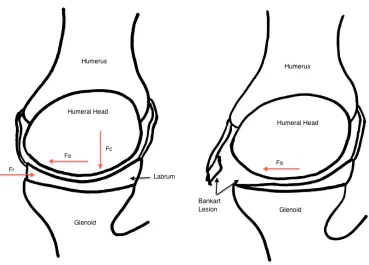
Figure 1-7. Glenohumeral Joint Concavity-Compression Effect.
Schematic diagram demonstrating the normal resistive force provided by the capsule and labrum (Fr) in response to an anterior translatory force applied to the humerus (Fa) (left). The concave shape of the glenoid and attached labrum help to contain the relatively spherical humeral head, providing resistance to translation. Additionally, the rotator cuff muscles and biceps provide a compression force (Fc) that keeps the humeral head centered in the glenoid concavity.
Limitations of the concavity-compression effect are seen in the circumstances of
weakness of the rotator cuff and injuries that further reduce the size of the glenoid
relative to the humeral head (24). Decreases in rotator cuff muscle strength of 50% have
been demonstrated to result in a 50% increase in anterior translation of the humeral head
(25). The glenoid size contributes to the scapulohumeral balance, which is the concept
that the glenoid must remain appropriately positioned to resist the forces applied through
the humeral head to allow joint stability. A larger glenoid will have a larger effective
1.3
Shoulder Instability
When the static and dynamic stabilizers fail and the balance between motion and
stability is disrupted, often by an applied external force, the result is typically an anterior
shoulder dislocation. While posterior and inferior dislocations can also occur, anterior
dislocations make up 85-95% of all shoulder dislocations and will thus be the focus of
this thesis (27,28).
The majority of dislocations occur in younger patients, with the average age of
injury being in the 20’s. Additionally, there is a male preponderance, representing
85-90% of all dislocations (27). The traditional mechanism of injury is often during sporting
events in the aforementioned ‘position of apprehension’ with the arm abducted to
approximately 90º and held in external rotation. A subsequent external rotation, and
often extension, force is applied and levers the humeral head out of the glenoid concavity.
However, additional patients may experience dislocations after a fall onto their
outstretched hand or onto their adducted shoulder, although these mechanisms are not as
common. Atraumatic dislocations can occur, particularly in those experiencing
multi-directional instability with predisposing factors, such as ligamentous laxity. These
dislocations represent the minority, and will not be discussed.
1.3.1
Pathophysiology
Typically, a shoulder dislocation will result from failure of one of the stabilizing
structures. Although initially soft-tissue injuries were thought to be the primary
pathology involved, it is now being recognized that bony restraints are equally involved
1.3.1.1
Soft-Tissue Pathology
Elderly patients frequently experience a disruption of the rotator cuff with their
initial dislocation (27). In younger patients, the majority of dislocations are associated
with tearing of the capsule and/or labrum – commonly called a ‘capsulolabral’ injury –
away from the anterior glenoid rim. This lesion, first described in 1923 by Bankart and
referred to by subsequent authors as a “Bankart lesion”, has been identified in 84-90% of
patients following their initial dislocation (27). The location of capsule and labrum
involved is typically at the insertion of the anterior band of the inferior GHL and middle
GHL at the anteroinferior portion of the glenoid rim, with injury to these structures often
part of the primary dislocation (Figure 1-7 above) (29). With these structures involved,
the shoulders ability to resist anterior translation, particularly with the arm abducted, is
dramatically reduced and the patient is predisposed to future dislocations. In fact,
resection of the labrum alone was found to reduce the resistance to anterior translation by
20% (24). An additional cadaveric study examined the effect of a chondrolabral lesion
on glenoid depth and shoulder stability, and found reductions of 80% and 65%
respectively, significantly decreasing shoulder stability in this setting (30).
1.3.1.2
Bony Pathology – Complex Instability
In addition to the soft-tissue pathology, anterior dislocations are frequently
associated with osseous lesions of the humeral head, glenoid, or both. When bony
lesions contribute to ongoing shoulder instability, it is appropriately known as ‘complex
instability’. In recent years, attention has been drawn to identification and management
of these osseous lesions because of their reported contribution to failure of soft-tissue
targeted stabilization procedures, with recurrence rates of up to 67% when these lesions
are not addressed (30–36)
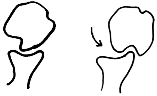
With disruption of the anterior capsulolabral structures, the humeral head is able
to translate anteriorly and inferiorly as it dislocates from the glenoid fossa. The
posterosuperolateral aspect of the humeral head then impacts on anterior glenoid rim
(37). The humeral head is largely made up of less dense, cancellous bone and
Sachs lesion, has been described throughout history (Figure 1-8). The earliest description
appeared in 1861 by Flowers, but it was not until 1940 when Hill and Sachs published a
concise review that the lesion adopted their names (26,37,38). This lesion has been
identified in up to 90% of patients following initial dislocations and 100% of patients
Figure 1-8. Bankart Lesion and Hill-Sachs Defect
Although the effect of the Hill-Sachs lesion is variable and depends largely on its
size, defects can ‘engage’ the anterior glenoid rim facilitating recurrent instability
(32,37,41). The term ‘engage’, described by Burkhart and De Beer, simply means that in
certain arm positions, mostly abduction and external rotation, the axis of the Hill-Sachs
lesion will match that of the anterior glenoid rim, allowing the humeral head to translate
anteriorly over the glenoid rim as the defect ‘engages’ the rim (Figure 1-9) (32,37). As a
result, the Hill-Sachs lesion has been recognized as a significant contributor to failed
Figure 1-9. Hill-Sachs Defect Engagement in External Rotation
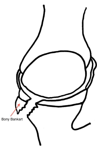
Glenoid defects may result from either attritional wear with bone loss from
repetitive dislocations, or an acute avulsion fracture of the anterior glenoid rim with the
attached capsulolabral complex (36,43). Because this is often seen as an extension of the
capsulolabral injury, it is frequently referred to as a ‘bony Bankart’ lesion (Figure 1-10)
(42,44). Glenoid defects are seen in 22% of patients following their initial dislocation
(39) and in up to 75% of those experiencing recurrent shoulder instability
(35,36,45)(35,36). Similar to the Hill-Sachs lesion, the effect of anterior glenoid bone
loss is variable, and often depends on the size of the segment involved. Loss of a
segment of the glenoid reduces the effective glenoid arc length and the
compressive-concavity restraint, reducing the glenoid’s ability to resist axial forces transmitted by the
humeral head (36,43). As a result, the ability to resist anterior translation is reduced,
especially with co-existing injury to the capsulolabral structures, and the shoulder is
Figure 1-10. Bony Bankart Lesion
Following a traumatic anterior shoulder dislocation, occasionally a more significant anterior injury can occur involving a portion of the glenoid rim. On this axial (overhead) diagram, a bony
1.4
Treatment Options
1.4.1.1
Immobilization
Initial management of an acute shoulder dislocation involves immediate
reduction. Following this, some have advocated for a period of immobilization of 3 or
more weeks in a sling, while others have only utilized a sling in the acute phase (1 week)
for patient comfort (46–50). Debate also existed over whether splinting in a position of
abduction and external rotation would improve Bankart healing against the glenoid neck.
Immobilization was also typically followed by physiotherapy focusing on range of
motion and strengthening of the shoulder and eventual return to sport/activities. While a
consensus was lacking early on for the appropriate duration of immobilization,
meta-analyses have shown a lack of benefit of sling immobilization beyond 1 week post-injury
and that the benefit of holding the limb in abduction and/or external rotation was not
reproducible among different populations (50).
1.4.1.2
Bankart Repair (Soft-tissue Stabilization)
Following initial immobilization, further debate existed as to when patients
should receive surgical referrals. Two theories existed, the first being an urgent referral
to consider stabilization following the initial dislocation, or alternatively, a
“wait-and-see” approach could be taken to see if recurrent instability developed, warranting referral
and eventual surgical stabilization (51). Stabilization procedures could then be
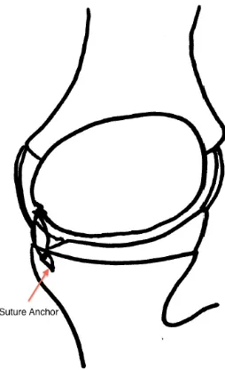
performed either open or arthroscopically, with the aim being to repair the Bankart lesion
by reattaching the torn labrum and capsule to the anterior glenoid rim, typically utilizing
suture anchors (Figure 1-11). Suture anchors, which are small threaded screws with
attached suture material, are placed into the glenoid rim in the region where the labrum
has been detached (Bankart lesion). The suture material is then passed around the labrum
and glenohumeral ligaments and used to pull these tissues back down to the bony glenoid
rim. This restores the “bumper effect” provided by the anterior labrum to resist
translation of the humeral head, re-tensions the glenohumeral ligaments in the at-risk
Several studies investigated acute versus delayed stabilization and found that the
procedures were equally effective in both groups, but that perhaps a certain subset of
patients (i.e. younger, with riskier sports like rugby or football) would benefit from
stabilization after their initial dislocation (51,53,54). Overall success rates for a Bankart
repair have been reported as 85 – 90% (55–57), with a recent long-term study indicating
no significant differences between recurrence rates for the arthroscopic and open
Figure 1-11. Bankart Stabilization
While the debate over which Bankart repair technique is best, open or
arthroscopic, has still not been convincingly settled, Burkhart and De Beer astutely noted
that failure rates of primary stabilization were substantially higher with both techniques
when associated osseous injuries of the glenoid or humeral head were not identified and
managed in conjunction with the Bankart lesion (32). When they retrospectively
analyzed their long-term outcomes following Bankart repair, they found 67% of patients
with osseous defects of either the glenoid or humeral head experienced a recurrent
episode of instability, compared to only 4% of those who did not have these associated
lesions (32). More recently, Balg and Boileau (2007) have confirmed the significance of
associated lesions of the humeral head or glenoid after retrospective review of a cohort of
patients following soft-tissue repair, noting increased rates of recurrent instability in those
with Hill-Sachs lesions or glenoid defects as well. They have included both injuries as
salient points on the ‘Instability Severity Index Score’, a tool they designed to help
identify patients that would benefit from open surgical stabilization to address bony
deficits (59).
The results of these studies have shifted attention towards managing these
associated osseous lesions that predispose to further episodes instability. However,
critical defect values and the corresponding standard of care for each are still being
defined, with numerous treatment options currently available.
1.4.1.3
Glenoid Arc Reconstruction
Around the time that Burkhart and De Beer noted the increased rate of recurrent
shoulder instability associated with osseous injuries, Itoi et al. (2000) (33) performed a
cadaveric study looking at the stability provided with a standard Bankart repair in the
setting of increasing glenoid defects. They found that once a critical defect value of 21%
of the width of the glenoid was surpassed, that an isolated Bankart repair was insufficient
in restoring stability and that alternative procedures to address the glenoid defect would
be required (33). Other studies have confirmed that defects >25% would benefit from
transfers. Additional procedures have included iliac crest bone graft reconstruction or
allograft bone reconstruction, although they are both not as common as the coracoid
transfers and will not be described here.
The Bristow coracoid transfer was initially described in 1958 by Helfet, but
named after his mentor Rowley Bristow (62). The initial description of the procedure
involved osteotomizing the distal half-inch of the coracoid maintaining its attached
conjoined tendon. A small vertical split was then made in the subscapularis tendon to
pass the coracoid segment, with its attached tendon, through this split and against the
anterior inferior glenoid rim (Figure 1-12). It was secured by incorporating the conjoined
tendon in the subscapularis repair (62–64). The primary effect of this, once healed, was
to allow the conjoined tendon to provide a sling-like buttress anteriorly, enhanced with
the arm in an abducted position, essentially mimicking the function of the middle and
inferior glenohumeral ligaments (64). Additionally, with the passage of the graft through
a split in the subscapularis, the inferior fibers of the subscapularis are also tensioned as
the arm is abducted to further resist anterior translation. The description of the Bristow
was later modified by May to include single screw fixation along the axis of the coracoid
fragment (“standing position”) with the undersurface of the coracoid sitting flush with the
glenoid face (65,66). In addition to the sling-effect of the conjoint tendon, this
modification added the benefit of an anterior bone block, which was more consistent with
descriptions of the Latarjet procedure, explained below. Early results of the Bristow
Figure 1-12. Bristow Coracoid Transfer
Latarjet (1954) had described a similar procedure to Helfet’s a few years prior,
involving a coracoid transfer and passing this segment with its attached conjoined tendon
through a split in the subscapularis as well (68). However, Latarjet’s technique involved
utilizing the entire horizontal component of the coracoid, and re-orienting the fragment,
fixing it such that the inferior surface of the coracoid was held against the scapular neck
with two screws (Figure 1-13) (63,64). Similarly, this allowed both a bony block effect
to increase the articular arc that is able to resist compressive forces by the humerus, while
also providing the sling-effect of the conjoined tendon and lower subscapularis in the
abducted and externally rotated positions to resist anteroinferior translation or dislocation
Figure 1-13. Latarjet Coracoid Transfer
A sagittal projection of the glenoid and coracoid process are shown (left) with their normal
1.4.1.4
Humeral Head Reconstruction
Similar to the glenoid defect, the significance of the humeral Hill-Sachs lesion
increases with its size. It has been commonly accepted that small lesions (<20-25% of
humeral head width) can generally be treated with benign neglect, simply managing the
associated soft-tissue Bankart lesion with either arthroscopic or open stabilization with
relatively good success (37,72,73). Alternatively lesions >40% are almost always seen as
clinically significant and at risk of “engaging” the glenoid rim, facilitating a dislocation.
Cadaveric studies have confirmed that defects of this size reproducibly lead to increased
shoulder instability (34,74). As a result, lesions of this size are generally treated
surgically with one of several options, including bone grafting (allograft) or rotational
osteotomies of the proximal humerus (37). Lesions between 20-40% represent a current
‘gray-zone’, although additional factors, such as the orientation of the defect and the
presence of additional capsulolabral injuries, may contribute to instability associated with
these lesions and often require them to be managed surgically (37,74,75). Common
treatment options for these slightly smaller defects include the remplissage procedure and
the Latarjet coracoid transfer, both which attempt to limit Hill-Sachs defect engagement.
Remplissage is a French term, which means simply ‘filling’. This procedure,
initially described by Connolly in 1972, involves imbricating the infraspinatus and
posterior capsule into the Hill-Sachs defect with suture anchors (Figure 1-14) (76,77).
This essentially makes the lesion extra-articular so that it can no longer engage the
anterior glenoid rim as a result of the “bumper” of soft-tissue created. Additionally, the
posterior soft tissues act as a tether to reduce the amount of anterior translation in the
shoulder, again preventing dislocation (77). Purchase et al. (2008) described an
arthroscopic version of this procedure, which was subsequently modified by Koo et al.
Figure 1-14. Remplissage Procedure
Regardless of the individual technique modifications, the effect remains the same,
which is to reduce the ability of the Hill-Sachs defect to engage the glenoid rim.
Biomechanical cadaveric studies have shown that in conjunction with a Bankart repair,
the addition of the remplissage procedure helped to significantly improve shoulder
stability with a 30% Hill-Sachs defect (80). Clinical studies have also confirmed the
success of the procedure with instability recurrence rates of only 2-8% in long-term
follow-up (77,78).
The Latarjet coracoid transfer, as described in the previous section, can also be
performed in the setting of an engaging Hill-Sachs defect. The goal of the procedure is to
provide the same sling-effect instilled by the conjoined tendon and subscapularis, but also
to increase the articular arc length to prevent the humeral head from engaging the glenoid
(37,42). The biomechanical and clinical effects of this procedure for an engaging
1.5
Study Rationale
While numerous treatment options have been described in the management of
complex shoulder instability, evidence to support their use has largely been limited to
reports involving small numbers of patients, while only occasional long-term reports have
been identified. Few comparative studies of these procedures exist, particularly focusing
on the biomechanics of the repairs.
As demonstrated above, glenoid defects following an anterior shoulder dislocation
with an associated capsulolabral injury have been clinically suspected, and cadaverically
demonstrated, to be a cause for recurrent dislocation when managed with a simple
Bankart repair (32,33). It has been accepted that glenoid defects >25% of the total width
should be addressed, with the recommended treatment being a coracoid transfer, often
referred to as a “Bristow-Latarjet” coracoid transfer (44,59,61,71,81–83). These
procedures have frequently been referred to synonymously, and their success collectively
has been well documented (66,67,84). However, their technical descriptions have
differed, as outlined above, while their biomechanical equivalence has never been
demonstrated.
Similarly, Hill-Sachs lesions of the humeral head have also been associated with
recurrent dislocations following soft-tissue stabilization procedures (31,32,35,41,76). As
identified, larger lesions, generally >20% of humeral head width, are best treated
surgically, although the best procedure for this size of defect has not been determined.
Presently, clinical focus has been on two of the described treatment options – the
remplissage procedure, and the Latarjet coracoid transfer, as detailed above. Both
reportedly have had reasonable success rates, although no comparative studies exist to
determine which is clinically or biomechanically superior.
Finally, while the Latarjet coracoid transfer potentially represents a treatment
option for both engaging Hill-Sachs lesions and large glenoid defects, the only procedural
complications usually listed are related to technical points, such as acute neurological
biomechanical effects of the procedure exist particularly on the effect that resection of the
coracoacromial ligament may have on superior shoulder stability.
1.6 Objectives
The objectives of this thesis were threefold:
1. To compare the stabilizing effect of the Bristow coracoid transfer and the
Latarjet coracoid transfer in the setting of an intact glenoid, and with 15% and 30%
glenoid defects,
2. To compare the stabilizing effect of the remplissage and Latarjet coracoid
transfer in the setting of an engaging 25% Hill-Sachs defect, and
3. To compare the degree of superior shoulder instability following Latarjet
coracoid transfer and alternative bone block procedures not requiring coracoacromial
ligament resection.
1.7 Hypotheses
The hypotheses for the above objectives were:
1. The Latarjet coracoid transfer will provide improved shoulder stability in
comparison to the Bristow coracoid transfer for all glenoid defect states.
2. The Latarjet coracoid transfer will provide improved shoulder stability,
and less restricted range of motion compared to the remplissage procedure in the
3. The Latarjet coracoid transfer will result in greater superior shoulder
migration compared to bone augmentation procedures that do not violate the
coracoacromial ligament.
1.8 Thesis Overview
Chapter 2 compares the Bristow and Latarjet coracoid transfers for various
glenoid defect states. Chapter 3 compares the remplissage and Latarjet procedures for
treatment of an engaging Hill-Sachs lesion. Chapter 4 compares the degree of superior
translation conferred by performing the Latarjet versus bone grafting procedures that
preserve the coracoacromial ligament. Chapter 5 provides a general discussion, summary
1.9 References
1. Tortora GJ, Grabowski SR. Principles of Anatomy and Physiology, Tenth Edition.
10th ed. Roesch B, editor. Hoboken, NJ: John Wiley & Sons, Inc.; 2003. p. 1104.
2. Swarm DL, Mahar AT, Weichel DW, Pedowitz RA. Shoulder Anatomy and
Biomechanics. In: Pedowitz RA, Johnson DH, editors. Practical Orthopaedic
Sports Medicine & Arthroscopy. Pennsylvania, PA: Lippincott Williams &
Wilkins; 2007. p. 145–56.
3. PrimalPicturesLtd. 3D Anatomy images. Riverwoods, Illinois: Primal Pictures
Ltd.; 2006.
4. Terry GC, Chopp TM. Functional Anatomy of the Shoulder. Journal of Athletic
Training. 2000;35(3):248–55.
5. Abboud JA, Soslowsky LJ. Interplay of the static and dynamic restraints in
glenohumeral instability. Clinical orthopaedics and related research. 2002
Jul;(400):48–57.
6. Hurov J. Anatomy and mechanics of the shoulder: review of current concepts.
Journal of hand therapy : official journal of the American Society of Hand
Therapists. 2009;22(4):328–42; quiz 343.
7. Plausinas D, Jazrawi LM, Zuckerman JD, Rokito AS. Anatomy and Biomechanics
of the Shoulder. In: Schepsis AA, Busconi BD, editors. Sports Medicine.
Pennsylvania, PA; 2006. p. 169.
8. Williams MM, Snyder SJ, Buford D. The Buford complex--the “cord-like” middle
glenohumeral ligament and absent anterosuperior labrum complex: a normal
anatomic capsulolabral variant. Arthroscopy : the journal of arthroscopic &
related surgery : official publication of the Arthroscopy Association of North
America and the International Arthroscopy Association. 1994 Jun;10(3):241–7.
9. Burkart AC, Debski RE. Anatomy and function of the glenohumeral ligaments in
anterior shoulder instability. Clinical orthopaedics and related research. 2002
10. Felli L, Biglieni L, Fiore M, Coviello M, Borri R, Cutulo M. Functional study of
glenohumeral ligaments. Journal of orthopaedic science : official journal of the
Japanese Orthopaedic Association. 2012 Sep;17(5):634–7.
11. Pope EJ, Ward JP, Rokito AS. Anterior shoulder instability - a history of
arthroscopic treatment. Bulletin of the NYU hospital for joint diseases. 2011
Jan;69(1):44–9.
12. Hunt SA, Kwon YW, Zuckerman JD. The rotator interval: anatomy, pathology,
and strategies for treatment. The Journal of the American Academy of Orthopaedic
Surgeons. 2007 Apr;15(4):218–27.
13. Hockman DE, Lucas GL, Roth CA. Role of the coracoacromial ligament as
restraint after shoulder hemiarthroplasty. Clinical orthopaedics and related
research. 2004 Feb;(419):80–2.
14. Moorman CT, Warren RF, Deng XH, Wickiewicz TL, Torzilli PA. Role of
coracoacromial ligament and related structures in glenohumeral stability: a
cadaveric study. Journal of surgical orthopaedic advances. 2012 Jan;21(4):210–7.
15. Hoppenfeld S. Physical Examination of the Shoulder. Physical Examination of the
Spine & Extremities. Upper Saddle River, NJ: Prentice Hall, Pearson Education;
1976. p. 1–34.
16. Labriola JE, Lee TQ, Debski RE, McMahon PJ. Stability and instability of the
glenohumeral joint: the role of shoulder muscles. Journal of shoulder and elbow
surgery / American Shoulder and Elbow Surgeons ... [et al.]. 2005;14(1 Suppl
S):32S–38S.
17. Curl LA, Warren RF. Glenohumeral joint stability. Selective cutting studies on the
static capsular restraints. Clinical orthopaedics and related research. 1996
Sep;(330):54–65.
18. Alexander S, Southgate DFL, Bull AMJ, Wallace AL. The role of negative
intraarticular pressure and the long head of biceps tendon on passive stability of
the glenohumeral joint. Journal of shoulder and elbow surgery / American
Shoulder and Elbow Surgeons ... [et al.]. 2013 Jan;22(1):94–101.
19. Kikuchi K, Itoi E, Yamamoto N, Seki N, Abe H, Minagawa H, et al. Scapular
2008 Jan;13(1):72–7.
20. Blasier RB, Guldberg RE, Rothman ED. Anterior shoulder stability: Contributions
of rotator cuff forces and the capsular ligaments in a cadaver model. Journal of
shoulder and elbow surgery / American Shoulder and Elbow Surgeons ... [et al.].
1992 May;1(3):140–50.
21. Lippitt SB, Matsen FA. Mechanisms of glenohumeral joint stability. Clinical
orthopaedics and related research. 1993 Jun;(291):20–8.
22. Itoi E, Motzkin NE, Morrey BF, An KN. Stabilizing function of the long head of
the biceps in the hanging arm position. Journal of shoulder and elbow surgery /
American Shoulder and Elbow Surgeons ... [et al.]. 1994 May;3(3):135–42.
23. Itoi E, Newman SR, Kuechle DK, Morrey BF, An KN. Dynamic anterior
stabilisers of the shoulder with the arm in abduction. The Journal of bone and joint
surgery. British volume. 1994 Sep;76(5):834–6.
24. Lippitt SB, Vanderhooft JE, Harris SL, Sidles JA, Harryman DT, Matsen FA.
Glenohumeral stability from concavity-compression: A quantitative analysis.
Journal of shoulder and elbow surgery / American Shoulder and Elbow Surgeons
... [et al.]. 1993 Jan;2(1):27–35.
25. Wuelker N, Korell M, Thren K. Dynamic glenohumeral joint stability. Journal of
shoulder and elbow surgery / American Shoulder and Elbow Surgeons ... [et al.].
1998;7(1):43–52.
26. Matsen FA, Thomas SC, Rockwood Jr CA, Wirth MA. Glenohumeral Instability.
In: Rockwood Jr CA, Matsen FA, Wirth MA, Harryman DT, editors. The
Shoulder. 2nd ed. Philadelphia, PA: W.B. Saunders Company; 1998. p. 611–754.
27. Liu SH, Henry MH. Anterior shoulder instability. Current review. Clinical
orthopaedics and related research. 1996 Feb;(323):327–37.
28. Mahaffey BL, Smith PA. Shoulder instability in young athletes. American family
physician. 1999 May 15;59(10):2773–82, 2787.
29. Bicos J, Mazzocca AD, Arciero RA. Anterior Instability of the Shoulder. In:
30. Lazarus MD, Sidles JA, Harryman DT, Matsen FA. Effect of a chondral-labral
defect on glenoid concavity and glenohumeral stability. A cadaveric model. The
Journal of bone and joint surgery. American volume. 1996 Jan;78(1):94–102.
31. Boileau P, Villalba M, Héry JY, Balg F, Ahrens P, Neyton L. Risk factors for
recurrence of shoulder instability after arthroscopic Bankart repair. The Journal of
bone and joint surgery. American volume. 2006 Aug;88(8):1755–63.
32. Burkhart SS, De Beer JF. Traumatic glenohumeral bone defects and their
relationship to failure of arthroscopic Bankart repairs: significance of the
inverted-pear glenoid and the humeral engaging Hill-Sachs lesion. Arthroscopy : the
journal of arthroscopic & related surgery : official publication of the Arthroscopy
Association of North America and the International Arthroscopy Association. 2000
Oct;16(7):677–94.
33. Itoi E, Lee SB, Berglund LJ, Berge LL, An KN. The effect of a glenoid defect on
anteroinferior stability of the shoulder after Bankart repair: a cadaveric study. The
Journal of bone and joint surgery. American volume. 2000 Jan;82(1):35–46.
34. Kaar SG, Fening SD, Jones MH, Colbrunn RW, Miniaci A. Effect of humeral head
defect size on glenohumeral stability: a cadaveric study of simulated Hill-Sachs
defects. The American journal of sports medicine. 2010 Mar;38(3):594–9.
35. Lynch JR, Clinton JM, Dewing CB, Warme WJ, Matsen FA. Treatment of osseous
defects associated with anterior shoulder instability. Journal of shoulder and elbow
surgery / American Shoulder and Elbow Surgeons ... [et al.]. 2009;18(2):317–28.
36. Piasecki DP, Verma NN, Romeo AA, Levine WN, Bach BR, Provencher MT.
Glenoid bone deficiency in recurrent anterior shoulder instability: diagnosis and
management. The Journal of the American Academy of Orthopaedic Surgeons.
2009 Aug;17(8):482–93.
37. Provencher MT, Frank RM, Leclere LE, Metzger PD, Ryu JJ, Bernhardson A, et
al. The Hill-Sachs lesion: diagnosis, classification, and management. The Journal
of the American Academy of Orthopaedic Surgeons. 2012 Apr;20(4):242–52.
38. Hill HA, Sachs MD. The Grooved Defect of the Humeral Head: A Frequently
Unrecognized Complication of Dislocations of the Shoulder Joint. Radiology.
Arthroscopic and physical examination findings in first-time, traumatic anterior
dislocations. The American journal of sports medicine. 1997;25(3):306–11.
40. Calandra JJ, Baker CL, Uribe J. The incidence of Hill-Sachs lesions in initial
anterior shoulder dislocations. Arthroscopy : the journal of arthroscopic & related
surgery : official publication of the Arthroscopy Association of North America
and the International Arthroscopy Association. 1989 Jan;5(4):254–7.
41. Burkhart SS, De Beer JF, Barth JRH, Cresswell T, Criswell T, Roberts C, et al.
Results of modified Latarjet reconstruction in patients with anteroinferior
instability and significant bone loss. Arthroscopy : the journal of arthroscopic &
related surgery : official publication of the Arthroscopy Association of North
America and the International Arthroscopy Association. 2007 Oct;23(10):1033–
41.
42. Chen AL, Hunt SA, Hawkins RJ, Zuckerman JD. Management of bone loss
associated with recurrent anterior glenohumeral instability. The American journal
of sports medicine. 2005 Jun;33(6):912–25.
43. Bollier MJ, Arciero R. Management of glenoid and humeral bone loss. Sports
medicine and arthroscopy review. 2010 Sep;18(3):140–8.
44. Bushnell BD, Creighton RA, Herring MM. Bony instability of the shoulder.
Arthroscopy : the journal of arthroscopic & related surgery : official publication
of the Arthroscopy Association of North America and the International
Arthroscopy Association. 2008 Sep;24(9):1061–73.
45. Bigliani LU, Newton PM, Steinmann SP, Connor PM, Mcllveen SJ. Glenoid rim
lesions associated with recurrent anterior dislocation of the shoulder. The
American journal of sports medicine. 1998;26(1):41–5.
46. Seybold D, Gekle C, Fehmer T, Pennekamp W, Muhr G, Kälicke T.
[Immobilization in external rotation after primary shoulder dislocation]. Der
Chirurg; Zeitschrift für alle Gebiete der operativen Medizen. 2006 Sep;77(9):821–
47. Scheibel M, Kuke A, Nikulka C, Magosch P, Ziesler O, Schroeder RJ. How long
should acute anterior dislocations of the shoulder be immobilized in external
rotation? The American journal of sports medicine. 2009 Jul;37(7):1309–16.
48. Liavaag S, Stiris MG, Lindland ES, Enger M, Svenningsen S, Brox JI. Do Bankart
lesions heal better in shoulders immobilized in external rotation? Acta
orthopaedica. 2009 Oct;80(5):579–84.
49. Dines DM, Levinson M. The conservative management of the unstable shoulder
including rehabilitation. Clinics in sports medicine. 1995 Oct;14(4):797–816.
50. Paterson WH, Throckmorton TW, Koester M, Azar FM, Kuhn JE. Position and
duration of immobilization after primary anterior shoulder dislocation: a
systematic review and meta-analysis of the literature. The Journal of bone and
joint surgery. American volume. 2010 Dec 15;92(18):2924–33.
51. Kirkley A, Werstine R, Ratjek A, Griffin S. Prospective randomized clinical trial
comparing the effectiveness of immediate arthroscopic stabilization versus
immobilization and rehabilitation in first traumatic anterior dislocations of the
shoulder: Long-term evaluation. Arthroscopy: The Journal of Arthroscopic &
Related Surgery. 2005 Jan;21(1):55–63.
52. Yamamoto N, Muraki T, Sperling JW, Steinmann SP, Itoi E, Cofield RH, et al.
Does the “bumper” created during Bankart repair contribute to shoulder stability?
Journal of shoulder and elbow surgery / American Shoulder and Elbow Surgeons
... [et al.]. 2012 Sep 27;
53. Grumet RC, Bach BR, Provencher MT. Arthroscopic stabilization for first-time
versus recurrent shoulder instability. Arthroscopy : the journal of arthroscopic &
related surgery : official publication of the Arthroscopy Association of North
America and the International Arthroscopy Association. 2010 Feb;26(2):239–48.
54. Owens BD, DeBerardino TM, Nelson BJ, Thurman J, Cameron KL, Taylor DC, et
al. Long-term follow-up of acute arthroscopic Bankart repair for initial anterior
shoulder dislocations in young athletes. The American journal of sports medicine.