Platelet Function during Platelet-Rich Plasma
Sequestration in Complex Cardiac Surgical
Procedures - Prospective Controlled Study
Slavik L*
1, Hajek R
2, Chaloupkova P
1, Ulehlova J
1and Lonsky V
21Department of Hemato-Oncology Faculty of Medicine and Dentistry Palacky University Olomouc and University Hospital Olomouc
2Department of Cardiac Surgery, Faculty of Medicine and Dentistry, Palacky University Olomouc; University Hospital Olomouc
Received: June 18, 2018; Published: June 28, 2018
*Corresponding author: Luděk Slavík, Department of Cardiac Surgery, Republic of Czech
Research Article
Open Access
ISSN: 2574-1241Introduction
Cardiopulmonary bypass (CPB) is associated with common activation of all four integral components of hemostasis, that is, the endothelium, plasma proteins, platelets and fibrinolysis. The causative factors include the presence of an artificial non-endothelial surface of the CPB system, non-pulsatile blood flow, hemodilution, hypothermia, surgical trauma and a systemic inflammatory response to CPB. Microthrombi formation, coagulation defects and hypercoagulability in the postoperative period may occur. The key pathway of activation is platelet binding to collagen via von Willebrand factor, platelet activation and aggregation and formation of an initial hemostatic plug. Thrombin generation (tF+fVIIa →fIXa →fXa →prothrombin →thrombin) activates platelets, fV, fVIII and fXI. Fibrinogen and fXIII stabilize the clot. Fibrinogen bound to the CPB circuit provides a strong binding site for platelets via the GP IIb/IIIa receptor. Bound platelets are activated, promoting further thrombin formation via the PAR-1 receptor. Shed blood, if reinfused, increases platelet activation. Plasmin erodes the clot and directly activates platelet consumption [1-15].
The use of antifibrinolytics preserves platelet activation and reduces platelet GPIb receptor cleavage [16]. Thrombocytopenia during and after CPB is common but an isolated platelet count below 50G/L is usually not associated with serious bleeding if no other hemostasis disorders are present [17]. Platelet function is slightly increased by mild hypothermia and decreased by severe hypothermia. Some commonly used drugs other than heparin;
protamine and platelet inhibitors reduce platelet activation or aggregation (NO donors such as nitrates or phosphodiesterase inhibitors such as milrinone). In summary, platelets are one of the most fragile components of hemostasis during CPB. Current cell-salvage techniques are capable of preserving platelets by autologous platelet-rich plasma (PRP) sequestration. The present study tested platelet function during and after PRP sequestration in cardiac surgery procedures using CPB. We hypothesized that platelet count and aggregability would be preserved by the method.
Methods
Patients were enrolled after their written informed consent and local ethics committee approval were obtained. The study comprised patients scheduled for elective cardiac surgery with an estimated CPB time of more than two hours (complex procedures such as multiple valvular, combined, redo and thoracic aortic surgery). The inclusion criterion was initial hematocrit >0.35 as 800ml of whole blood had to be collected and processed before CPB. Excluded were patients with hematocrit <0.35, on active antiplatelet agents or known to have hematological disorders. The CPB method included the Capiox RX 25 membrane oxygenator with rheoparin coating, Stöckert S5 centrifugal pump, crystalloid/colloid priming (Plasmalyte1000mL, HAES 6% 500mL, Mannitol 20% 200mL) and St. Thomas cold blood cardioplegia. Anticoagulation was provided with unfractionated heparin (UFH) as follows: 3 mg/ kg i.v. bolus + 1mg/kg into the priming volume, a targeted activated Keywords: Fibrinolysis; Hypothermia; Hypercoagulability; Non-Pulsatile; Hemostasis; Hemodilution; Trauma; Coagulation; Thrombocytopenia; Hemostatic Plug
coagulation time (ACT) (Hemochron, kaolinactivated) >400s, with possible additional bolus doses of UFH. Heparin reversal after CPB termination was accomplished with protamine sulphate at a 1:1 weight ratio to heparin.
Prophylactic administration of tranexamic acid 30mg/kg i.v. bolus and 15mg/kg into CPB was mandatory. After induction of general anesthesia, 800mL of whole blood was collected from a large-bore (min. 8 Fr) central venous catheter and replaced with an adequate volume of a balanced crystalloid solution. Blood was processed in the Sorin Xtra cell saver (LivaNova, Italy). The manufacturer’s protocol was used (175ml Latham bowl, 2-port sequestration system, CPD-A bags for whole blood collection, Vacutainer EDTA tubes, prime flow 100ml/min with manual reduction to 70mL/min after complete filling, PRP spill flow 20 ml/ min). Red blood cell (RBC) and PRP sequestration was performed simultaneously. PRP was stored at room temperature in the operating theater under an anesthetist’s supervision and always retransfused immediately after CPB before the end of surgery. RBCs were retransfused in the theater if necessary or postoperatively in the ICU according to hematocrit and the clinical situation. The transfusion protocol was guided by the center’s experiences and thromboelastography (TEG 5000, Haemonetics, USA); kaolin-activated plain and heparinase cups were used for each test. Blood samples were collected from an arterial line and processed in the laboratory before surgery, from PRP, after PRP retransfusion and at the end of surgery.
Blood Sampling
Blood was withdrawn from the antecubital vein using a 21-gauge butterfly needle (0.8 x 19mm; Greiner Bio- One, Kremsmünster, Austria) 72hours after percutaneous intervention. After the initial 3mL of blood had been discarded, to reduce procedurally induced platelet activation, blood was drawn into a 3.8% sodium citrate Vacuette tube (Greiner Bio-One; 9 parts of whole blood, 1 part of sodium citrate 0.129M/L) for evaluations by light transmission aggregometry, into a 3.2% sodium citrate Vacuette tube (Greiner Bio-One; 9 parts of whole blood, 1 part of sodium citrate 0.109M/L), and into a Vacuette tube containing hirudin (15IU/ml) for determination by multiple electrode platelet aggregometry. To avoid procedural deviations, all blood samples were taken by the same team using the same method. The blood samples were mixed adequately by gently inverting the tubes. To avoid investigator-related variation of results, each of the different tests was performed by just one single blind operator. The results of all assays were available to all patients.
Light Transmission Aggregometry (LTA)
LTAwas performed on the APACT4004 aggregometer (LABiTec, Ahrensburg, Germany). Citrate-anticoagulated whole blood was centrifuged at 150 x g for 10minutes at room temperature to obtain platelet-rich plasma (PRP). Platelet-poor plasma (PPP) was obtained from the remaining specimen by re-centrifugation at 2,000 x g for10 minutes. Platelet counts were not adjusted with a median platelet count of 250 x 1012/L (range 225 - 278 x 1012/L). The baseline optical density was set with PPP. Aggregation was
5μM), ristocetin (final conc. 1 mg/mL)and epinephrine (final conc. μm) (Helena Biosciences, United Kingdom) at a final concentration of 10. Optical density changes were recorded photoelectrically for 6 minutes as platelets began to aggregate. The maximal aggregation response was registered and used to differentiate between patients with and without residual ADP-inducible platelet aggregation.
Results
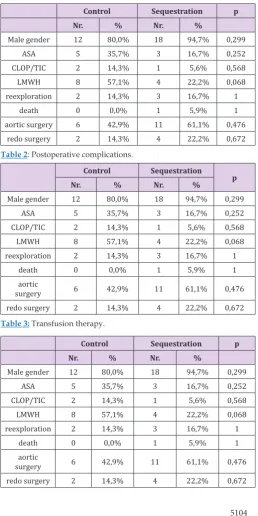
Enrolled in the study were 18 patients for a PRP sequestration group and 12 controls. The demography of both groups was similar (Table 1). There were no differences in major postoperative complications (Table 2) frequency of transfusion therapy (Table 3) CPB parameters, the only exception being a slightly longer maximum ACT on CPB in the PRP group which is clinically insignificant (Table 4) perioperative laboratory parameters (Table 5) and patients’ clinical outcome (Tables 6-8).
Table 1: Demography.
Control Sequestration p
Nr. % Nr. %
Male gender 12 80,0% 18 94,7% 0,299
ASA 5 35,7% 3 16,7% 0,252
CLOP/TIC 2 14,3% 1 5,6% 0,568
LMWH 8 57,1% 4 22,2% 0,068
reexploration 2 14,3% 3 16,7% 1
death 0 0,0% 1 5,9% 1
aortic surgery 6 42,9% 11 61,1% 0,476
redo surgery 2 14,3% 4 22,2% 0,672
Table 2: Postoperative complications.
Control Sequestration
p
Nr. % Nr. %
Male gender 12 80,0% 18 94,7% 0,299
ASA 5 35,7% 3 16,7% 0,252
CLOP/TIC 2 14,3% 1 5,6% 0,568
LMWH 8 57,1% 4 22,2% 0,068
reexploration 2 14,3% 3 16,7% 1
death 0 0,0% 1 5,9% 1
aortic
surgery 6 42,9% 11 61,1% 0,476
redo surgery 2 14,3% 4 22,2% 0,672
Table 3: Transfusion therapy.
Control Sequestration p
Nr. % Nr. %
Male gender 12 80,0% 18 94,7% 0,299
ASA 5 35,7% 3 16,7% 0,252
CLOP/TIC 2 14,3% 1 5,6% 0,568
LMWH 8 57,1% 4 22,2% 0,068
reexploration 2 14,3% 3 16,7% 1
death 0 0,0% 1 5,9% 1
aortic
Table 4: Parameters of CPB.
Control Sequestrtaion
p
Mean SD Median Mean SD Median
ACTstart (s) 135,8 16,6 133,5 131,3 14,4 133,5 0,423
ACTmax (s) 504,0 80,4 480,0 587,8 104,9 614,0 0,019
ACTend (s) 131,8 16,4 128,0 125,1 12,7 128,0 0,274
heparin (mg) 357,9 57,5 330,0 383,9 70,1 375,0 0,270
protamin (mg) 395,0 54,5 375,0 413,9 56,4 400,0 0,262
TT min (°C) 32,8 2,7 34,2 33,8 1,1 33,9 0,909
TT end (°C) 36,6 0,5 36,9 36,7 0,3 36,9 0,743
blood loss
periop.(ml) 564,3 253,0 500,0 605,6 336,9 500,0 0,705
blood loss 24h
(ml) 652,1 468,8 510,0 711,7 382,0 530,0 0,425
Euroscore (%) 3,62 2,05 2,96 3,37 2,84 2,00 0,077
Table 5: Laboratory.
Control Sequestrtaion p
Mean SD Median Mean SD Median
ACTstart (s) 135,8 16,6 133,5 131,3 14,4 133,5 0,423
ACTmax (s) 504,0 80,4 480,0 587,8 104,9 614,0 0,019
ACTend (s) 131,8 16,4 128,0 125,1 12,7 128,0 0,274
heparin (mg) 357,9 57,5 330,0 383,9 70,1 375,0 0,270
protamin (mg) 395,0 54,5 375,0 413,9 56,4 400,0 0,262
TT min (°C) 32,8 2,7 34,2 33,8 1,1 33,9 0,909
TT end (°C) 36,6 0,5 36,9 36,7 0,3 36,9 0,743
blood loss
periop.(ml) 564,3 253,0 500,0 605,6 336,9 500,0 0,705
blood loss 24h
(ml) 652,1 468,8 510,0 711,7 382,0 530,0 0,425
Euroscore (%) 3,62 2,05 2,96 3,37 2,84 2,00 0,077
Table 6: Outcome.
Control Sequestration
p
Mean SD Median Mean SD Median
ICULOS (day) 3,7 0,9 3,5 7,1 8,5 4,0 0,165
HLOS (day) 12,9 4,6 10,5 14,6 10,8 11,5 1
Age (years) 68,3 6,8 67,0 64,2 10,0 67,0 0,329
Table 7: Agregation of platelet before surgery (1).
Controls/patient
Mann-Whitney U test p
Controls Patients
Median Minimum Maximum Median Minimum Maximum
COL 1 43,0 2,0 99,0 5,3 1,0 28,0 0,0005
ADP 1 79,0 21,0 98,0 4,0 1,0 86,0 <0,0001
RISTO 1 95,0 70,0 99,0 99,0 72,0 100,0 0,010
Table 8A: Aggregation of platelet after the end of the surgical procedure (2).
Controls/patient
Mann-Whitney U test p
Controls Patients
Median Minimum Maximum Median Minimum Maximum
COL 2 43,0 2,0 99,0 5,3 1,0 28,0 0,0005
ADP 2 79,0 21,0 98,0 4,0 1,0 86,0 <0,0001
RISTO 2 95,0 70,0 99,0 99,0 72,0 100,0 0,010
EPI 2 37,0 7,0 99,0 5,0 1,0 94,0 <0,0001
Table 8B: Aggregation of platelet after retransfusion of platelet (3).
Controls/patient
Mann-Whitney U test p
Controls Patients
COL 3 Median Minimum Maximum Median Minimum Maximum
ADP 3 90,0 26,0 99,0 27,0 4,3 91,0 0,0002
RISTO 3 97,0 71,0 99,0 77,0 40,0 100,0 0,049
EPI 3 98,0 85,0 99,0 91,5 51,0 100,0 0,203
COL 3 91,0 30,0 99,0 26,0 8,0 94,0 0,0001
Discussion
There is little evidence about platelet preservation using PRP sequestration perioperatively. A recent study from the Texas Heart Institute compared a standard protocol with the use of PRP during aortic arch repair [18]. Early mortality, stroke and respiratory complications were similar between the groups. Only acute renal failure was reduced in the PRP group (7% vs 0%;p < 0.014). The mean transfusion rate of packed red blood cells was reduced by 34%, fresh frozen plasma by 52.8%, cryoprecipitate by 70% and platelets by 56.7% in the PRP group (p < 0.02). Hospital length of stay (9.4 ± 5.3 days vs 12.7 ± 6.3 days; p < 0.014) and transfusion costs ($1,396 ± $1,755 vs $2,762 ± $2,267; p < 0.004) were reduced in the PRP group. Unfortunately, no clinical impact on outcome and transfusion rate was observed in the present study.
We are fully aware of the fact that the present study is only preliminary, including a small number of patients and reporting our first experience with PRP sequestration. To the best of our knowledge, this was the first study focusing on platelet aggregation in this setting; however, the results are controversial. The technique of PRP sequestration probably did not considerably harm platelets because ristocetin-mediated aggregation was preserved, reaching about 90% of baseline levels. After stimulation of thromboxane A2 (TXA2) and ADP receptors, however, aggregation was rather decreased. This cannot be explained by the effect of antiplatelet therapy since most patients scheduled for valvular surgery received no antiplatelet agents and if yes, it was withdrawn according to the ESA/ESC guidelines. Retransfusion of PRP was associated with mildly increased aggregation after TXA2 receptor stimulation. Even hematologists in our group could not explain the phenomenon of selective preservation of ristocetinmediated platelet aggregation. Further tests of both morphological and functional integrity of platelets will be needed to determine their potential damage.
Conclusion
PRP sequestration may be a safe and useful method in complex cardiac surgery. It preserves platelet count and ristocetin-mediated platelet aggregation and partly restores aggregation mediated by other activators after CPB. Additional future randomized controlled trials may confirm these pilot results.
Acknowledgment
The study was supported by institutional grant of MH CZ – DRO (FNOl, 00098892) and LF-2018-004.
References
1. Emani S, Sleeper LA, Faraoni D, Mulone M, Diallo F, et al. (2018) Thromboelastography is Associated with Surrogates for Bleeding after Pediatric Cardiac Surgery. Ann Thorac Surg S0003-4975(18)30649-30650.
2. Di Dedda U, Ranucci M, Porta A, Bari V, Ascari A, et al. (2018) The combined effects of the microcirculatory status and cardiopulmonary bypass on platelet count and function during cardiac surgery. Clin Hemorheol Microcirc.
3. Fleming K, Redfern RE, March RL, Bobulski N, Kuehne M, et al. (2017) TEG-Directed Transfusion in Complex Cardiac Surgery: Impact on Blood Product Usage. J Extra Corpor Technol 49(4): 283-290.
4. Mongero L, Stammers A, Tesdahl E, Stasko A, Weinstein S (2018) The effect of ultrafiltration on end-cardiopulmonary bypass hematocrit during cardiac surgery. Perfusion 33(5): 367-374.
5. Rizza A, Di Felice G, Luciano R, Porzio O, Panizzon O, et al. (2017) Calibrated automated thrombogram values in infants with cardiac surgery before and after cardiopulmonary bypass.Thromb Res160:91-96.
Submission Link: https://biomedres.us/submit-manuscript.php
Assets of Publishing with us • Global archiving of articles
• Immediate, unrestricted online access
• Rigorous Peer Review Process
• Authors Retain Copyrights
• Unique DOI for all articles
https://biomedres.us/ This work is licensed under Creative
Commons Attribution 4.0 License
7. Olsson A, Alfredsson J, Ramström S, Svedjeholm R, Kenny D, (2018) Better platelet function, less fibrinolysis and less hemolysis in re-transfused residual pump blood with the Ringer’s chase technique - a randomized pilot study. Perfusion33(3):185-193.
8. Ranucci M, Baryshnikova E, Ciotti E, Ranucci M, Silvetti S (2017) Surgical and Clinical Outcome REsearch (SCORE) Group. Hemodilution on Cardiopulmonary Bypass: Thromboelastography Patterns and Coagulation-Related Outcomes. J Cardiothorac Vasc Anesth31(5):1588-1594.
9. Suehiro S, Shimizu K, Imai K, Niii A, Akeho K, et al. (2017) Polymer-coated cardiopulmonary bypass circuit attenuates upregulation of both proteases/protease inhibitors and platelet degranulation in pigs. Perfusion32(8):645-655.
10. Sivapalan P, Bäck AC, Ostrowski SR, Ravn HB, Johansson PI (2017) Transfusion requirements in elective cardiopulmonary bypass surgery patients: predictive value of Multiplate and Thromboelastography (TEG) Platelet Mapping Assay. Scand J Clin Lab Invest77(5):345-351.
11. Gruzdeva O, Uchasova E, Fanaskova E, Akbasheva O, Penskaya T, et al. (2017) Use of thrombin generation test for monitoring hemostasis in coronary bypass surgery. Clin Hemorheol Microcirc66(1):57-66.
12. Van Hout FM, Hogervorst EK, Rosseel PM, Van Der Bom JG, Bentala M, et al. (2017) Does a Platelet Transfusion Independently Affect Bleeding and Adverse Outcomes in Cardiac Surgery. Anesthesiology126(3):441-449.
13. Fabbro M, Gutsche JT, Miano TA, Augoustides JG, Patel PA (2016) Comparison of Thrombelastography-Derived Fibrinogen Values at Rewarming and Following Cardiopulmonary Bypass in Cardiac Surgery Patients. Anesth Analg123(3):570-577.
14. Mahla E, Prueller F, Farzi S, Pregartner G, Raggam RB, et al. (2016) Does Platelet Reactivity Predict Bleeding in Patients Needing Urgent Coronary Artery Bypass Grafting During Dual Antiplatelet Therapy. Ann Thorac Surg102(6):2010-2017.
15. Höfer J, Fries D, Solomon C, VelikSalchner C, Ausserer J (2016) A Snapshot of Coagulopathy After Cardiopulmonary Bypass. Clin Appl Thromb Hemost22(6):505-511.
16. Weber C, Klages M, Zacharowski K (2013) Perioperative coagulation management during cardiac surgery. Curr OpinAnest 26(1): 60-64. 17. Karkouti K, OFarrel R, Yau TM, Beattie WS (2006) Prediction of massive
blood transfusion in cardiac surgery. Can J Anaesth53(8):781-794.