R E V I E W
Management of patients with calciphylaxis: current
perspectives
This article was published in the following Dove Press journal: Chronic Wound Care Management and Research
Cornelia Erfurt-Berge Regina Renner
Department of Dermatology, University Hospital Erlangen, Erlangen 91054, Germany
Video abstract
Point your SmartPhone at the code above. If you have a QR code reader the video abstract will appear. Or use:
https://www.youtube.com/watch?v=_BtBinSIqLk
Abstract: Calciphylaxis (CP) is a rare disorder presenting with painful ischemic skin ulcerations. Its association with end-stage renal disease, but also non-uremic cases have been described. Several risk factors, like metabolic syndrome, calcium-phosphate imbal-ance, anticoagulative medication and female gender have been discussed. Multidisciplinary therapeutic approaches are necessary since evidence-based guidelines are missing due to lack of pathophysiological understanding of the disease and the absence of studies with large patient cohorts. However, strategies for local wound therapy, reduction of risk factors and systemic therapies are being developed. Changes in duration, frequency and method of hemodialysis and specific medication to lower the calcium upload can be effective in uremic CP cases. Systemic treatments with cinacalcet, sodium thiosulfate or bisphosphonates have been applied successfully in CP patients, but large placebo-controlled randomized trials are still needed to gather better insight into this fatal disease.
Keywords:calciphylaxis, calcific uremic arteriolopathy, non-uremic calciphylaxis, calcification, treatment, sodium thiosulfate
Calciphylaxis (CP) is a rare vasculopathic disorder associated with necrotic skin
ulcers and high mortality due to calcification of small cutaneous vessels. It has to be
discussed as differential diagnosis when unusual and recalcitrant chronic wounds appear. Since the exact pathogenesis remains unclear, different entities might be
subsumed under the term CP. Well known is the synonym calcific uremic
arteriolo-pathy (CUA) when the typical necrotic ulcers appear together with end-stage renal disease, a frequent observed co-morbidity. Around 1% of patients depending on
hemodialysis suffer from CUA.1 Most of the studies dealing with CP were
per-formed in patients with CUA. Typically, in the histological analysis of a deep
scalpel biopsy with skin and subcutaneous tissue one canfind the trias of calcifi
ca-tion of the media layer of small cutaneous vessels, intimal hyperplasia/fibrosis and
thrombotic occlusion. The stenosis and occlusion of small dermal and pannicular
arterioles leads to the typical clinical presentation of a skin infarction.2Additional
stainings like van Kossa might help to confirm the calcium deposits.3But on the
other hand, one can also find lesions with the typical histological pattern without
the abovementioned associated renal disease. These cases are described as
non-uremic CP and are even harder to verify and to treat.4However, the serum calcium
and phosphate levels can be affected by a large number of possible underlying
disorders beyond chronic kidney disease and these have to be clarified. Among
Correspondence: Cornelia Erfurt-Berge Department of Dermatology, University Hospital Erlangen, Ulmenweg 18, Erlangen 91054, Germany
Tel + 49 9 131 853 5000 Email Cornelia.Erfurt-Berge@uk-erlangen.de
Chronic Wound Care Management and Research
Dove
press
open access to scientific and medical research
Open Access Full Text Article
Chronic Wound Care Management and Research downloaded from https://www.dovepress.com/ by 118.70.13.36 on 20-Aug-2020
these disorders are hyperparathyroidism, tumors,
medica-tion or vitamin imbalances.2
For lack of alternative options, treatment options known from CUA are also transferred to non-uremic cases despite their off-label use and missing recommenda-tions for a particular therapeutic approach. The systemic therapies and the local wound management for CP will be discussed below. The high mortality described for CUA patients is the impetus for an early diagnosis and treat-ment. In a case-control study, a 1-year survival rate of 45%
was found for CP patients.5 A monocentric retrospective
study in Spain showed a mortality rate higher in CUA than
in non-uremic cases of CP.6It has been observed that the
risk for death is eightfold higher in patients with CP and associated end-stage renal disease compared to other
hemodialysis patients.7
Diagnosis
CP is considered when necrotic ulcers with surrounding
livedo racemosa or indurated plaques occur–particularly
in the lower extremities – and additionally when the
patient´s history is positive for chronic renal disease or even hemodialysis. However, the presentation of the cuta-neous lesions can highly vary concerning their clinical appearance with subcutaneous induration, hemorrhagic patches or stellate-shaped ulcers with thick necrosis as well as the associated comorbidities with likewise described cases of non-uremic CP. An unusual localization
can even hinderfinding a clear diagnosis.8
A review about CP would not be complete without
discussion of differential diagnoses, specifically when the
association with renal disease as a diagnostic clue is miss-ing. When the ulcers appear on the lower leg, frequent
causes of leg ulcer like vascular ulcers have to be clarified.
Another important and highly discussed diagnosis with some overlapping patterns to CP is the hypertensive
ischemic leg ulcer Martorell.9Here, we also find rapidly
progressive, painful necrotic ulcers, preferentially on the dorsal calf in association with a history of long-lasting
arterial hypertension. Histological findings show also
cal-cification in small cutaneous arterioles. A clear
differentia-tion from CP is often hard to find and some authors
postulate a common origin for these ulcers.
Thrombophilic disorders also have to be differentiated when thrombosis of small vessels can be seen in the histological investigation. Livedoid vasculopathy also with painful, necrotic ulcers of the lower leg, livedo race-mosa of the surrounding skin and occlusion of small
cutaneous vessels can be mentioned as a thrombotic
dis-ease which can appear clinically like ulcers from CP.10
Some authors advocate strict anticoagulative therapy for patients with CP, but the choice of anticoagulative agent is highly restricted since warfarin has been implicated in the
pathogenesis of CP as described below.11Other causes of
cutaneous vessel occlusion like cholesterin embolization, cryoglobulinemia or medication-induced ulcerations have
to be differentiated, too.12 In a case-control study of CP
patients with concomitant renal disease, the authors found higher rates of hypercoagulability in the CP patients than
in controls with same stages of renal diseases.13
To differentiate other underlying diseases with the presen-tation of skin ulcers parameter for collagenoses or vasculitis, as well as cryoglobulines and antiphospholipid antibodies
should be controlled.12Taken together, there is some hint of
a correlation between CP and hypercoagulability.14
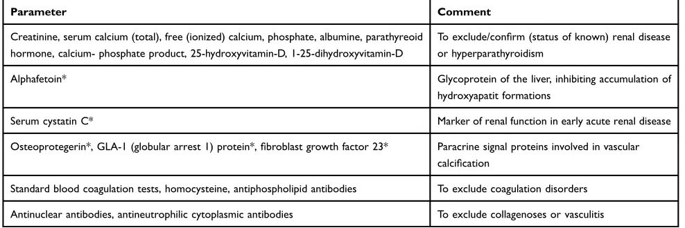
Calcium-phosphate levels, stage of renal disease, hypercoagulability and exclusion of differential diagnoses can mainly be performed via an orientating blood analysis.
More specific markers of calcification can be added when
necessary. Table 1 gives an overview of possible blood
markers. To confirm the diagnosis of CP, a single elevated
parameter (eg, an increased value of calcium-phosphate product) alone is not evidentiary enough.
Risk factors and co-morbidities
A number of risk factors for the development of CP have been described in case-control studies, mainly for patients with end-stage renal disease. But most studies are still limited by small number of patients or selection bias and true causality has not been determined. In most cases, no data about direct affectation of CP by the described
co-morbidity are available.15 Table 2shows an overview of
highly acknowledged risk factors. Next to end-stage renal disease and its co-morbidities like anemia and cardiovas-cular diseases, associations of CP with diabetes mellitus
have been described.16 Moreover, smaller numbers of
reports have been published about possible associations between CP and autoimmune disorders, infectious or alco-holic hepatitis, antiphospholipid syndrome or duration of
dialysis.15 Chronic inflammatory states are discussed as
possible risk factors for vascular calcification as TNF-a
induces an osteogenic phenotype of human smooth-muscle
cells in vitro.17Other cytokines are also linked to calcifi
-cation. Aluminum excess has also been discussed to be at least a co-factor in the pathogenesis of some CP cases by
Chronic Wound Care Management and Research downloaded from https://www.dovepress.com/ by 118.70.13.36 on 20-Aug-2020
involvement in the NFkB pathway which itself might
influence vascular calcification.2
Special emphasis has been placed on the discussion
about induction of CP by drugs.18Patients with end-stage
renal disease are often under multiple medications and should be subject to an explicit review of drugs possibly contributing to CP. Warfarin is one of the best known factors associated with CP and the course of disease. It
has been shown that warfarin can negatively influence
vitamin K-dependent proteins which prevent vascular
cal-cification. Although this could be shown in animal models,
the role of warfarin in human vascular calcification remains
unclear.19 Some authors even postulated distinguishing
between warfarin-associated and -unassociated CP.20 To
explore the effect of vitamin K supplementation in patients with uremic CP, a randomized controlled study trial is
currently recruiting (ClinicalTrials.gov Identifier:
NCT02278692). However, the identification and treatment
of underlying coagulative disorders in any case of CP is
essential.21Stopping warfarin, vitamin D, calcium
supple-ments and iron intake is recommended by the majority
when treating CP patients.18,22,23Direct thrombin inhibitors
have been shown to be safe and well tolerated in smaller
case series and lead to improvement of CP.24Another case
series with 15 patients observed a positive effect for tissue plasminogen activator as an adjunctive treatment option in
CP.25,26 However, these therapeutic decisions require an
interdisciplinary approach regarding the individual patient and his underlying morbidities.
Treatment
Clear therapeutic guidelines for the treatment of CP are still missing which is caused by the missing insights into
CP pathophysiology so far. Efficient therapeutic
proce-dures and evidence-based recommendations are needed especially for non-uremic CP, since therapeutic strategies can so far only be transferred from CUA treatment without
a pathophysiological correlation.27
Concepts of a modern wound management should be followed with special emphasis on debridement and pre-vention or early treatment of wound infections which are involved in the high mortality of CP patients due to sepsis. Non-adhesive modern wound dressings or products with silicone layer can be applied to the wound to enable an
Table 2 Overview of risk factors observed for development of CP (data from5,7)
Chronic kidney disease, hemodialysis, kidney transplant recipient
Hyperphosphatemia
Hypercalcemia
High levels of alkaline-phosphatase Calcium-phosphate product >70 mg2/dL2
Secondary hyperparathyroidism
Female gender
BMI >30 kg/m2
Liver disease
Systemic corticosteroid use
Hypalbuminemia
Warfarin therapy
Serum aluminium >25 ng/mL
Table 1Recommendations for possible blood analysis in patients with suspicion of CP. The values marked with * might be restricted to cases with further research interest
Parameter Comment
Creatinine, serum calcium (total), free (ionized) calcium, phosphate, albumine, parathyreoid hormone, calcium- phosphate product, 25-hydroxyvitamin-D, 1-25-dihydroxyvitamin-D
To exclude/confirm (status of known) renal disease or hyperparathyroidism
Alphafetoin* Glycoprotein of the liver, inhibiting accumulation of
hydroxyapatit formations
Serum cystatin C* Marker of renal function in early acute renal disease
Osteoprotegerin*, GLA-1 (globular arrest 1) protein*,fibroblast growth factor 23* Paracrine signal proteins involved in vascular calcification
Standard blood coagulation tests, homocysteine, antiphospholipid antibodies To exclude coagulation disorders
Antinuclear antibodies, antineutrophilic cytoplasmic antibodies To exclude collagenoses or vasculitis
Chronic Wound Care Management and Research downloaded from https://www.dovepress.com/ by 118.70.13.36 on 20-Aug-2020
atraumatic dressing change and reduce pain. Antiseptics like octenidin, polihexanide or hypochloric acids as well as antimicrobial effective substances are part of the local treatment regimens. Removal of necrosis and wound deb-ris is essential but often hard to implement due to pain or the patient´s multimorbidity. While some authors advocate
an early, consequent surgical debridement,9 others prefer
less invasive treatment like maggot therapy.28
Debridement can also be combined with negative pressure
wound therapy and–after achieving a satisfying
granula-tion tissue–split skin grafting.29In a retrospective study
of 64 patients, an estimated 1-year survival rate of 61.6% was observed for 17 patients receiving surgical debride-ment compared with 27.4% for the 46 who did not
(p=0.008).5 However, there is even discussion whether
performing deep skin biopsy may worsen the clinical
course30,31and too intensive manipulation is not advised.1
Hyperbaric oxygen therapy (HBOT) might be an adjunctive topical treatment option, but is only available in a small number of specialised centres. In a retrospective analysis of 34 patients with uremic CP, HBOT was very effective with 50% complete wound healing and 58%
improvement.32 Patients have to be selected according to
availability and their overall health condition for such
specific and extensive local wound treatments.
In addition to modern wound management, a supportive analgesic medication is essential for CP patients. The course of disease is often prolonged or chronic and pain is a frequent symptom which has to be addressed according to
its characteristics and specificity with adequate medication
according to the analgesic ladder and the patient´s needs. In summary, local wound therapy in CP patients has to be performed in a professional manner and where available in
specialized wound care centers.33A clear therapeutic
recom-mendation beyond intensive and antimicrobial local wound care cannot be given according to the currently available literature. Individual therapeutic decisions have to be made and an evaluation by experienced wound carers has to be ensured.
Systemic treatment
While the cessation of warfarin medication and vitamin D or calcium intake has already been discussed above, ther-apeutic approaches with systemic medication shall be addressed in the following section. As the presented ther-apeutic strategies mainly derive from those for uremic CP,
more efficient therapeutic measures and evidence-based
recommendations are needed for non-uremic cases.27 A
clear diagnostic classification as CP is mandatory.
Searching for clinical trials in CP patients does not reveal many hits in research databases. Due to the multi-morbidity and limited mobility of CP patients, randomized controlled trials in specialized centers are highly restricted
in terms of patient acquisition.1
Systematic data collection might be supported by clin-ical registers like the German Calciphylaxis Network
(www.calciphylaxis.net) (NCT02635373).34
In CP patients with hemodialysis, the calcium-phosphate
product might be positively influenced by changes in
fre-quency, duration or method of bloodfiltration. Additionally,
vitamin D intake or calcium supplements have to be restricted. In a small pilot study, four dialysis patients with CP were successfully treated with lanthanum carbonate, a calcium-free phosphate binder inhibiting the intestinal absorption of
phosphate.35 Results from the EVOLVE trial showed that
patients with end-stage renal disease and uncontrolled hyper-parathyroidism, a risk factor for CP, showed a lowered inci-dence of CP over an observation period of 5 years when treated with cinacalcet, a calcimimetic substance activating the calcium-sensing receptor and thereby reducing parathyroid
hormone production.36Parathyroidectomy is also discussed as
a treatment option for patients with CP and secondary
hyperparathyroidism.37Early treatment is recommended for
these cases.38However in a comprehensive review, the use of
cinacalcet showed a reduction of parathyroid hormone within
a period of 1–2 years,39so some authors only recommend
parathyroidectomy in cases with no response to medication
with cinacalcet.12A new substance (SNF472) to inhibit
pro-gressive calcification in patients with hemodialysis may be
promising and future study results have to be awaited to assess
its efficacy in CP patients (NCT02790073). The substance
inhibits the development of ectopic calcification by blocking
the formation and growth of hydroxyapatite crystal.40When it
comes to treatments focusing on inhibition of calcification,
bisphosphonates were also successfully applied in CP patients with and without renal diseases. While the use of bispho-sphonates in patients with severe renal disease is usually avoided due to long-term adverse events concerning bone-turnover effects, their effect in a potentially life-threatening disease like CP is promising. Newly proposed pathways
dis-cuss the influence of bisphosphonates on different calcification
pathways like enhancing osteoprotegerin, inhibiting calcifi
ca-tion of vascular smooth-muscle cells or the formaca-tion of
calcium-phosphate crystals.41Even anti-inflammatory effects
Chronic Wound Care Management and Research downloaded from https://www.dovepress.com/ by 118.70.13.36 on 20-Aug-2020
are discussed.41Several case series showed successful use of
bisphosphonates also in non-uremic CP patients.42,43
Another approach with a growing number of reported cases of successful treatment is the intravenous use of sodium thiosulfate (STS) in CP patients. The proposed mechanism behind this therapy is its ability to enhance the solubility of calcium in deposits and therefore its
availabil-ity for dialysis.41 Other effects may be antioxidative or
vasodilatory.31Currently, two clinical studies dealing with
STS are recruiting in France and the United States. One multicentre, Phase III, randomized placebo-controlled study is focusing on the potential of STS to reduce pain in CP patients (NCT03150420) next to the clinical improve-ment of skin lesions while another retrospective follow-up
study comprises one of the largest global cohorts of CP
patients treated with STS and investigates the influence of
STS on patients´ mortality. More than 350 cases with the use
of STS in CP patients have been reported so far,44either with
STS alone or as part of a combination therapy. Positive
effects were seen in CP patients with renal disease/dialysis45
as well as in non-uremic CP cases46where the therapeutic
effect cannot be explained so far. Even intralesional
injec-tions have been described occasionally47and showed a good
clinical improvement but pain during injections. Due to the renal clearance of STS, dosages and application frequency for intravenous application have to be adapted to renal
function and the presence of dialysis. Specific side effects
are rare and can be handled by supportive therapy.41Table 3
Table 3Therapeutic options for the treatment of CP
Local wound therapy
Debridement Adapted to extent of necrosis, wound surface area, pain and available method
Modern wound dressings To maintain atraumatic wound dressing
Antiseptic and antimicrobial effective wound dressings For prevention or therapy of wound infection
HBOT Adapted to availability
Systemic (supportive) therapy
Pain medication Adapted to degree of pain and individual patient´s requirement
Antibiotics For therapy of wound infection
Cessation of vitamin K-antagonists Supplement of vitamin K can be considered
Anticoagulation with direct thrombin inhibitors or tissue plasminogen activator
Especially if clues for underlying coagulative disorder can be found.
Systemic therapy (in case of calcium overload)
Optimization of the underlying renal disease
Changes in frequency or duration of dialysis, change offiltration/dialysis system
Calcium-free phosphate binders
Reduction of vitamin D intake
Cinacalcet When secondary hyperparathyroidism is confirmed
Parathyreoidectomy When hyperparathyroidism is confirmed
Systemic therapy to reduce calcification
Sodium thiosulfate Adapted to renal disease or dialysis
Bisphosphonates Attention should be paid to possible side effects or contraindications
SNF472 So far only in study protocols
Abbreviation:HBOT, hyperbaric oxygen therapy.
Chronic Wound Care Management and Research downloaded from https://www.dovepress.com/ by 118.70.13.36 on 20-Aug-2020
summarizes therapeutic attempts made in CP patients while
Table 4 gives an overview of currently recruiting clinical
studies.
Conclusion
The diagnosis of CP is often obvious with the typical necrotic ulcers and a history of chronic renal disease or
dialysis. Several risk factors have been identified and
should be addressed early in affected patients. Different treatment approaches for local and systemic therapy have been acknowledged although randomized controlled stu-dies are still missing. Next to an atraumatic and antiseptic wound management, systemic approaches like improve-ment of calcium and phosphate levels as well as treatimprove-ment of secondary hyperparathyroidisms are among the thera-peutic considerations. The systemic application of STS or
bisphosphonates is confirmed as successful in several case
studies. Randomized controlled studies on larger cohorts are needed to clear upcoming issues about dosage, treat-ment duration and long-term outcomes. Diagnosis and treatment are hindered for the non-uremic forms of CP where so far the concepts of CUA can only be transferred in lack of distinct recommendations.
Disclosure
The authors report no conflicts of interest in this work.
References
1. Brandenburg VM, Martin H, Sohn CM, et al. [Calciphylaxis].
Dtsch Med Wochenschr. 2015;140:347–351. doi:10.1055/s-0041-100834
2. Weenig RH. Pathogenesis of calciphylaxis: hans Selye to nuclear factor kappa-B. J Am Acad Dermatol. 2008;58:458–471. doi:10.1016/j.jaad.2007.12.006
3. Mochel MC, Arakaki RY, Wang G, et al. Cutaneous calciphylaxis: a retrospective histopathologic evaluation. Am J Dermatopathol. 2013;35:582–586. doi:10.1097/DAD.0b013e31827c7f5d
4. Nigwekar SU, Wolf M, Sterns RH, Hix JK. Calciphylaxis from nonuremic causes: a systematic review. Clin J Am Soc Nephrol. 2008;3:1139–1143. doi:10.2215/CJN.00530108
5. Weenig RH, Sewell LD, Davis MD, et al. Calciphylaxis: natural history, risk factor analysis, and outcome. J Am Acad Dermatol. 2007;56:569–579. doi:10.1016/j.jaad.2006.08.065
6. Fernandez M, Morales E, Gutierrez E, et al. Calciphylaxis: beyond CKD-MBD. Nefrologia. 2017;37:501–507. doi:10.1016/ j.nefro.2017.02.006
7. Mazhar AR, Johnson RJ, Gillen D, et al. Risk factors and mortality associated with calciphylaxis in end-stage renal disease.Kidney Int. 2001;60:324–332. doi:10.1046/j.1523-1755.2001.00803.x
8. Garcia-Lozano JA, Ocampo-Candiani J, Martinez-Cabriales SA, et al. An update on calciphylaxis.Am J Clin Dermatol.2018;19:599– 608. doi:10.1007/s40257-018-0361-x
9. Hafner J. Calciphylaxis and martorell hypertensive ischemic leg ulcer: same pattern - one pathophysiology. Dermatology. 2016;232:523–533. doi:10.1159/000448245
10. Kerk N, Goerge T. Livedoid vasculopathy - current aspects of diag-nosis and treatment of cutaneous infarction.J Dtsch Dermatol Ges. 2013;11:407–410. doi:10.1111/ddg.12064
11. El-Azhary RA, Hickson L, McBane RD. Calciphylaxis. N Engl J Med.2018;379:397–398. doi:10.1056/NEJMc1807324
12. Hoff NP, Homey B. [Calciphylaxis. Pathogenesis and therapy].
Hautarzt.2011;62:509–515. doi:10.1007/s00105-010-2111-8
Table 4Overview of recruiting studies worldwide (data survey on clinicaltrials.gov on 05/06/2019)
ClinicalTrials. gov identifier
Title Study type Study design Study
substance
NCT03150420 A Phase 3 Clinical Trial of Intravenous Sodium Thiosulfate in Acute Calciphylaxis Patients (CALISTA)
Interventional ● Multicentre
● Randomized
● Double-blind
● Placebo-controlled
● 111 patients planned
Sodium thiosulfate
NCT02278692 Evaluation of Vitamin K Supplementation for Calcific Uremic Arteriolopathy (VitK-CUA)
Interventional ● Single center
● Randomized
● Placebo-controlled
● 20 patients planned
Vitamin K
NCT02635373 European Calciphylaxis Registry Network (EuCalNet) Observational ● Prospective
● Registry
● 1000 patients planned
N.A.
NCT03032835 Observational ● Prospective
● Registry
● 300 patients planned
● Observation for 20 years N.A.
Chronic Wound Care Management and Research downloaded from https://www.dovepress.com/ by 118.70.13.36 on 20-Aug-2020
13. Dobry AS, Ko LN, St JJ, et al. Association between hypercoagulable conditions and calciphylaxis in patients with renal disease: a case-control study. JAMA Dermatol. 2018;154:182–187. doi:10.1001/ jamadermatol.2017.4920
14. Harris RJ, Cropley TG. Possible role of hypercoagulability in calci-phylaxis: review of the literature.J Am Acad Dermatol.2011;64:405– 412. doi:10.1016/j.jaad.2009.12.007
15. Nigwekar SU, Kroshinsky D, Nazarian RM, et al. Calciphylaxis: risk factors, diagnosis, and treatment. Am J Kidney Dis. 2015;66:133– 146. doi:10.1053/j.ajkd.2015.01.034
16. Renner R, Dissemond J, Goerge T, et al. Analysis of the German DRG data for livedoid vasculopathy and calciphylaxis.J Eur Acad Dermatol Venereol.2017;31:1884–1889. doi:10.1111/jdv.14190 17. Tintut Y, Patel J, Parhami F, Demer LL. Tumor necrosis factor-alpha
promotes in vitro calcification of vascular cells via the cAMP pathway.
Circulation.2000;102:2636–2642. doi:10.1161/01.cir.102.21.2636 18. Portales-Castillo I, Kroshinsky D, Malhotra CK, et al.
Calciphylaxis-as a drug induced adverse event.Expert Opin Drug Saf.2019;18:29– 35. doi:10.1080/14740338.2019.1559813
19. Danziger J. Vitamin K-dependent proteins, warfarin, and vascular
calci-fication. Clin J Am Soc Nephrol. 2008;3:1504–1510. doi:10.2215/ CJN.00770208
20. Yu WY, Bhutani T, Kornik R, et al. Warfarin-associated nonuremic calciphylaxis. JAMA Dermatol. 2017;153:309–314. doi:10.1001/ jamadermatol.2016.4821
21. Lehman JS, Chen TY, Lohse CM, et al. Evaluating the validity of subclassifying warfarin-associated nonuremic calciphylaxis: a retrospec-tive cohort study. Int J Dermatol. 2018;57:572–574. doi:10.1111/ ijd.13884
22. Seethapathy H, Brandenburg VM, Sinha S, et al. Review: update on the management of calciphylaxis. QJM. 2019;112:29–34. doi:10.1093/qjmed/hcy234
23. Zacharias JM, Fontaine B, Fine A. Calcium use increases risk of calciphylaxis: a case-control study.Perit Dial Int.1999;19:248–252. 24. King BJ, El-Azhary RA, McEvoy MT, et al. Direct oral anticoagulant medications in calciphylaxis. Int J Dermatol. 2017;56:1065–1070. doi:10.1111/ijd.13685
25. El-Azhary RA, Arthur AK, Davis MD, et al. Retrospective analysis of tissue plasminogen activator as an adjuvant treatment for calciphylaxis.
JAMA Dermatol.2013;149:63–67. doi:10.1001/2013.jamadermatol.5 26. Sewell LD, Weenig RH, Davis MD, et al. Low-dose tissue
plasmino-gen activator for calciphylaxis.Arch Dermatol.2004;140:1045–1048. doi:10.1001/archderm.140.9.1045
27. Gomes F, La FP, Costa C, et al. Non-uremic calciphylaxis: a rare diagnosis with limited therapeutic strategies.Eur J Case Rep Intern Med.2018;5:000986.
28. Tittelbach J, Graefe T, Wollina U. Painful ulcers in calciphylaxis -combined treatment with maggot therapy and oral pentoxyfillin. J Dermatolog Treat.2001;12:211–214. doi:10.1080/09546630152696035 29. Sattler DR, Preiss S, Altmann S, et al. [The plastic surgical treatment of progressive skin lesions caused by calciphylaxis].Zentralbl Chir. 2011;136:621–624. doi:10.1055/s-0031-1271458
30. Latus J, Kimmel M, Ott G, et al. Early stages of calciphylaxis: are skin biopsies the answer? Case Rep Dermatol. 2011;3:201–205. doi:10.1159/000333007
31. Ross EA. Evolution of treatment strategies for calciphylaxis.Am J Nephrol.2011;34:460–467. doi:10.1159/000332221
32. An J, Devaney B, Ooi KY, et al. Hyperbaric oxygen in the treatment of calciphylaxis: a case series and literature review. Nephrology (Carlton).2015;20:444–450. doi:10.1111/nep.12433
33. Dado DN, Huang B, Foster DV, et al. Management of calciphylaxis in a burn center: a case series and review of the literature.Burns. 2019;45:241–246. doi:10.1016/j.burns.2018.09.008
34. Brandenburg VM, Kramann R, Rothe H. et al. Calcific uraemic arteriolopathy (calciphylaxis): data from a large nationwide registry.
Nephrol Dial Transplant.2016. gfv438. doi:10.1093/ndt/gfv438 35. Chan MR, Ghandour F, Murali NS, et al. Pilot study of the effect of
lanthanum carbonate (Fosrenol(R)) in patients with calciphylaxis: a Wisconsin Network for Health Research (WiNHR) study.J Nephrol Ther.2014;4:1000162. doi:10.4172/2161-0959.1000162
36. Floege J, Kubo Y, Floege A, Chertow GM, Parfrey PS. The effect of cinacalcet on calcific uremic arteriolopathy events in patients receiv-ing hemodialysis: the EVOLVE trial. Clin J Am Soc Nephrol. 2015;10:800–807. doi:10.2215/CJN.10221014
37. Girotto JA, Harmon JW, Ratner LE, Nicol TL, Wong L, Chen H. Parathyroidectomy promotes wound healing and prolongs survival in patients with calciphylaxis from secondary hyperparathyroidism.
Surgery.2001;130:645–650. doi:10.1067/msy.2001.117101 38. Duffy A, Schurr M, Warner T, Chen H. Long-term outcomes in
patients with calciphylaxis from hyperparathyroidism. Ann Surg Oncol.2006;13:96–102. doi:10.1245/ASO.2006.03.042
39. Deen J, Schaider H. The use of cinacalcet for the treatment of calciphylaxis in patients with chronic kidney disease: a comprehen-sive review.Australas J Dermatol.2019. doi:10.1111/ajd.12992 40. Ferrer MD, Perez MM, Canaves MM, et al. A novel
pharmacody-namic assay to evaluate the effects of crystallization inhibitors on calcium phosphate crystallization in human plasma. Sci Rep. 2017;7:6858. doi:10.1038/s41598-017-07203-x
41. Ross EA. What is the role of using sodium thiosulfate or bispho-sphonates in the treatment for calciphylaxis? Semin Dial. 2011;24:434–436. doi:10.1111/j.1525-139X.2011.00954.x
42. Schliep S, Schuler G, Kiesewetter F. Successful treatment of calci-phylaxis with pamidronate. Eur J Dermatol. 2008;18:554–556. doi:10.1684/ejd.2008.0499
43. Torregrosa JV, Duran CE, Barros X, et al. Successful treatment of calcific uraemic arteriolopathy with bisphosphonates. Nefrologia. 2012;32:329–334. doi:10.3265/Nefrologia.pre2012.Jan.11137 44. Peng T, Zhuo L, Wang Y, et al. Systematic review of sodium
thio-sulfate in treating calciphylaxis in chronic kidney disease patients.
Nephrology (Carlton).2018;23:669–675. doi:10.1111/nep.13081 45. Devey S, Valois A, Cazajous G, et al. [Calciphylaxis in hemodialysis
patients: 8 cases treated with sodium thiosulfate]. Ann Dermatol Venereol.2018;145:288–292. doi:10.1016/j.annder.2018.02.002 46. Bourgeois P, De HP. Sodium thiosulfate as a treatment for
calciphy-laxis: a case series. J Dermatolog Treat. 2016;27:520–524. doi:10.3109/09546634.2016.1163316
47. Isoherranen K, Bouchard L, Kluger N. Benefits of intralesional injections of sodium thiosulfate in the treatment of calciphylaxis.
Int Wound J.2017;14:955–959. doi:10.1111/iwj.12738
Chronic Wound Care Management and Research
Dove
press
Publish your work in this journal
Chronic Wound Care Management and Research is an international, peer reviewed, open access, online journal publishing original research, reviews, editorials, and commentaries on the causes and management of chronic wounds and the major issues related to chronic wound management. Topics also include chronic wounds as comorbidities to
other conditions, patient adherence to therapy, and the economic burden of chronic wounds. The manuscript management system is completely online and includes a very quick and fair peer review system, which is all easy to use. Visit http://www.dovepress.com/ testimonials.php to read real quotes from published authors.
Submit your manuscript here:https://www.dovepress.com/chronic-wound-care-management-and-research-journal
Chronic Wound Care Management and Research downloaded from https://www.dovepress.com/ by 118.70.13.36 on 20-Aug-2020