_____________________________________________________________________________________________________
*Corresponding author: E-mail: dangelogatil@terra.com.br;
Analysis of Bone-implant Interfacial Stress during
Disocclusion in Complete Implant-supported
Prosthesis: A Finite Element Study
Gustavo Diniz Greco
1, Carolina Nemesio de Barros Pereira
2,
Giovani Lana Peixoto de Miranda
3, Guilherme Carvalho Silva
3,
Cláudia Silami de Magalhães
3and Allyson Nogueira Moreira
3*1
Department of Restorative Dentistry, Sistema Integrado de Ensino de Minas Gerais Ltda., Belo Horizonte, MG, Brazil.
2
Private Practice,Belo Horizonte, MG, Brazil.
3
Department of Restorative Dentistry, Faculdade de Odontologia, Universidade Federal de Minas Gerais, Pampulha, Belo Horizonte, MG, Brazil.
Authors’ contributions
This work was carried out in collaboration between all authors. Author GDG designed the study, performed the study wrote the protocol and the first draft of the manuscript. Authors CNBP, GLPM and GCS managed the literature searches, analyses of the study and English translation. Authors CSM and ANM managed the literature searches, designed the study and final review. All authors read and approved the final manuscript.
Article Information
DOI: 10.9734/BJMMR/2017/33607 Editor(s): (1) Emad Tawfik Mahmoud Daif, Professor of Oral & Maxillofacial Surgery, Cairo University, Egypt. Reviewers: (1) Roberta Gasparro, University of Naples Federico II, Italy. (2)Takahiro Kanno, Shimane University Faculty of Medicine, Japan. (3)Anirudh Bhattacharya, Narayana Hospital, Jaipur, India. (4)Koji Watanabe, Meikai University School of Dentistry, Japan. Complete Peer review History:http://www.sciencedomain.org/review-history/19170
Received 22nd April 2017 Accepted 17th May 2017 Published 23rd May 2017
ABSTRACT
Aim: Little is known about the biomechanical behavior of disocclusion patterns in
implant-supported prostheses. Thus, the aim of the present study was to analyze the stresses generated at bone-implant interface during different patterns of disocclusion in an implant-supported lower complete denture without free distal ends using the three-dimensional (3-D) finite element method.
Study Design: Finite element method.
Methods: A 3-D model of a complete denture supported by five inter-foraminal implants and two
distal was developed (CAE software Abaqus). A canine guide disocclusion (CG) was simulated applying a nodal load of 15 N at an angle of 45° on the canine tooth prosthesis, while to a bilateral balanced occlusion (BBO) a similar load pattern was applied at four distinct points, bilaterally. Linear elastic static analysis was used to compare the magnitude of maximum and minimum principal stresses at bone-implant interface for each simulation.
Results: The disocclusion pattern generated during CG exhibited a greater stress concentration at
the bone-implant interface of the distal implant on the working side. BBO showed a more homogeneous stress distribution pattern at the bone-implant interface of the two distal implants. Anterior implants showed lower stress concentration.
Conclusions: Bilateral balanced occlusion (BBO) resulted in a more favorable stress distribution
in this complete implant-supported prosthesis model.
Keywords: Dental occlusion; dental implant; biomechanics; dental stress analysis.
1. INTRODUCTION
Prosthetic rehabilitation using dental implants has shown long-term predictability primarily in completely edentulous patients [1,2]. However, biological and mechanical failures can still occur. Understanding the biomechanical phenomena of the bone-implant-prosthesis system is essential for the maintenance and stability of the osseointegrated rehabilitation [2].
Due to the presence of anatomical jaw structures such as the mandibular canal, implant-supported complete-fixed prostheses with cantilevers have been used frequently and have been found to be reliable [1]. The placement of four to six implants in the inter-foraminal region allows complete mandibular rehabilitation by means of a complete fixed denture with distal cantilever extension, which may restore aesthetics, phonetics, and function of the stomatognathic system [1,3]. This prosthesis configuration has been shown to have long-term predictability in clinical evaluation [1].
Although prostheses with free ends on both sides are acceptable, the cantilever length should not be more than 1.5 times the distance between the most distal and the most medial implant. Increased cantilever length may compromise
stress distribution in implant, bone, and
prosthetic components [4,5]. When the free end is loaded, the implant closest to the load experiences compressive stress, while the adjacent implants experience tensile stress [5].
This extended length can result in unfavorable stress distribution even if more implants are used for support [6]. Thus, it is important to reduce or even eliminate the cantilever extension in implant-supported prostheses. In edentulous patients, if anatomy is favorable, implants should
be placed distal to the mental foramen. This allows the construction of prostheses with shorter cantilevers or even without them [6].
The occlusal pattern can be considered a critical factor for the longevity of any oral rehabilitation, including the implant-supported prostheses. The periodontal ligament of the natural dentition exhibits a fully different behavior from that observed in the implant-bone interface [4,7]. Thus, stresses generated in the bone-implant interface and in the prosthesis components differ from those observed in the natural dentition. If the occlusal forces exceed the capacity of the prosthesis system, oral rehabilitation may fail due to overloading or unfavorable load distribution [4,5,7-9]. Complete dentures may present canine guide disocclusion (CG) or bilateral balanced occlusion (BBO). Besides having different effects on occlusal balance and stability, they may influence the stress generated at the
bone-implant interface. Thus, the pattern of
disocclusion may influence the preservation of the osseointegration since occlusal overload on implants may increase the incidence of marginal bone loss [10].
The aim of the present study was to compare the stress generated at the bone-implant interface in
implant-supported complete mandibular
prosthesis without free distal ends by simulating two patterns of disocclusion using the finite element method, in a linear three-dimensional simulation.
2. MATERIALS AND METHODS
The 3-D model was modified from a previous study [8], with the insertion of two distal short implants. The mesh of each component of the model was set separately and these meshes were subsequently joined to obtain the complete model. The interface between each component
pair (bone-implant, implant-metallic
infrastructure, and metallic infrastructure-artificial teeth) was generated using the CAE software (Abaqus, Dassault Systemes, Waltham, MA, USA). Each node in the contact surface of one component was constrained to move together with the adjacent node of the other component. This model contained 148,399 elements and 33,964 nodes. The mesh was tested and the areas of interest were refined until the response did not change significantly.
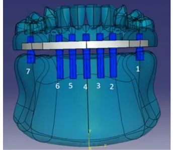
The model simulated a mandible with seven titanium implants, five of them installed between the mental foramina, with a distance of 4 mm between their platforms. These inter-foraminal implants were cylindrical and possessed a height and diameter of 13 and 3.75 mm respectively (Brånemark System Mk III, Nobel Biocare, Gothenburg, Sweden). Two posterior implants, located at the midpoint of the occlusal surface of the first molar, were cylindrical, and possessed a height and diameter of 5 and 5 mm (Titamax WS, Neodent, Curitiba, PR, Brazil). The height of the simulated prosthetic components was 3 mm and were composed of titanium (Multi-unit Abutment, Nobel Biocare, Gothenburg, Sweden). A distance of 3 mm was provided between the base of the prosthesis infrastructure and the bone surface (Fig. 1).
An implant-supported complete denture was designed with a nickel-chromium infrastructure (Wiron, BEGO, Bremen, Germany); the denture was 6 mm thick, 112 mm long, and a height of 4 mm. Over this structure, twelve acrylic resin teeth and acrylic gingival tissue were designed (Fig. 2). Implants 1 and 7 were short and most distally located. Implants 2 and 6 were situated close to the canine on the working and balancing sides
respectively. Implants 3, 4, and 5 were
situated in the mental area between implants 2 and 6.
Fig. 1. Three-dimensional (3-D) CAD mandible model including implants, abutments, and the proposal prosthesis: complete metal-acrylic
(hybrid) denture supported by 5 inter-foraminal and 2 distal dental implants
Fig. 2. Transparent frontal view of the 3-D CAD model of mandible and prosthesis. Note
the numbered implants inserted into bone, and the abutments linking the implants into
metal (nickel-chromium) prosthetic infrastructure
Poisson's ratio and elastic modulus of different materials used in the model were in accordance with the literature (Table 1) [3,4,816,17].
molar on the non-working side (Fig 3). As the stress distribution in the artificial teeth was not relevant for the analysis, no special precaution was taken regarding the local stress concentration at the point of load application.
Table 1. The Poisson's ratio and elasticity modulus of different materials used in the model were determined according to the
literature
Material Young´s modulus (MPa)
Poisson´s ratio
Cortical bone 13700 0.3
Trabecular bone 1370 0.3
Titanium 110000 0.35
Acrylic resin 2700 0.35
Nickel-chromium 188000 0.28
The results were based on a linear elastic static analysis and were used to compare the magnitude of the principal stresses for each simulation.
3. RESULTS
The results of the stress distribution analysis at
the bone-implant interface during CG
disocclusion are shown in Figs. 3, 4, 5, and 6. During CG, areas of high tensile stress concentration were detected in the mesial region at bone-implant interface of implant 1, and compressive stresses were visualized at bone-implant interfaces of the distal regions of implants 1 and 2. Tensile and compressive stress patterns of implants 1 and 2 (canine area on the working side) differed, with compressive stresses greater than the tensile stresses. In implant 7 on the non-working side, the maximum
and minimum stresses were lower and
proportional.
Fig. 3. 3-D CAD models simulating canine guide (CG): A nodal load of 15 N at an angle of 45° to the canine near implant 2. Simulation of bilateral balanced occlusion (BBO): the points were
distributed among the canine on the working side, in the region of the canine guide (CG) simulation, in the external portion of the mesio-buccal and distal vestibule of the first molar on
the working side, and in the internal aspects of the mesio-buccal portion of the atrium and the distal portion of the first molar on the non-working side.
Fig. 4. Occlusal view of the 3-D mesh. CG stresses on the bone surface: Maximum principal stresses. Note the highest stress (tensile) in the mesial aspect of periimplant bone around
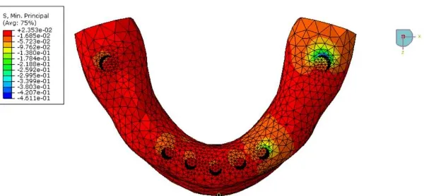
Fig. 5. Occlusal view of the 3-D mesh. CG stresses on the bone surface: minimum principal stresses. Note the highest stress (compressive) in the distal aspect of periimplant bone
around implants number 1
Fig. 6. Graphic distribution of maximum and minimum principal stresses in the bone-implant interface at implants 1, 2 and 7 in CG. These data confirm higher compression stresses around
implant 1 and greater tensile stress at implants 2 and 7
Fig. 7. Occlusal view of the 3-D mesh. BBO stresses on the bone surface: Maximum principal stresses. Note the highest stress (tensile) in the buccal aspect of periimplant bone around
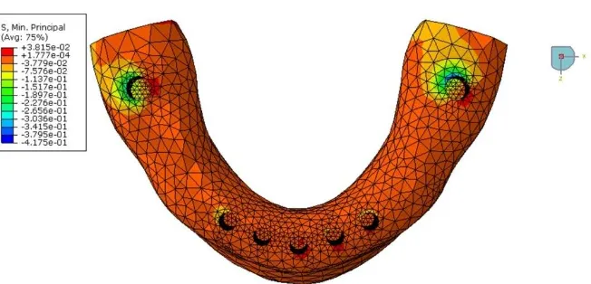
Fig. 8. Occlusal view of the 3-D mesh. BBO stresses on the bone surface: minimum principal stresses. Note the highest stress (compressive) in the lingual aspect of periimplant bone
around implant number 1 and in the buccal aspect around implant number 7
Fig. 9. Graphic distribution of maximum and minimum principal stresses in the bone-implant interface at implants 1, 2 and 7 in BBO. These data confirm that both implant 1 and 7 generate
tensile and compression stresses, minimizing the bone loss around these implants and decreasing the stresses around implant 2.
The results of stress distribution analysis during BBO are shown in Figs. 7, 8, and 9. During BBO, stress concentration occurred in distal implants in a bucco-lingual direction. Tensile stresses were observed in a direction opposite to that of the force vector, in the buccal and disto-lingual regions of implants 1 and 7 respectively. Compressive stresses were observed at the bone-implant interface in the same direction of the force vector, in the lingual and buccal regions of implants 1 and 7 respectively.
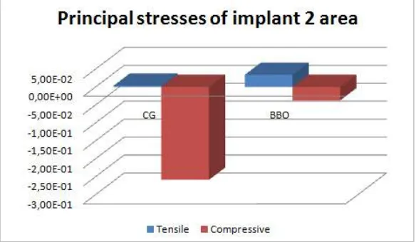
Fig. 10 shows the distribution of maximum and minimum principal stresses at the bone-implant interface of implant 2 corresponding to the canine area on the working side. During BBO,
the tensile and compressive stresses were similar and lower. During CG, compressive stresses were greater than the tensile stresses. Both patterns of disocclusion showed low levels of tensile stresses in the surrounding areas.
Regardless the patterns of disocclusion, stresses were lower in the anterior implants (Figs. 3, 4, 5, and 6).
4. DISCUSSION
Fig. 10. Maximum and minimum stresses in the region of the implant 2 in bone-implant interface.It is observed a counterbalanced stresses in the BBO pattern and a higher
concentration of stresses in the CG pattern.
will not be dissipated by the periodontal ligament, as occurs in natural teeth [4,7]. Functional loads generate stresses, and these will be transferred through all materials that compose a
implant-supported prosthesis, from the veneering
material to the peri-implant bone. The type and amount of stresses reaching each component of the implant-bone-prosthesis complex are critical for the clinical longevity of an oral rehabilitation [5,14,16]. Biomechanical stress may result on biological or prosthetic complications in a dental
implant-prosthesis complex [16]. These
complications include implant crestal bone loss
[10], early implant failure, fracture of
the prosthesis, abutment or prosthetic screw loosening, and problems with overdenture attachments [16]. An engineering approach to
resolve biomechanical problems involves
determining the nature of complications and then designing an approach to eliminate their underlying causes. Treatment planning should incorporate methods to reduce stress and minimize its initial and long-term effects. The treatment plan is altered when forces are greater or bone is less dense than usual to minimize the negative impact of stress on the implant, bone, and restoration. The goal is to decrease the amount of force, or increase the implant-bone surface area, to decrease the chance of implant-restoration complications [5,16]. One factor that may contribute to the control of undesirable stresses is the disocclusion pattern of the prosthesis.
The concepts and philosophies underlying disocclusion patterns were developed during the initial phase of the last century when Christensen
[17-22] advocated the use of BBO for rehabilitation with removable dentures for achieving greater stability and masticatory efficiency. Using the concepts of mutually protected occlusion, Nagao [23] in 1919 recommended CG for natural dentition and fixed prostheses [24-26]. Nowadays, still there is no unanimous consensus on the primary choice of disocclusion pattern for complete fixed dentures, since both concepts have been supported mainly by simple clinical observation and personal experience [27]. A comprehensive literature review found no difference in masticatory efficiency or patience satisfaction in conventional complete dentures adjusted either with CG or BBO [27]. However, for a complete implant-supported prosthesis with a distal free end, CG disocclusion has been shown to be more favorable, as it involves generation of lower stresses in the peri-implant interface when compared with BBO [8]. When considering disocclusion patterns in complete implant-supported prostheses without cantilevers, no data is available.
A complete implant-supported prosthesis, without free distal ends, should be the first choice of treatment for fully edentulous patients. Force
magnifiers, such ascantilevers, offset loads, and
monumental forces to the implant body
a minimum [5,6,28,30,31] or elimination in the
prescription to the dental technician in
implant-supported prostheses should be addressed when possible [31]. However, the height of the posterior mandibular bone after tooth loss, and the anatomical position of the inferior alveolar
nerve may compromise or even impair
conventional implant placement. With the advent of short implants, that have been proved to be successful mainly when they are splinted to other implants [32,33], the cantilever can be eliminated in some cases. On the other hand, implant length does affect the overall surface area through geometry [34]. Longer implants has been suggested to provide greater stability under lateral loads as same as teeth with longer roots represent a better biomechanical system to resist [34], although the aforementioned anatomical conditions often impair the placement of long implants in the mandibular posterior region of fully edentulous patients. Anyway, it can be addressed that, whenever possible, short or long implants may be placed in the posterior areas to avoid cantilevers.
In the present study, a non-cantilevered complete implant-supported prosthesis with short implants placed in the posterior mandibular region, representing a viable form of oral rehabilitation, was designed and analyzed using the 3-D finite element method. Numerical
modeling of peri-implant structures for
biomechanical studies presents the advantage of being non-invasive, and also allows identification
and calculation of stresses, strains and
displacements at bone-implant interface
[3,4,7,8,11-15]. In the present simulation, the principal maximum and minimum stresses were analyzed. The Von Mises stresses were not analyzed, since they are suitable only for ductile materials such as metal. The selected method has proved to be adequate for bone analysis
because it allows visualization of areas
experiencing compressive and/or tensile
stresses, and evaluation of a possible bone remodeling behavior. Identification of the nature of the stresses, tensile or compressive, is essential in biomechanical analysis, since different stresses result on distinct actions on bone. Interfacial stresses to an implant should be compressive in nature due to bone’s ability to
better resist under compressive stresses;
consequently tensile stresses are more
deleterious [35].
In the current analysis, CG showed concentration of compressive stresses at the implant-bone
interface of the distal region of implants 1 and 2. Remarkable tensile stresses were also observed in the implant 1 region. Lower the tensile stress at the bone-implant interface, better would be the treatment prognosis since these stresses at high intensity can promote bone resorption [10,35],
putting the prosthetic rehabilitation at
biomechanical risk. During the BBO simulation, stress concentration was observed in the bucco-lingual direction. Implants 1 and 7 showed areas of tensile stress concentration in the opposite direction of loading and areas of compressive stress in same direction of loading. Although the compressive and tensile stress values during BBO were greater than that during CG, they were similar, better distributed, and alternated between the working and non-working sides. This could result in balanced bone remodeling. Bone remodeling refers to the sequential and combined action of osteoblasts and osteoclasts, which remove and replace old bone with new tissue [35]. Balanced mechanical remodeling occurs in a situation wherein bone is resorbed and deposited simultaneously. The adaptation of bone to an applied load corresponds to the physiologic strain, which allows the bone to gain density and strength [35]. This phenomenon occurs after the balance of forces has been re-established [16,35]. Both patterns of disocclusion exhibited low tensile stresses at the bone-implant interfaces in the anterior implants, which was expected, since these areas are subjected to lower masticatory functional forces. Also, the presence of distal implants contributed to a more equilibrated stress distribution, regardless the disocclusion pattern, preserving the anterior implants. Since there are no clinical studies comparing both patterns of disocclusion in implant-supported complete prostheses, the results of the present study could not be linked to a clinical reality. Nevertheless, the present findings may suggest the BBO as a better
disocclusion scheme considering the
biomechanical aspect of the present prosthesis design. Obviously, besides the biomechanical factor, other aspects such as laboratorial technical ability and clinical experience should be considered when choosing between considering the disocclusion patterns.
5. CONCLUSION
Within the limitations of this study, it can be concluded that the bilateral balanced occlusion
(BBO) pattern was more favorable
implants and exhibited a more homogenous tensile and compressive stress distribution at the bone-implant interface.
CONSENT
It is not applicable.
ETHICAL APPROVAL
It is not applicable.
COMPETING INTERESTS
Authors have declared that no competing interests exist.
REFERENCES
1. Ekelund JA, Lindquist LW, Carlsson GE,
Jemt T. Implant treatment in the
edentulous mandible: A prospective study on Branemark system implants over more
than 20 years. Int J Prosthodont.
2003;16:602-28.
2. Misch CE, Perel ML, Wang
HL, Sammartino G, Galindo-Moreno
P, Trisi P, et al. Implant success, survival, and failure: The International Congress of
Oral Implantologists (ICOI) Pisa
Consensus Conference. Implant Dent. 2008;17:5-15.
3. Greco GD, Las Casas EB, Cornacchia TP,
Magalhaes CS, Moreira AN. Standard of
disocclusion in complete dentures
supported by implants without free distal ends: analysis by the finite elements method. J Appl Oral Sci. 2012;20:64-9.
4. Lin CL, Wang JC, Kuo YC. Numerical
simulation on the biomechanical
interactions of tooth/implant-supported
system under various occlusal forces with rigid/non-rigid connections. J Biomech. 2006;39:453-63.
5. Rangert B, Jemt T, Jorneus L. Forces and
moments on branemark implants. Int J Oral Maxillofac Implants. 1989;4:241-7.
6. Becker CM, Kaiser DA. Implant-retained
cantilever fixed prosthesis: where and when. J Prosthet Dent. 2000;84:432-5.
7. Eskitascioglu G, Usumez A, Sevimay M,
Soykan E, Unsal E. The influence of occlusal loading location on stresses
transferred to implant-supported
prostheses and supporting bone: A
three-dimensional finite element study. J
Prosthet Dent. 2004;91:144-50.
8. Greco GD, Jansen WC, Landre Junior J,
Seraidarian PI. Biomechanical analysis of
the stresses generated by different
disocclusion patterns in an implant-supported mandibular complete denture. J Appl Oral Sci. 2009;17:515-20.
9. Taylor TD, Agar JR, Vogiatzi T. Implant
prosthodontics: Current perspective and future directions. Int J Oral Maxillofac Implants. 2000;15:66-75.
10. Misch CE, Suzuki JB, Misch-Dietsh FM,
Bidez MW. A positive correlation between occlusal trauma and peri-implant bone loss: Literature support. Implant Dent. 2005;14:108-16.
11. Akca K, Iplikcioglu H. Finite element stress
analysis of the effect of short implant usage in place of cantilever extensions in mandibular posterior edentulism. J Oral Rehabil. 2002;29:350-6.
12. Himmlova L, Dostalova T, Kacovsky A,
Konvickova S. Influence of implant length and diameter on stress distribution: A finite
element analysis. J Prosthet Dent.
2004;91:20-5.
13. van Staden RC, Li X, Guan H, Johnson
NW, Reher P, Loo YC. A finite element study of short dental implants in the posterior maxilla. Int J Oral Maxillofac Implants. 2014;29:e147-54.
14. Silva GC, de Andrade GM, Coelho RC,
Cornacchia TP, de Magalhães CS, Moreira AN. Effects of screw- and cement- retained implant-supported prostheses on bone: a non-linear 3-D finite element analysis. Implant Dent. 2015;24:464-71.
15. Kim S, Kim S, Choi H, Woo D, Park
YB, Shim JS, et al. A three-dimensional finite element analysis of short dental implants in the posterior maxilla. Int J Oral Maxillofac Implants. 2014;29:e155-64.
16. Misch CE. Consideration of biomechanical
stress in treatment with dental implants. Dent Today. 2006;25:80,82,84-5,quiz 85.
17. Christensen. The problem of the bite.
DentCosmos. 1905;47:1184-95.
18. Granger ER. Functional relations of the
same stomatognathic system. J Am Dent Assoc. 1954;4:638-47.
19. Kurt LE. Balanced Occlusion. J Am Dent
Assoc. 1954;4:150-67.
20. Pound E. Lost-fine arts in the fallacy of the
ridges. J Prosthet Dent. 1954;4:6-16.
21. Landa JS. Biologic significance of
balanced occlusion and balanced
22. Woda A, Vigneron P, Kay D. Nonfunctional and functional occlusal contacts: A review
of the literature. J Prosthet Dent.
1979;42:335-41.
23. Nagao M. Comparative studies on the
curve of spee in mammals, with a discussion of its relation to the form of the
fossa mandibularis. J Dent Res.
1919;1:159-202.
24. Nairn RI. Lateral and protrusive occlusions.
J Dent. 1973;1:181-7.
25. D'amico A. Functional occlusion of the
natural teeth of a man. J Prosthet Dent. 1961;11:899-915.
26. Sheppard IM. Denture base dislodgment
during mastication. J Prosthet Dent. 1963;13:462-8.
27. Farias-Neto A, Carreiro Ada F. Complete
denture occlusion: an evidence-based approach. J Prosthodont. 2013;22:94-7.
28. Misch CE, Bidez MW. Implant-protected
occlusion. Int J Dent Symp. 1994;2:32-7.
29. Mesquita AM, Silva JH, Saraceni CH,
Kojima AN, Ozcan M.Effect of different abutments and connections in deformation crestal bone. Implant Dent. 2016;25: 328-34.
30. Padhye OV, Herekar M, Patil V, Mulani
S, Sethi M, Fernandes A. Stress
distribution in bone and implants
in mandibular 6-implant-supported
cantilevered fixed prosthesis: A 3D finite element study. Implant Dent. 2015;24: 680-5.
31. Misch CE, Bidez MW. Implant-protected
occlusion: A biomechanical rationale. Compendium. 1994;15:1330-2.
(34 passim; quiz 44).
32. Mezzomo LA, Miller R, Triches D, Alonso
F, Shinkai RS.Meta-analysis of single crowns supported by short (<10 mm) implants in the posterior region. J Clin Periodontol. 2014;41:191-213.
33. Anitua E, Pinas L, Begona L, Orive G.
Long-term retrospective evaluation of short implants in the posterior areas: Clinical
results after 10-12 years. J Clin
Periodontol. 2014;41:404-11.
34. Misch CE. Implant design considerations
for the posterior regions of the mouth. Implant Dent. 1999;8:376-86.
35. Frost HM. A 2003 update of bone
physiology and Wolff's Law for clinicians. Angle Orthod. 2004;74:3-15.
_________________________________________________________________________________
© 2017 Greco et al.; This is an Open Access article distributed under the terms of the Creative Commons Attribution License (http://creativecommons.org/licenses/by/4.0), which permits unrestricted use, distribution, and reproduction in any medium, provided the original work is properly cited.
Peer-review history: