Clinical Ophthalmology
Dove
press
open access to scientific and medical researchOpen Access Full Text Article O R i g i n A L R e s e A R C h
Differences between the combination of the
25-gauge vitrectomy with phacoemulsification versus
20-gauge vitrectomy and phacofragmentation
Pere Romero-Aroca1
Matias Almena-garcia1
Marc Baget-Bernaldiz1
Juan Fernández-Ballart2
isabel Méndez-Marin1
Angel Bautista-Perez1
1Ophthalmic service, hospital Universitario sant Joan, iisPV, Universidad Rovira i Virgili, Reus, spain; 2Department of Basic sciences, University Rovira i Virgili,
Tarragona, spain
Correspondence: Pedro Romero C/Ample 55 1º 43202
Reus (Tarragona), spain Tel 0034977310300 ext 5308 Fax +34 9 7732 3754 email romeropere@gmail.com, promero@grupsagessa.com
Introduction: In the present study we determine the differences observed between 25-gauge-vitrectomy combined with phacoemulsification, and the 20-gauge-vitrectomy combined with pars plana phacofragmentation.
Methods: A prospective study of a sample of 987 eyes of 661 patients randomly divided into two groups. 25-gauge-vitrectomy plus phacoemulsification included 486 eyes, and 20-gauge-vitrectomy plus phacofragmentation 501 eyes. We evaluated the differences at the time of the surgery, the intra- and postoperative complications, and the variations in intraocular pressure.
Results: The final visual outcome was similar in both groups. The most important differences between groups were: surgical time was faster in group 1 than in group 2, (mean time: 35.16 ± 3.49, 44.74 ± 5.30 minutes). Intraoperative complications were more numerous in group 1. In group 1, postoperative low levels of intraocular pressure are present in all patients with 2.77% of patients with hypotension (8 mmHg), and three choroidal effusion. In group 2, intraocular lens decentration and retinal detachment are more frequent (2.38% and 1.39%, respectively).
Conclusions: In the present study, both techniques have a similar number of complications and have a similar postoperative outcome, and are valid for the management of the pathologies selected.
Keywords: transconjunctival sutureless vitrectomy, sutureless vitrectomy, 25-gauge, 20-gauge, cataract, phacoemulsification, phacofragmentation, hypotony, complications
Introduction
Vitrectomy has been used to treat vitreoretinal disorders for more than 25 years.1,2 The management of the lens during vitrectomy has come full circle since the first vitrectomy interventions.3,4 The lens is removed frequently during surgery even if clear, and the lens status affects the management of retinal pathologies by pars plana vitrectomy. In many cases, it is advantageous to remove the lens, such as in patients with proliferative diabetic retinopathy, which permits better dissection of the anterior vitreous.5
If the lens is not removed during the vitrectomy, one of the most likely outcomes is cataract formation. In young patients, a cataract may take many years to develop, but in older patients, especially those aged over 60 years, with pre-existing nuclear sclerosis, visual loss may develop from a cataract much sooner, forcing cataract extraction in a short time after vitrectomy.6–8 If the cataract develops after vitrectomy, subsequent cataract surgery is more challenging than when the lens is removed at the time of com-bined surgery. Difficulties and risks involved cataract extraction as zonular dehiscence,
Clinical Ophthalmology downloaded from https://www.dovepress.com/ by 118.70.13.36 on 21-Aug-2020
For personal use only.
Number of times this article has been viewed
This article was published in the following Dove Press journal: Clinical Ophthalmology
Romero-Aroca et al Dovepress
increased mobility of the posterior capsule, extremely deep anterior chamber during phacoemulsification, and loss of the nuclear fragments inside the vitreous cavity.9–13
Classically, lens removal in conjunction with vitrectomy can be managed with pars plana lensectomy by phacofragmatome, the retention of the anterior lens capsule and the introduction of a posterior chamber lens in front of the capsule. This technique is available with 20-gauge vitrectomy, and the combined surgery hastens and simplifies the visual recovery of patients in whom cataracts will predictably develop after vitrectomy.
However, there is a new vitrectomy technique, trans-conjunctival sutureless vitrectomy (TSV 25), introduced in 2001, which has become widely adopted for vitreoretinal surgery. With this new technique there is no 25-gauge phacofragmentation tip, so a combination of phacoemul-sification and 25-gauge vitrectomy is needed in these cases.14,15
The aim of the present study is to determine the differences observed between two groups of patients, the first submitted to 25-gauge vitrectomy combined with clear corneal phacoemulsification, and the second submitted to 20-gauge vitrectomy combined with pars plana phacofragmentation (lensectomy).
Methods
Design
A prospective study of a sample of 987 eyes of 661 patients who needed a pars plana vitrectomy in combination with lens extraction, selected according to the inclusion criteria, and randomly divided into a two groups; the first including 486 eyes of 331 patients submitted to 25-gauge transconjunc-tival vitrectomy combined with clear corneal phacoemulsi-fication, and the second including 501 eyes of 320 patients submitted to 20-gauge vitrectomy and phacofragmenta-tion via pars plana. The recruitment dates extended from January 1st, 2003 to December 31st, 2008.
setting
The Hospital St. Joan is the only public ophthalmic center in Reus, Spain, with a dependent population of 389,471 inhabitants.
ethical adherence
The study was carried out in keeping with local legal require-ments (approval by the local ethical committee of Hospital Universitario Sant Joan – Reus) and in accordance with the revised guidelines of the Declaration of Helsinki. The nature
of the study was explained and all patients gave written consent to participate.
Diagnostic criteria
The time for phacoemulsification/lensectomy is defined as the time that the surgeon needed between opening the lens and the end of cataract extraction. We divided the time of vitrectomy into three steps. The time for surgical opening was defined as the interval between the first instrument contacting the conjunctiva through to the placement of all cannulae and the infusion line. The closing time was defined as the time required for removing the cannulae and infusion line. The operative time was defined as the interval between the opening and closing times.
The main outcome measures were the incidence of intraoperative or postoperative complications. Persistent vitreous postoperative hemorrhage was defined as the presence noted within the first postoperative week. Hypotony was defined as an intraocular pressure of less than 8 mmHg. Lens opacities were classified according to the lens opacities classification system III.16 Postoperative ocular irritation has been evaluated subjectively, by questionnaire, at 24 hours, seven days, and 15 days. All patients were asked the following question: “Do you have a sensation of foreign body in your eye?” The answer could be none or mild, moderate, or severe.
Also, an objective evaluation of the conjunctival hyperemia was performed in the following way:
• None or mild hyperemia, if none or minimal conjunctival vessels are dilated.
• Moderate, if vessels are more ingurgitated.
• Severe, if hemorrhages are present.
Inclusion criteria for the cataract extraction.
• Patients with a cataract that makes retina visualization difficult, with a nuclear opacity NO3, NC3, and/or cortical opacity C3, and/or subcapsular opacity P3
• All patients aged 60 years. Exclusion criteria for the study.
• Patients that had previously undergone intraocular surgery.
• Patients who presented surgical diagnosis of proliferative vitreoretinopathy associated detachments, extensive diabetic retinal proliferation or tractional detachment.
• Patients with retinal detachment, or giant retinal tears.
• Patients with glaucoma (chronic open angle, neovascular, or previous closed-angle glaucoma).
• Patients who needed silicone oil infusion at the end of surgery.
The distribution of diagnosis indicating surgical interven-tion for both surgical groups is listed in Table 1.
Clinical Ophthalmology downloaded from https://www.dovepress.com/ by 118.70.13.36 on 21-Aug-2020
Different combination of cataract surgery and vitrectomy
Dovepress
Patient examination
Visual acuity was measured using the standard Snellen methods. All visual acuity values were converted to logarithm of the minimum angle of resolution (logMAR) scores, where each 0.1 logMAR unit represents one line of visual acuity. Off the chart visual acuity designated as counting fingers, hand motions or light perception were assigned logMAR values of 1.6, 1.9, or 2.2, respectively, according to previously established conventions.17 Intraocular pressure (IOP) values were measured the day before surgery by Goldman tonometer.
Anterior segment photography was obtained in order to evaluate the lens opacity, according to the LOCS III system.
Before cataract surgery all patients underwent a fundus retinography, if possible, using a fundus camera. The macular area was examined with a noncontact 70-diopter fundus lens. Fluorescein angiography and optical coherence tomography was performed in patients with macular epiretinal membrane, macular diabetic edema and macular hole if the lens are clear enough. The interpretation of these explorations was made by an independent investigator.
There was a follow-up control of patients after surgery at 24 hours, one week, 15 days, one month, three months, and every six months thereafter. As a control, patients were submitted to the visual acuity and IOP measures, anterior segment biomicroscopic exploration, and fundus exploration with retinography and non contact lens macular exploration.
surgery
Both 25-gauge + phacoemulsification and 20-gauge +
phacofragmentation surgery were performed by one of two consultants (PR-A, MB-B) in standard fashion at a single centre, both surgeons had previously performed at least 50 25-gauge vitrectomies. At the end of the study
the differences between the number and type of procedure performed by the surgeons were not statistically significant.
Preoperative dilating regimen included, for both techniques, a mixture of 2.5% phenylephrine and 0.5% tropicamide, instilled three times at 30-minute intervals two hours before surgery. All patients received sedation and local anesthetic consisting of 5 ml of a retrobulbar injection of a 50:50 mixture of 2% mepivacine and 0.75% bupivicaine. Supplementary anaesthetic was administered as necessary. Patients’ skin and lashes and the ocular surface were prepared with 5% povidine-iodine solution.
Group 1 patients submitted to 25-gauge vitrectomy combined to phacoemulsification. In the first step, the trocar of the infusion sclerotomy was located 3.5 mm behind the limbus, and the infusion is turned off. The second step was the phaco-emulsification surgical procedure, a technique that included a superior limbal 2.8 mm incision, phacoemulsification of the cataract and in the bag implantation of a hydrophilic acrylic lens (Akreos®). The wound was sutured with a point of 10-0 nylon, and viscoelastic (Amvisc Plus®). The third step was the three port 25-gauge vitrectomy, a technique that consisted of the lateral displacement of conjunctiva in the two sites where the trocars would be placed at the conclusion of the procedure. The trocars were withdrawn and the conjunc-tiva was pushed laterally with a cotton bud, applying pressure for few seconds over the sclera. Finally the viscoelastic was removed from the anterior chamber by aspiration.
Group 2 patients submitted to 20-gauge pars plana vitrectomy combined with lensectomy (phacofragmentation). A 4 mm infusion cannula was placed through a sclerotomy located 3.5 mm behind the limbus. Once the infusion had been started, two additional sclerotomies were made at the 2:30 and 9:30 clock positions; from each of these Table 1 Description of previous pathology of the eyes submitted to 25-gauge and 20-gauge vitrectomy combined with cataract extrac-tion
Diagnostic Number of eyes
(25-gauge)
Percentage Number of eyes (20-gauge)
Percentage
Vitreous hemorrhage due to diabetic retinopathy
210 43.20% 212 42.31%
Vitreous hemorrhage due to posterior vitreous detachment
32 6.83% 34 6.78%
Vitreous hemorrhage due to CRVO or branch-CRVO
16 3.29% 19 3.79%
Macular epiretinal membrane 105 21.60% 97 19.36%
Tractional-diabetic macular edema 57 11.73% 65 12.97%
Macular hole 53 11.11% 59 11.77%
Uveitis 13 3.67% 15 2.99%
Abbreviation: CRVO, central retinal vein occlusion.
Clinical Ophthalmology downloaded from https://www.dovepress.com/ by 118.70.13.36 on 21-Aug-2020
Romero-Aroca et al Dovepress
sclerotomies, the blade was directed into the nucleus lens through the equator, a fragmentation tip was passed through the nucleus and a classic lensectomy was performed, the anterior capsule was preserved safely. The second step was the posterior vitrectomy and intravitreous manoeuvres that needed the patient. The sclerotomies were sutured at the end of surgery by 7-0 vicryl. Finally a sulcus lens (Meridian HP60M®), was inserted via clear corneal 3.2 mm incision, the corneal wound was sutured by a point of 10-0 Nylon.
Gas-fluid exchange was performed only in the patients with macular hole. A Bausch and Lomb (Rochester, NY) Millennium system was utilized in all cases.
statistical analysis
For the statistical analysis, two groups of patients were formed:
• Group 1 patients submitted to 20-gauge pars plana vitrec-tomy combined with lensecvitrec-tomy (phacofragmentation).
• Group 2 patients submitted to 25-gauge vitrectomy com-bined with phacoemulsification.
Descriptive statistics were created using SPSS statistical software (version 17.0; SPSS Inc, Chicago, IL, USA). Values are expressed as mean ± standard deviation, and statistical significance was determined using the Student’s t-test for paired data. Student’s paired test was used to compare the quantitative data as:
• Visual acuity.
• Current age.
• Postoperative IOP.
• Surgical time.
Best-corrected acuity was converted to LogMAR values to allow for statistical analysis.
When possible, intraoperative and postoperative complications were assessed by statistical analysis. Various statistical tests such as paired Student t-test for quantitative data and chi-squared analysis for qualitative data were used to compare means and assess contingencies between groups.
In all cases, P-values of less than 0.05 were considered statistically significant. We also determined the values of the odds ratio with a confidence interval of 95%.
Results and discussion
Results
Demographic data
All 987 patients completed the study and the mean follow-up was 18.35 ± 1.65 months (7–34 months) for group 1, and 18.17 ± 1.72 for group 2 (8–36 months). The preoperative demographic data of the patients is listed in Table 1. Mean age in group 1 was 68.29 ± 9.59 (53–88) years and for group 2
was 69.34 ± 9.21 (55–90) years. Group 1 comprised 54.93% men and 46.07% women, and group 2 comprised 52.87% men and 47.13% women.
Visual acuity
The best preoperative corrected visual acuity was 0.20 ± 0.13 (hand motion to 0.40) in group 1 and 0.22 ± 0.18 (hand motion to 0.40) in group 2.
Mean visual acuity in group 1 at the end of the study was 0.39 ± 0.26 (hand motion to 0.80), LogMAR: +0.41 ±+0.67 (+1.7–0).
Mean visual acuity in group 2 at the end of the study was 0.38 ± 0.28 (hand motion to 0.80), LogMAR: +0.43 ±+0.61 (+1.7–0.1).
Final visual acuity for both groups was well correlated to the diagnostic pathology, and the differences between the two groups are not statistically significant.
Variations in the surgery time (Table 2)
The time taken for cataract extraction was similar in both groups of patients and the differences were not significant, but the vitrectomy time was less in patients submitted to 25-gauge vitrectomy. The total time of the surgery was less in group 1 (35.16 ± 3.49 minutes) than in group 2 (44.74 ± 5.33 minutes). These differences were more important in the initial time (sclerotomies versus insertions of trocars) and in the closing time because the extractions of the trocars was faster than closing the sclerotomies by sutures. It is important to observe that the operation time was longer in 25-gauge vitrectomy (23.17 ± 0.97 minutes) than in 20-gauge vitrectomy (20.88 ± 0.45 minutes). These findings were obvious because the aspiration-cutting time in 25-gauge was inferior to the 20-gauge vitrectomy, differences which were significant in the statistical analysis with more advantage for 20-gauge system.
intraoperative complications (Table 3)
The more frequent intraoperative complication is the 9.26% alteration rate in the depth of the anterior chamber in group 1. These changes may be attributed to the fact that the anterior chamber was not completely closed, because the phacoemul-sification had been performed before the vitrectomy, despite a 10-0 nylon suture being leased in the wound and the ante-rior chamber being filled with viscoelastic. The techniques used during vitrectomy, and possible changes in the vitreous pression may have produced variations in the contents of the anterior chamber. In addition, these variations are the cause of the miosis that has been more frequently observed in group 1. An important finding is that an intraoperative number of
Clinical Ophthalmology downloaded from https://www.dovepress.com/ by 118.70.13.36 on 21-Aug-2020
Different combination of cataract surgery and vitrectomy
Dovepress
retinal tears were observed more frequently in group 2. Perhaps a major traction of the vitreous base by 20-gauge instruments was the cause of this, although we have to bear in mind that the intraoperative tears were more observed in patients with macular hole as diagnostic pathology.
Postoperative complications (Table 3)
The most important differences in postoperative complica-tions between the two groups of patients correlated well
with the major intraoperative complications, such as the alterations of the deep chamber in group 1 and the fact that the sclerotomies in 25-gauge vitrectomy were not sutured at the end of the surgery. As we may observe in group 1, corneal edema is more frequent, such as the postoperative hypotony, and choroidal effusion (which was observed in 1 patient in group 1). These findings in the group 2 presented a more frequent development of retinal detachment, retinal tears, and decentration of the lens.
Table 2 Surgical time: Mean time employed in the 25-gauge vitrectomy plus phacoemulsification versus 20-gauge vitrectomy plus
phacofragmentation
Surgical step Phacoemulsification +
25-gauge vitrectomy n = 486
Phacofragmentation + 20-gauge vitrectomy n = 501
Phacoemulsification in 25-gauge/phacofragmentation in 20-gauge
10.90 ± 0.50 minutes 11.30 ± 0.40 minutes P= 0.174
surgical opening time 1.97 ± 0.13 minutes 5.20 ± 0.30 minutes P 0.0001
Operative time 23.17 ± 0.97 minutes 20.88 ± 0.45 minutes P 0.001
Closing time 2.10 ± 0.29 minutes 8.24 ± 0.40 minutes P 0.0001
Total 35.16 ± 3.49 minutes 44.74 ± 5.33 minutes P= 0.01
Table 3 intraoperative and postoperative complications in the two group of patients
Complication Group 1
(25-gauge)
Group 2 (20-gauge)
Signification
intraoperative Miosis 29 (6.17%) 9 (1.79%) P= 0.001
Changes in the anterior chamber depth 45 (9.26%) 0 P 0.001
Anterior or posterior capsule rupture 11 (2.35%) 18 (3.59%) P= 0.087
Retinal tears 9 (1.86%) 17 (3.59%) P= 0.020
Postoperative Wound leak 6 (1.23%) 0 P 0.001
Retinal tears 12 (2.47%) 11 (2.19%) P= 0.201
Retinal detachment 0 7 (1.39%) P= 0.020
hypotony 13 (2.77%) 0 P 0.001
Choroidal effusions 1 (0.02%) 0 P 0.001
Opacification of the anterior or posterior capsule at six months/one year
45 (9.25%)/132 (27.16%)
51 (10.17%)/141 (28.14%)
P= 0.143/P= 0.178
Postoperative vitreous hemorrhage 45 (9.26%) 48 (9.58%) P= 0.221
Corneal edema 23 (4.91%) 6 (1.19%) P= 0.020
Uveitis 5 (1.06%) 4 (0.79%) P= 0.301
iris neovascularitzation 2 (0.42%) 3 (0.59%) P= 0.203
intraocular lens decentration 0 12 (2.38%) P= 0.001
CMe after six months of the cataract surgery
35 (7.47%) 34 (6.78%) P= 0.113
Abbreviation: CMe, cystoid macular edema.
Clinical Ophthalmology downloaded from https://www.dovepress.com/ by 118.70.13.36 on 21-Aug-2020
Romero-Aroca et al Dovepress
The other postoperative complications were not statistically different between groups, with a similar rate of postoperative vitreous hemorrhage, rubeosis iridis, and postoperative cystoid macular edema.
Changes in the iOP (Table 4, Figure 1)
The changes in the of postoperative IOP are greater in the group of patients after 25-gauge vitrectomy, as shown in Table 4 and after the seventh day we can consider that the IOP has been recovered, and its values are normal. The differences between the two groups are significant in the statistical analysis.Comfort and inflammation (Table 5)
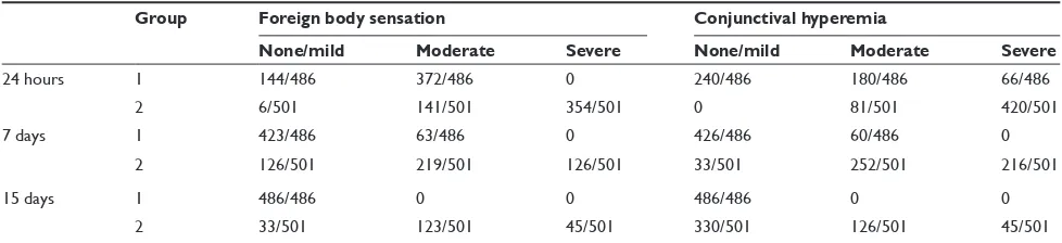
The patients in group 1 had poor sensation of foreign body since the first day postsurgery. These findings agree with the hyperemia visualization of the conjunctiva. Group 2 presented a more frequent sensation of foreign body.
Discussion
The big advantage of combined surgery is that a single intervention can repair all current and likely significant future intraocular diseases for the patient. If surgery is done sequentially, a likely scenario is for the patient to gradu-ally lose vision due to a formation of cataracts possibly up to 80%.6–8 On the other hand the drawbacks to combined surgery is in young patients with intact accommodation. Other disadvantages include increased opening time, and a tendency to cause more inflammation, especially in patients who require gas and laser.
The introduction of 25-gauge vitrectomy in 2001 repre-sented a revolutionary advance in posterior segment surgery, that simplified opening and closing of the eye, minimized sur-gical trauma for the patient, and made postsursur-gical recovery faster and less painful.18–20 Despite these advantages, if we
want to combine lens extraction surgical phacofragmentation, the tips do not exist. In order to perform lens extraction via pars plana, the phacoemulsification is the technique of choice,21,22 but there are many difficulties in combining the 25-gauge vitrectomy with phacoemulsification.
In the cataract extraction step, we observed that the mean time of the surgery between the two groups is similar, and the difference was not significant. In the intervention technique it is important to note that in group 1 the insertion of the first trocar (the trocar of the infusion canula, in the inferotemporal) is important in order to facilitate the posterior insertion of the two other trocars after the phacoemulsification step if it hadn’t been implanted before. Insertion after phacoemulsification is difficult because the eyes are hypotense. In group 2, the pars plana lensectomy may lead to loose fragments of the lens entering the vitreous, but in this event in our series this fragments has not made the vitrectomy difficult.
In group 1, the location of the intraocular lens (IOL) implant is important, and the insertion is into the capsular bag, but in group 2 the location of the IOL was in the sulcus. This implantion has caused the decentration observed in 2.38% of the series. This problem can be avoided if the lensectomy is substituted for phacoemulsification by clear cornea in group 2, as described in the literature.23–27
There are many differences between groups in respect to the time taken for the vitrectomy. The most important advantage of group 1 is the reduction of the mean time of the vitrectomy phase. This reduction occurred in the initial and closing phases of the vitrectomy, despite the vitrectomy operation time being greater in group 2. In the operation time the authors should be indicate that one of the major problem with 25-gauge vitrectomy is the flexibility of the instruments that hampers thorough removal of most of the peripheral vitreous.28–30 The introduction of the new 23-gauge technique may avoid this problem.31–35
Table 4 The intraocular pressure values according to the diagnosis of patients
Diagnostic Phacoemulsification + 25-gauge vitrectomy Phacofragmentation + 20-gauge vitrectomy Before surgery 24 hours after
surgery
7th day 15th day Before surgery
24 hours after surgery
7th day 15th day
Vitreous hemorrhage 16.46 ± 1.98* 12.11 ± 2.06 15.70 ± 1.59 15.71 ± 1.68 16.87 ± 1.94 15.87 ± 1.53 16.06 ± 1.36 16.46 ± 1.72 Tractional-diabetic
macular edema
15.50 ± 12.13 11.80 ± 2.13 15.20 ± 1.87 15.80 ± 1.87 15.90 ± 0.28 15.80 ± 2.07 15.60 ± 2.22 16.20 ± 1.91
Macular epiretinal membrane
15.50 ± 1.37 12.15 ± 1.57 14.33 ± 1.63 15.17 ± 1.47 15.83 ± 1.16 15.50 ± 1.51 15.33 ± 1.21 16.50 ± 1.22
Macular hole 16.45 ± 1.12 12.18 ± 1.67 14.36 ± 1.68 15.45 ± 0.68 16.81 ± 1.66 15.45 ± 1.36 15.54 ± 1.12 16.45 ± 0.82 Uveitis 17.00 ± 1.00 11.34 ± 1.14 15.33 ± 1.15 17.66 ± 1.52 15.66 ± 2.08 15.33 ± 1.52 15.33 ± 1.53 15.67 ± 1.76
Note: *Values in mmhg.
Clinical Ophthalmology downloaded from https://www.dovepress.com/ by 118.70.13.36 on 21-Aug-2020
Different combination of cataract surgery and vitrectomy
Dovepress
In our series one of the major problems during the vitrectomy was the change observed in the depth of the anterior chamber. Despite the presence of viscoelastic, these variations worsened the vision of the retina, and may be the cause of postoperative corneal edema observed in 4.93% in group 1 against 1.19% of patients in group 2.
Also the anterior chamber depth may worsen mydriasis. An excellent pupillary dilatation is an essential prerequisite to safe and successful vitreoretinal surgery. An inadequate mydriasis makes the vitrectomy difficult, as we have observed in this series. The patients submitted to phacoemulsifica-tion plus 25-gauge vitrectomy had more frequent pupillary constriction (6.17%) than patients of group 2 (1.79%). This finding is probably due to the changes in the depth of the anterior chamber observed in group 1.
One important finding in postoperative time is the pres-ence of IOL decentration observed in patients of group 2. The implantation of a lens in the sulcus and not in the capsular bag is disadvantageous and causes the decentration. Compared with pars plana lensectomy, phacoemulsification offers the benefit of allowing the IOL to be placed in the capsular bag.
Hypotony in our series was experienced by 2.47% of the patients and choroidal effusion appeared in one
patient (0.02%). We defined hypotony as the presence of IOP 8 mmHG in the postoperative period. The majority of the patients recovered normal IOP without any interven-tion; only one required a suture of the sclerotomies. Only one sclerotomy patient presented a wound leakage in the superotemporal sclerotomy after the surgery, this patient was also the patient with choroidal effusion, which recovered after the suture of sclerotomy. In our series there is a greater reduction in the IOP after the 25-gauge vitrectomy than after 20-gauge vitrectomy, as shown in Table 4 and Figure 1. For all diagnostic pathology, the IOP decreased more in the first group of patients. The IOP got back to normal after one month and the differences between the groups were not sig-nificant.19,20,30,36 Hypotony, even if transient, is not a benign condition and may increase the risks of serious complica-tions postoperation, including retinal or vitreal incarceration, suprachoroidal hemorrhage, and endophthalmitis.18,37 There is evidence in the literature that 25-gauge sclerotomy frequently does not self-seal.
No endophthalmitis has been observed in our series, despite there being a theoretical possibility that sutureless sclerotomy may serve as a conduit for the entry of bacteria into the eye postoperation. Covering the sites of sclerotomies 0
2 4 6 8 10 12 14 16 18
Preop
Intraocular pressure (IOP)
25-gauge
20-gauge
15 days 7 days
24 hours
Figure 1 Evolution of the mean intraocular pressure during the first month.
Table 5 Comfort and conjunctival hyperemia in the patients
Group Foreign body sensation Conjunctival hyperemia
None/mild Moderate Severe None/mild Moderate Severe
24 hours 1 144/486 372/486 0 240/486 180/486 66/486
2 6/501 141/501 354/501 0 81/501 420/501
7 days 1 423/486 63/486 0 426/486 60/486 0
2 126/501 219/501 126/501 33/501 252/501 216/501
15 days 1 486/486 0 0 486/486 0 0
2 33/501 123/501 45/501 330/501 126/501 45/501
Clinical Ophthalmology downloaded from https://www.dovepress.com/ by 118.70.13.36 on 21-Aug-2020
Romero-Aroca et al Dovepress
by conjunctiva should serve as a barrier to the entry of these germs.38–41 In the literature there is a description of a high rate of endophthalmitis in 25-gauge against 20-gauge vitrectomy.42 Kunimoto and colleagues reported one case of endophthalmitis in 5,498 eyes (0.018%) vitrectomized by 20-gauge against seven cases in 3,103 eyes (0.23%) who underwent 25-gauge vitrectomy. In the literature, the presence of postoperative retinal tears or retinal detachment has more frequently been reported in patients submitted to 25-gauge vitrectomy. Ibarra and colleagues43 reported a prevalence of 4.4% in their series, and Fujii and colleagues15 reported a prevalence of 2%. In spite of these published studies,15,43–45 in our series the presence of intraoperative and postoperative retinal tears are not more frequent in 25-gauge vitrectomy. In fact we may observe a major presence of retinal tears in the 20-gauge vitrectomy group. The authors think that minor trac-tion over the vitreous base carried out in the 25-gauge system may prevent the appearance of retinal tears, and the small size of the postoperative sclerotomy performed in 25-gauge may be the cause of the minor postoperative contraction of the vitreous base around the wound. It is important to indicate that in the literature a high number of postoperative retinal tears and detachments have been observed in patients diag-nosed with macular hole, in agreement with our series.43,44
The results of the present study show that visual acuity outcomes are similar in both groups. Preoperative and post-operative complications are not excessively distinct in both groups, and the differences may be attributed to the technique used in each case. Thus group 1 presents more complications attributed to the extraction of cataract via clear cornea, with the consequent loss of the anterior chamber depth. Group 2 has more frequent complications in the IOL decentration. It is important that in our series no cases of endophthalmitis have been observed, but this may be because of the small sample size, in other series with more patients the endo-phthalmitis appeared and is more frequent than in 20-gauge vitrectomy.45,46 In postoperative retinal tears, we did not show an increase in group 1 as we may expect according to the published data. It is important to remark that the mean time of surgery and postoperative inflammation are advantages for group 1. The authors think that careful patient selection is critical to a successful combined surgery. In the present study, both techniques have a similar number of complica-tions and have similar postoperative outcomes, and are valid for the management of the pathologies selected.
This study is limited because it is a case series study, despite its prospective and randomized technique, with a limited number of eyes that may become a handicap.
This factor may affect the low prevalence of some complications such as, endophthalmitis or retinal detach-ment. This study was not designed to assess comparative visual change nor standardized best-corrected ETDRS measurements made. Finally, long-term complications may appear and may not be detected with follow-up. In the present study, the follow-up of 18.35 ± 1.65 months may have been insufficient depending on the postoperative complication. Further studies including a cumulative multicenter analy-sis is called for. A case for selection of surgical pathology remains important in order to select the best surgical method for each patient.
Conclusion
In the present study, both techniques have a similar number of complications and have a similar postoperative outcome, and are valid for the management of the pathologies selected. These complications may be attributed to the different systems employed in each group of patients.
Disclosures
The authors report no conflicts of interest in this work.
References
1. Machemer R, Parel JM, Norton EW. Vitrectamy: a pars plana approacn. Technical improvements and further results. Trans Am Acad Ophthalmal
Otolaryngol. 1972;76:462–466.
2. Machemer R. A new concept for vitreous surgery. Two instrument techniques in pars plana vitrectomy. Arch Opnthalmal. 1974;92: 407–412.
3. Dowas NG. Pars plana vitrectomy microsurgical pars plana lensecomy.
Trans Sect Ophthalmol Am Acad Ophthalmol Otorlaryngol. 1976;81:
371–381.
4. Blankenship GW, Cortez R, Machemer R. The lens and pars plana vitectomy for diabetic retinopathy complications. Arch Ophthalmol. 1979;97:1263–1267.
5. Blankenship GW. The lens influence on diabetic vitrectomy results. Report of a prospective randomized study. Arch Ophthalmol. 1980; 98:2196–2198.
6. Blankenship GW, Machemer R. Long-term diabetic vitrectomy results; report of 10 year follow-up. Ophthalmology. 1985;92:503–506. 7. de Bustros S, Thompson JT, Michels RG, et al. Nuclear sclerosis after
virectomy for idiopatic epiretinal membranes. Am J Ophthalmol. 1988; 7:160–164.
8. Novak MA, Rice TA, Michels RG, Auer C. The crystalline lens after vitrectomy for diabetic retinopathy. Ophthalmology. 1984;91: 1480–1484.
9. Biro Z, Kovacs B. Results of cataract surgery in previously vitrectomized eyes. J Cataract Refract Surg. 2002;28:1003–1006.
10. McDermott ML, Puklin JE, Abrams GW, Eliott D. Phacoemulsification for cataract following pars plana vitrectomy. Ophthalmic Surg Lasers. 1997;28:558–564.
11. Grusha YO, Masket S, Miller KM. Phacoemulsification and lens implantation after pars plana vitrectomy. Ophthalmology. 1998; 105:287–294.
12. Ahfat FG, Yuen CH, Groenewald CP. Phacoemulsification and intra-ocular lens implantation following pars plana vitrectomy: a prospective study. Eye. 2003;17:16–20.
Clinical Ophthalmology downloaded from https://www.dovepress.com/ by 118.70.13.36 on 21-Aug-2020
Clinical Ophthalmology
Publish your work in this journal
Submit your manuscript here: http://www.dovepress.com/clinical-ophthalmology-journal Clinical Ophthalmology is an international, peer-reviewed journal covering all subspecialties within ophthalmology. Key topics include: Optometry; Visual science; Pharmacology and drug therapy in eye diseases; Basic Sciences; Primary and Secondary eye care; Patient Safety and Quality of Care Improvements. This journal is indexed on
PubMed Central and CAS, and is the official journal of The Society of Clinical Ophthalmology (SCO). The manuscript management system is completely online and includes a very quick and fair peer-review system, which is all easy to use. Visit http://www.dovepress.com/ testimonials.php to read real quotes from published authors.
Different combination of cataract surgery and vitrectomy
Dovepress
Dove
press
13. Braunstein RE, Airiani S. Cataract surgery results after pars plana vitrectomy. Curr Opin Ophthalmol. 2003;14:150–154.
14. Fujii GY, de Juan E Jr, Humayun MS, et al. A new 25-gauge instrument system for transconjunctival sutureless vitrectomy surgery. Ophthalmology. 2002;109:1807–1812.
15. Fujii GY, de Juan E Jr, Humayun MS, et al. Initial experience using the transconjunctival sutureless vitrectomy system for vitreoretinal surgery.
Ophthalmology. 2002;109:1814–1820.
16. Chylack LT Jr, Wolfe JK, Singer DM, et al.The lens classification system III. The longitudinal study of cataract study group. Arch
Ophthalmol. 1993;111:831–836.
17. Westheimer G. Scaling of visual acuity measurements. Arch
Ophthal-mol. 1979;97:327–330.
18. Chen E. 25-gauge transconjunctival sutureless vitrectomy. Curr Opin
Ophthalmol. 2007;18:188–193.
19. Lakhanpal RR, Humayun MS, de Juan E Jr, et al. Outcomes of 140 consecutive cases of 25-gauge transconjunctival surgery for posterior segment disease. Ophthalmology. 2005;112:817–824.
20. Ibarra MS, Hermel M, Prenner JL, Hassan TS. Longer-term outcomes of transconjunctival sutureless 25-gauge vitrectomy. Am J Ophthalmol. 2005;139:831–836.
21. Moreno-Montañes J, Barrio-Barrio J, Garcia-layana A. Combined cataract surgery and 25-gauge sutureless vitrectomy for posterior lentiglobus. J Cataract Refract Surg. 2007;33:380–382.
22. Hwang JU, Yoon YH, Kim DS, Kim JG. Combined phacoemulsification, foldable intraocular lens implantation, and 25-gauge transconjunctival sutureless vitrectomy. J Cataract Refract Surg. 2006;32:727–731. 23. Chang CJ, Chang YH, Chiang SY, Lin LT. Comparison of clear corneal
phacoemulsification combined with 25-gauge transconjunctival suture-less vitrectomy and standard 20-gauge vitrectomy for patients with cataract and vitreoretinal diseases. J Cataract Refract Surg. 2005;31: 1198–1207.
24. Demetriades AM, Gottsch JD, Thomsen R, et al. Combined phacoemul-sification, intraocular lens implantation, and vitrectomy for eyes with coexisting cataract and vitreoretinal pathology. Am J Ophthalmol. 2003; 135:291–296.
25. Schorwey K, Pavlovic S, Jacobi KW. Combind clear corneal phaco-emulsification, vitreoretinal surgery, and intaocular lens implantation.
J Cataract Refract Surg. 1999;25:693–698.
26. Lahey JM, Francis RR, Kearney JJ. Combining phacoemulsification with pars plana vitrectomy in patients with proliferative diabetic retinopathy: a series of 223 cases. Ophthalmology. 2003;110:1335–1339. 27. Lahey JM, Francis RR, Kearney JJ, Cheung M. Combining
phaco-emulsification and vitrectomy in patients with proliferative diabetic retinopathy. Curr Opin Ophthalmol. 2004;15:192–196.
28. Amato JE, Akduman L. Incidence of complications in 25-gauge trans-conjunctival sutureless vitrectomy based on the surgical indications.
Ophthalmic Surg Lasers Imaging. 2007;38:100–102.
29. Bahar I, Axer-Siegel R, Weinberger D. Pars plana vitrectomy: comparison of three techniques for the treatment of diabetic vitreous hemorrhage. Ophthalmic Surg Lasers Imaging. 2006;37:364–369. 30. Byeon SH, Chu YK, Lee SC, et al. Problems associated with the
25-gauge transconjunctival sutureless vitrectomy system during and after surgery. Ophthalmologica. 2006;220:259–265.
31. Romero P, Salvat M, Almena A, et al. Experience with 25-gauge trans-conjunctival vitrectomy compared to a 20-gauge system. Analysis of 132 cases. J Fr Ophtalmol. 2006;29:1025–1032.
32. Eckardt C. Transconjunctival sutureless 23-gauge vitrectomy. Retina. 2005;25:208–211.
33. Hubschman JP, Gonzalez CR, Bourda DH, et al. Combined 25- and 23-gauge surgery: a new sutureless vitrectomy technique. Ophthalmic
Surg Lasers Imaging. 2007;38:345–348.
34. Hubschman JP. Comparison of different vitrectomy systems (Comparaison des différents systèmes de vitrectomie). Jr Fr Ophtalmol. 2005;28: 606–609.
35. Oliveira LB, Reis PA. Silicone oil tamponade in 23-gauge transcon-junctival sutureless vitrectomy. Retina. 2007;27:1054–1058. 36. Patelli F, Radice P, Zumbo G, et al. G.25-gauge macular surgery: results
and complications. Retina. 2007;27:750–754.
37. Liu DT, Chan CK, Fan DS, et al. Choroidal folds after 25 gauge trans-conjunctival sutureless vitrectomy. Eye. 2005;19:825–827.
38. Acar N, Unvear YB, Altan T, Kabran Z. Acute endophthalmitis after 25-gauge sutureless vitrectomy. Int Ophthalmol. 2007;27:361–363. 39. Taylor SR, Aylward GW. Endophtalmitis following 25-gauges
vitrectomy. Eye. 2005;19:1228–1229.
40. Taban M, Ufret-Vicentry RL, Sears JE. Endophthalmitis after 25-gauge transconjunctival sutureless vitrectomy. Retina. 2006;26:830–831. 41. Stewart JM. Wound integrity and the conjunctiva in prevention of
endophthalmitis following sutureless 25-gauge vitrectomy. Eye. 2006; 20:1490.
42. Kunimoto DY, Kaiser RS. Wills Eye Retina Service. Incidence of endophthalmitis after 20- and 25-gauge vitrectomy. Ophthalmology. 2007;114:2133–2137.
43. Ibarra MS, Hermel M, Prenner JL, Hassan TS. Longer-term outcomes of transconjunctival sutureless 25-gauge vitrectomy. Am J Ophthalmol. 2005;139:831–836.
44. Okuda T, Nishimura A, Kobayashi A, Sugivama K. Postoperative retinal break after 25-gauge transconjunctival sutureless vitrectomy: report of four cases. Graefes Arch Clin Exp Ophthalmol. 2007;245:155–157. 45. Kunimoto DY, Kaiser RS; Wills Eye Service. Incidence of
endo-phthalmitis after 20- and 25-gauge vitrectomy. Ophthalmology. 2007; 114:2133–2137.
46. Scott IU, Flynn HW Jr, Dev S, et al. Endophthalmitis after 25-gauge and 20-gauge pars plana vitrectomy: incidence and outcomes. Retina. 2008;28:138–142.
Clinical Ophthalmology downloaded from https://www.dovepress.com/ by 118.70.13.36 on 21-Aug-2020