_____________________________________________________________________________________________________ *Corresponding author: E-mail: [email protected];
www.sciencedomain.org
Prevalence and Pattern of Impacted Teeth in the
North-East China
Nyimi Bushabu Fidele
1*, Sekele Isouradi Bourley
2, Em Kalala Kazadi
3,
Duan Feng
1, C. Mfutu Mana
4, P. Bobe Alifi
4, J. Bolenge Ileboso
3,
P. Muyembi Muinaminayi
4, Guan Jian
1and A. Mantshumba Milolo
21
Department of Oral and Maxillofacial Surgery, School of Stomatology, Second Affiliated Hospital of Jiamusi University, Jiamusi, China.
2
Prosthodontics and Orthodontics Service, Affiliated Hospital of Kinshasa University, DR. Congo.
3
Stomatology and Maxillofacial Service Unit of Periodontology Surgery, Affiliated Hospital of Kinshasa University, DR. Congo.
4
Unit of Oral Surgery, Affiliated Hospital of Kinshasa University, DR. Congo.
Authors’ contributions
This work was carried out in collaboration between all authors. Authors NBF, SIB, EKK and DF designed the study, wrote the protocol and the first draft of the manuscript. Authors CMM, PBA and JBI managed the literature searches and analyses statistics data. Authors PMM, GJ and AMM reviewed the manuscript and managed the final version by all verification. All authors read and approved the final manuscript.
Article Information
DOI: 10.9734/BJMMR/2016/27134 Editor(s): (1) Ibrahim El-Sayed M. El-Hakim, Ain Shams University, Egypt and Riyadh College of Dentistry and Pharmacy, Riyadh,
Saudi Arabia. Reviewers: (1) Rodrigo Lorenzi Poluha, State University of Maringá, Brazil. (2)Parveen Akhter Lone, Indira Gandhi Government Dental College Jammu, University of Jammu, Jammu, India. Complete Peer review History:http://sciencedomain.org/review-history/15388
Received 20th May 2016 Accepted 28th June 2016 Published 14th July 2016
ABSTRACT
Aims: To investigate the prevalence and pattern of impacted teeth in the sample of North-East China.
Study Design: Descriptive and Retrospective study.
Place and Duration of Study: School of Stomatology, Department of oral and maxillofacial surgery, Second Affiliated Dental Hospital of Jiamusi University Between Jun 2013 to October 2015.
the impacted teeth in Five thousand seven hundred and eighty four randomly selected patients. All of 5784 patients were examined (3754 males, 2030 females), with an age range of 7-76 years and a mean age of 23±4 years. The minimum age for inclusion was 7 years. The data was entered into the computer and analyzed using the Statistical Package for Social Sciences (version 20. Inc. Chicago, USA). The Pearson’s Chi‑square was used to determine the differences in the distribution of impacted teeth between genders. The significant level was tested at the 5%.
Results: Out of 5784 patients, a total of 1342 (23.2%) presented an impacted tooth, 701 (52.2%) were male and 641(47.8%) were female. Among them, 1485 were the number of impacted teeth. The prevalence of impacted teeth was 23.2%; third molars were the most common (11.70%; n=677), followed by canines (5.55%; n=321), incisor and premolars (2.92%; n=169 and 2.82%; n=163). The impacted teeth were mostly seen in the age group between 17-26 years old (43.8%; n=774). No significant relationship between impacted teeth among the gender was found (p=0.22). Conclusion: The prevalence of impacted teeth was 23.2% in this research and the patients aged between 17 to 26 years were most affected. The minimum age of 7 years must be an inclusion criteria study for to assess the real prevalence of incisor impaction.
Keywords: Prevalence; pattern; impacted teeth.
1. INTRODUCTION
Impacted teeth or unerupted teeth are those teeth that have failed to erupt completely or partially in the dental arch according to clinical and radiographic evaluation. Any permanent tooth can become impacted. The main causal factors are local like supernumerary teeth, dense overlying bone, deciduous tooth retention, arch-length deficiency, odontogenic tumors, cleft lip and palate. Also have systemic and genetic disorders such as Cleidocranial dysostosis syndrome, Down, Gardner’, and Gorlin-Sedano syndrome were reported [1-3].
The prevalence of impacted teeth in different populations and ethnic groups has been the subject of several studies. However, there is a discrepancy in the prevalence of teeth impaction in different population and ethnic groups, as well as variation in the prevalence and distribution of impacted teeth in different regions of the jaw itself [4-9]. The selected age group, eruption time of teeth, the racial differences and difference methodology of the study and radiographic criteria are some of the factors that affect the prevalence. According to literature, the most pattern impacted teeth are the mandibular third molars followed by the maxillary third molars, the maxillary canines, the mandibular premolars and mandibular incisors [9,10,11,12,13].
Assessing the prevalence of impacted teeth in population is important for the establishment of parameter data as well as for the planning of preventive and therapeutic strategies aimed at this population and with a direct influence on the management of the patient and clinical decision- making [1].
There are currently no study found on the prevalence and pattern of impacted teeth in Chinese patients of the North–East China. The purpose of this study was to investigate the prevalence and pattern occurrence of impacted teeth in the sample of North- East China.
2. MATERIALS AND METHODS
The Orthopantomogram (OPG) radiographs and clinical dentals records of 5784 Chinese patients attending the department of Oral and Maxillofacial Surgery, in the School of Stomatology, Second Affiliated Dental Hospital of Jiamusi University, Between Jun 2013 to October 2015 were examined for this retrospective study. All OPG were taken with the Dents ply Gendex Orthoralix 9200 (Dentsply Asia, Milford, US), and the magnification factor was 1.23.
A tooth was considered to be impacted when it was obstructed on its path of the eruption by an adjacent tooth, bone, or soft tissue and/or failed to erupt fully into the oral cavity. According to the mean eruption time, the teeth were considered as impacted when they remained in the jaw for a minimum of 2 years after the corresponding mean age of eruption [14]. Therefore, the minimum age for inclusion was 7 years because the generally accepted view is that the first series of permanent teeth have erupted and remained 2 years in the jaw. Patients’ clinical dental records and OPG radiographs were examined in order to detect the impacted incisors, canines, premolars and impacted molars.
viewer to determine the number and the pattern of impacted tooth. If an impacted third molar was identified in a patient, the eruption of his/her’s remaining third molars were also assessed. The depth and orientation of impacted third molars were assessed without it’s associated pathologies. The depth of impacted teeth or third molar was documented based on Winter’s lines classification while the angulation of impacted teeth was measured using the long axis of the impacted and adjacent teeth as described by Schersten [15].
Even though the Orthopantomogram (OPG) radiograph is very simple and intuitive, but it cannot provide all the information regarding the impacted teeth. For to ensure diagnosis validity in the recent study, radiographic findings were verified with clinical dental records, which were collected on standard forms as part of the routine examination process. The patients who had participated in this study were essentially the Chinese persons from the Jiamusi city and from the small city around Jiamusi. All patients presenting with any pathological conditions including trauma or fracture of the jaw or any hereditary diseases or syndromes that might affect the normal growth of dentition were excluded from the study.
The data was entered into the computer and analyzed using the Statistical Package for Social Sciences (version 20. Inc. Chicago, USA). The Pearson’s Chi‑square was used to determine the differences in the distribution of impacted teeth between genders. The significant level was tested at the 5%. Ethical approval was not received from the School of stomatology of Jiamusi University for the retrospective study because the patients were not exposed to additional radiation and not subjected to additional treatment. But a consent was obtained for to use the patients’ medical record data.
3. RESULTS AND DISCUSSION
In this analysis data, 1342 patients (23.2%) had impacted teeth; 701 (52.2%) were male and 641(47.8%) were female (Table 1) with an age range of 7-76 years. No statistically significant was found between impacted teeth and gender (P=0.22).
In Table 2, the prevalence of impacted teeth was 23.2% and impacted third molars was the most prevailing (11.7%; n=677), followed by impacted canines (5.55%; n=321), impacted incisors
(2.92%; n=169) and impacted premolars (2.82%; n=163). Impacted first and second molars were the least prevailing teeth (0.21%; n=12). Impacted deciduous teeth were not noted in the present study. The pattern of impacted teeth were more occurred in the mandible than maxilla (51.9%; n=772 and 48%; n=713) with a ratio of 1.08:1 and the third molars was the most commonly impacted teeth (52.3%). The canine, incisor, first and second molar were mostly noted in the maxilla than in the mandible (22.2% and 1.5% for canines; 10.7% and 0.8% for incisor, and 0.6% and 0.2% for first-second molars) respectively. However, the Third molar and premolars were mostly occurred in the mandible than the maxilla with 42.6% and 9.7% for third molars; and 6.7% and 4.3% for premolars. The impacted teeth mostly occurred in the age group between 17-26 years (43.8%; n=774) (Table 3).
Table 4, showed that, impaction of impacted teeth were diagnosed in all permanent teeth. The impacted teeth were routinely classified according to the direction of the crown of the tooth and a vertical impaction was a common pattern of impaction (34.34%), followed by mesio-angular impaction (29.36%) and by horizontal impaction (24.44%). The distal-angular impaction, buccal-lingual-angular and inverted impaction occurrence were less by 6.46%, 2.82%, and 2.55% respectively.
3.1 Discussion
Unerupted teeth are those teeth that have failed to erupt completely or partially in the dental arch. The main causes are local factors and also systemic and genetic disorders were reported [1-3]. The prevalence of impacted teeth is a discrepancy in different population and ethnic groups, as well as, variation in the prevalence and distribution of impacted teeth in different regions of the jaw [4-9]. Depending to some studies, the most pattern impacted teeth are the mandibular third molars followed by the maxillary third molars, the maxillary canines, the mandibular premolars and mandibular incisors [9,10,11,12,13].
North-East China studies). But, inclusion criteria for minimum age was different like some other previous studies that have investigated specific
age-groups and specific type of impacted teeth only, thus justify this difference [8,9,10,11].
Table 1. Distribution of patients and teeth number according to gender
Gender Number
of patients
Had impacted teeth P. value
Number of patients Number of teeth
Male 3754 701(52.2%) 731 (49.2%) 0.22
Female 2030 641(47.8%) 754 (50.8%)
Total 5784 1342(100%) 1485 (100)
Table 2. Prevalence, number of patients, localization and pattern of impacted teeth
Impacted tooth Pattern of impacted teeth N. of
patients N=1342
Prevalence %=23.2 Maxilla (%)
N=713(48)
Mandible (%) N=772(51.9)
Total (%) N=1485
Incisors 160(10.7) 13 (0.8) 173 (11.6) 169 2.92
Canines 331 (22.2) 22 (1.5) 353 (23.7) 321 5.55
Premolars 68 (4.3) 100 (6.7) 168 (11.3) 163 2.82
First
second molars
9 (0.6%) 4 (0.2) 13 (0.8) 12 0.21
Third molars 145 (9.7) 633 (42.6) 778 (52.3) 677 11.70
Legend: N= number; %= percent
Table 3. Distribution of prevalence impacted teeth according to age group
Age group (years) Number of patients Patients and mpacted
No. (%)
7- 16 1548 385 (24.8)
17- 26 1765 774 (43.8)
27- 36 1184 147 (12.4)
37- 46 742 24 (3.2)
47- 56 290 8 (2.7)
57- 66 175 3 (1.7)
67- 77 80 1 (1.2)
Total 5784 1342 (23.2)
Table 4. Impacted teeth orientation
Tooth position
Horizontal M-A Vertical D-A B-L-A Inverted Total
Upper/ lower jaw
U1/L1 9/0 24/0 26/10 14/0 16/0 35/0 124/10
U2/L2 0/0 16/0 18/3 2/0 0/0 0/0 36/3
U3/L3 38/2 122/6 162/10 0/4 9/0 0/0 331/22
U4/L4 0/1 7/5 3/9 0/0 0/0 0/0 10/15
U5/L5 7/3 12/18 28/55 6/9 5/0 0/0 58/85
U6/L6 0/0 3/0 0/0 0/0 0/0 0/0 3/0
U7/L7 0/0 2/3 4/1 0/0 0/0 0/0 6/4
U8/L8 0/303 55/163 87/94 3/58 0/12 0/3 145/633
Total 54/309 241/195 328/182 25/71 30/12 35/3 713/772
Global Total U
363 (24.44)
436 (29.36)
510 (34.34)
96 (6.46)
42 (2.82)
38 (2.55)
1485 (100)
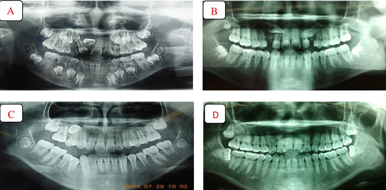
Fig. 1. Radiographs image of impacted tooth (A: Maxillary incisal impacted tooth; B: Maxillary canines impacted teeth; C: Maxillary second premolar impacted tooth and D: maxillary and
mandibular molar impacted teeth)
More than 27% of patients in the current study were aged between 7 and 18 years (Table 3). This may reflect increased prevalence of
impacted incisor tooth which as reported that an impacted maxillary incisors and canines teeth are most often occur in the patients with 8 to 12 years and 10 to 14 years old respectively
[8]. The pattern of impacted tooth (Table 2) seen with the most common being mandibular third molars, followed by upper canines, upper incisors and mandibular premolars were unlike to
those reported by some authors [4-7], but similar to study of Hou, et al. [8]. Also, the finding of an impacted third molars occourred in 52.3% of all impacted third molars teeth, and impacted
mandibular third molars occurred in 42.6% were not close of those found by others authors [7,12,13], may difference due to the
methodology used and selection of age group.
The canine tooth has a second complicated
eruption pattern and is one of the last teeth to erupt in the dental arch. Due to these
conditions, this tooth may not erupt naturally [16]. The maxillary canine was the second most
pattern impacted teeth (Table 2) in the recent
research, which is close to another study [17]. The results of others studies indicate that the
incidence of impacted canine may be higher in some populations whereas less in another population such as study of Fardi, et al. [4] that canines were the most commonly impacted teeth with 8.8% in the Greek population, 3.6% for study of Zahrani AA, [18] in Saudi population, 3.58% for study of Aydin et al. [19] and 0.92 for study of Dachi SF, et al. [20].
The different results may be attributed to the racial differences, Selection of age group, eruption time of teeth and differences in the methodology of the study. Maxillary canines impaction are believed to occur 10-20 times more frequently than mandibular canine impaction and there are limited numbers of studies revealing its frequency of occurrence [21]. In a study of Shah, et al. [22] 8 unerupted mandibular canines were found in 7886 individuals, while in the study of Grover PS, et al. [23] 11 impacted mandibular canines were found in 5000 individuals which resulted in an incidence of 0.10%. These results are quite similar with our finding (Table 2).
Some studies have reported that the impaction of the maxillary central incisor is almost as prevalent as impacted canines but its etiology is different [24]. Chaushu et al. [25] provided evidence of a significant environmental influence from the impacted maxillary incisor in delaying and altering the eruption path of the ipsilateral maxillary canine. The recent study found that the incidence of maxillary incisor was seen in one hundred sixty of the Chinese patients. However, the prevalence of maxillary incisor impaction (10.7%) was much less than that of canine impaction (22.2%). Our results are similar with to those found by Hou, et al. and also for those found by FSC Zhu, et al. [7,8]. But in order of prevalence, the most impacted teeth in the recent study was third molars followed by canines, incisors, and premolars which are also similar to the study of Hou, et al. [8] but different
A B
with the study of FSC Zhu, et al. [4-9] who reported that the premolars were seen in third order of prevalence, followed by incisors. According to those results, we can conclude that those studies will only included patients above 17 years old and not have included patient aged between 6 or 7 years, as the patients old than 17 years may have already consulted the dental practitioner for orthodontic treatment or extraction before the study, and hence the prevalence of impacted incisors found will not correlate to reality of that patients. The correct prevalence of impacted incisors must include the patients of minimum age of 7 years in the study.
Very few studies have been done regarding impacted premolars. It has been concluded from the results of previous studies that premolars impaction are rare [14,26], with the prevalence ranging from 2.1 to 2.7%, alike our recent research. The mandibular premolars were the frequently impacted than maxillary premolars, analogous to Hou, et al. [8]. Impacted mandibular first and second molars and impacted maxillary first and second molars are relatively rare with few cases reported and their prevalence ranged from 0.03% to 0.3%, with a slight predilection for males [27,28]. This affirmation was similar to our study and also to the study of Matheus Bandeca et al. [5].
It is difficult to estimate the 3-dimensional direction of impacted teeth from the x-ray films. Therefore, we have only performed a 2-dimensional investigation of the impaction orientation using the OPG radiographs in our present research. In this research, the vertical impaction was the most common, occurring in nearly 1/3 of all the impacted teeth, followed by Mesio-angular impaction and by a horizontal impaction who impacted mandibular third molars are more usual (Table 4). This result was different to the study of Hou R, et al. [8], who the most common angulation of impaction in both maxillary was vertical and mesio-angular impaction orientation. This dissimilarity could be explained by the fact that all of the type of impacted teeth were included in our study unlike the study of Hou, et al. [8] who the third molars impaction were excluded. About 27.84% impacted maxillary central incisors had inverted impaction. This affirmation is similar to our study and the study of Hou R, et al. [8].
4. CONCLUSION
The prevalence of impacted teeth was 23.2%. The minimum age of 7 years must be included in
the study for to assess the real prevalence of incisor impaction. The impacted teeth were mostly encountered in Chinese patients aged between 17 to 26 years old. All dentist, oral and maxillofacial surgeons should know this prevalence and pattern to perform a thorough evaluation, interceptive treatments and valid support in planning suitable treatment to be provided.
ACKNOWLEDGEMENTS
The authors thank Pr. Dr. Meng Cun Fang; Head of the department of imaging and nuclear medicine, Second Affiliated Hospital of Jiamusi University for his help with the interpretation of radiographs.
COMPETING INTERESTS
Authors have declared that no competing interests exist.
REFERENCES
1. Yildirim D, Yilmaz HH, Aydin U. Multiple impacted permanent and deciduous teeth. Dentomaxillofac Radiol. 2004;33(2):133-135.
2. Ghapanchi J, Haghnegahdar A, Khodadadzadeh S, Pourshahidi S, Ebrahimi H. Prevalence of taurodontism, missing and impacted teeth in South of Iranian population. Australiant Journal of Basic and Applied Sciences. 2011; 5(9):430-434.
3. D’alessadro G, Tagariello T, Piana G. Cleidocranial dysplasia: Aetiology and stomatognathic and craniofacial abnormalities. Minerva Stomatol. 2010; 59:117-127.
4. Fardi A, Kondylidou-Sidira A, Bachour Z, Parisis N, Tsirlis A. Incidence of impacted and supernumerary teeth- a radiographic study in a North Greek population. Med Oral Patol Oral Cir Bucal. 2011;16:e56-61. 5. Pedro FLM, Bandeca MC, Volpato LER,
Marques ATC, Borba AM, de Musis CR, Borges AH. Prevalence of impacted teeth in a Brazalian subpopulation. J Contemp Dent Pract. 2014;15(2):209-213.
6. Patil S, Maheshwari S. Prevalence of impacted and supernumerary teeth in the North Indian population. J Clin Exp Dent. 2014;6(2):e116-20.
Available:http://www.medicinaoral.com/odo /volumenes/v6i2/jcedv6i2p116.pdf
of impacted teeth and associated pathologies—a radiographic study of the Hong Kong Chinese population. Hong Kong Med J. 2003;9(3).
8. Hou R, Kong L, Ao J, Liu G, Zhou H, Qin R, et al. Investigation of impacted permanent teeth except the third molars in Chinese patients through an x-ray study. J Oral Maxillofac Surg. 2010;68(4):762-767. 9. Ahlqwist M, Grondahl HG. Prevalence of
impacted teeth and associated pathology in middle-aged and older Swedish women. Community Dent Oral Epidemiol. 1991;19: 116-9.
DOI: 10.1111/j.1600-0528.1991.tb00124.x 10. Hattab FN, Rawashdeh MA, Fahmy MS. Impaction status of third molars in Jordanian students. Oral Surg Oral Med Oral Pathol Oral Radiol Endod. 1995; 79(1):24-9.
11. Peltola JS. A panoramatomographic study of the teeth and jaws of Finnish university students. Community Dent Oral Epidemiol. 1993;21:36-9.
12. Msagati, et al. Pattern of occurrence and treatment of impacted teeth at the Muhimbili National Hospital, Dares Salaam, Tanzania. BMC Oral Health. 2013;13:37.
13. Ould Mohameden A. Étude des inclusions et enclavements des troisièmes molaires dans une population mauritanienne. Thèse Chir Dent Dakar; 2008. n° 6. French. 14. Wedl JS, Danias S, Schmelzle R, Friedrich
RE. Eruption times of permanent teeth in children and young adolescents in Athens (Greece). Clin Oral Investig. 2005;9:131-4. 15. Schersten E, Lysell L, Rohlin M.
Prevalence of impacted third molars in dental students. Swed Dent J. 1989;13(1-2):7-13.
16. Bishara SE. Impacted maxillary canines: A review. Am J Orthod Dentofacial Orthop. 1992;101:159-71.
17. Rozsa N, Fabian G, Szadeczky B, Kaan M, Gabris K, Tarjan I. Prevalence of impacted permanent upper canine and its treatment in 11-18-year-old orthodontic patients. Fogorv Sz. 2003;96:65-9.
18. Zahrani AA. Impacted cuspids in a Saudi population: Prevalence, aetiology and complications. Egypt Dent J. 1993;39:367-74.
19. Aydin U, Yilmaz HH, Yildirim D. Incidence of canine impaction and transmigration in a patient population. Dentomaxillofac Radiol. 2004;33:164-9.
20. Dachi SF, Howell FV. A survey of 3,874 routine full-mouth radiographs. II. A study of impacted teeth. Oral Surg Oral Med Oral Pathol. 1961;14:1165-9.
21. Sharma G, Nagpal A. Transmigration of mandibular canines- report of four cases and review of literature. Case Rep Dent. 2011;2011:381382.
22. Shah RM, Boyd MA, Vakil TF. Studies of permanent tooth anomalies in 7,886 Canadian individuals. I: Impacted teeth. Dent J. 1978;44:262-4.
23. Grover PS, Lorton L. The incidence of unerupted permanent teeth and related clinical cases. Oral Surg Oral Med Oral Pathol. 1985;59:420-5.
24. Bayram M, Ozer M, Sener I. Maxillary canine impactions related to impacted central incisors: Two case reports. J Contemp Dent Pract. 2007;8(6):72-81. 25. Chaushu S, Zilberman Y, Becker A.
Maxillary incisor impaction and its relationship to canine displacement. Am J Orthod Dentofac Orthop. 2003;124(2): 144-50.
26. Kramer RM, Williams AC. The incidence of impacted teeth. A survey at Harlem hospital. Oral Surg Oral Med Oral Pathol. 1970;29:237-41.
27. Sawicka M, Racka-Pilszak B, Rosnowska-Mazurkiewicz A. Uprighting partially impacted permanent second molars. Angle Orthod. 2007;77(1):148-154.
DOI:http://dx.doi.org/10.2319/010206-461R.1
28. Baccetti T. Tooth anomalies associated with failure of eruption of first and second permanent molars. Am J Orthod Dentofacial Orthop. 2000;118:608-10. _________________________________________________________________________________ © 2016 Fidele et al.; This is an Open Access article distributed under the terms of the Creative Commons Attribution License (http://creativecommons.org/licenses/by/4.0), which permits unrestricted use, distribution, and reproduction in any medium, provided the original work is properly cited.
Peer-review history: