___________________________________________________________________________________________ 4(12): 2431-2445, 2014
SCIENCEDOMAINinternational
www.sciencedomain.org
Radiation-induced Testicular Injury and Its
Amelioration by
Telfairia occidentalis
S. A. Adejuwon
1*, J. O. Omirinde
2, A. P. Ebokaiwe
3, O. O. Aina
2A. Adenipekun
4and E. O. Farombi
31Department of Anatomy, College of Medicine, University of Ibadan, Ibadan, Nigeria. 2Department of Veterinary Anatomy, University of Ibadan, Ibadan, Nigeria.
3Drug Metabolism and Toxicology Research Laboratories, Department of Biochemistry,
College of Medicine, University of Ibadan, Ibadan, Nigeria. 4Department of Radiotheraphy, College of Medicine, University of Ibadan, Ibadan, Nigeria.
Authors’ contributions
This work was carried out in collaboration between all authors. Author SAA designed the study, wrote the protocol and performed the statistical analysis. Authors JOO and APE managed the analyses of the study, managed the literature searches and wrote the first draft of the manuscript. Author OOA read the histological slides. Author AA facilitated the radiation of experimental animals. Author EOF provided the reagents used for antioxidant assays. All authors read and approved the final manuscript.
Received 6thDecember 2013
Accepted 18thJanuary 2014
Published 7thFebruary 2014
ABSTRACT
Aims: The radioprotective effect of Telfairia occidentalis aqueous extract (TOE) against gamma irradiation induced oxidative damage and altered sperm parameters in rats.
Study Design:Randomized controlled experiment
Place and Duration of Study: Experimental Animal Unit and Department of Anatomy, University of Ibadan between January and April, 2012.
Methodology: Male Wistar strain rats were divided into six groups and pretreated with TOE (400, 800 and 1600 mg/kg of b.wt.) and vitamin E (VE) 50 mg/kg of b.wt prior to whole body gamma irradiation exposure of 2Gy. Sections of the testes were taken for the assessments of biochemical markers of oxidative stress: Lipid peroxidation (LPO), Superoxide dismutase (SOD), Catalase (CAT) and Glutathione (GSH) and histopathological variations.
Results: Exposure of animals to 2Gy gamma radiation resulted in decreased sperm
characteristics and increased sperm morphological abnormalities; significant elevation in lipid peroxidation (LPO) and hydrogen peroxides, and decreased activities of superoxide dismutase (SOD), glutathione-s-transferase (GST), glutathione peroxidase (GSH-Px) and levels of reduced glutathione (GSH) in testes; distorted architecture of seminiferous tubules from day 1 to 30 post-irradiation. The rats pretreated with TOE showed normal sperm parameters, normal testicular histo-architecture and normal antioxidant enzymatic activities.
Conclusion:Aqueous extract ofTelfairia occidentalishas radioprotective activities.
Keywords: Telfairia occidentalis; testes; gamma irradiation; radioprotection; antioxidant.
1. INTRODUCTION
Plants have evolved the ability to produce chemical compounds that help them to protect against predators such as insects, fungi and herbivorous mammals. Some of these compounds whilst being toxic to plant predators turn out to have beneficial effects when used to treat human diseases; these secondary metabolites have antioxidants, immunostimulants, cell proliferation stimulators, anti-inflammatory and microbial agents that can protect against radiation-induced reactive oxygen species (ROS)-mediated damage [1]. They have been exploited successfully for the treatment of free radical-mediated diseases in humans such as rheumatoid arthritis, atherosclerosis, cancer, Alzheimer’s disease, aging and several other conditions [2,3]. Telfairia occidentalis (family Cucurbitacea) is a tropical vine grown in West Africa and highly reputed in traditional medicine [4]. The leaves are rich in essential and non-essential amino acids, flavonoids and other antioxidants, vitamins and minerals. The diet preparation of the air-dried leaves of the plant significantly increased red blood cell count, white blood cell count, packed cell volume and haemoglobin concentration in rats [5].
The interaction of ionizing radiation with the biological system results in the generation of reactive oxygen species (ROS) like superoxide anion (O2•─), hydroxyl radical (•OH),
hydrogen peroxide (H2O2), etc, which are highly reactive, short lived and known to cause
damage to cellular and molecular components including lipid, protein, carbohydrate and DNA, which may lead to mutations and chromosomal aberrations [6]. Radiation induced free radicals and increase in membrane lipid peroxidation (LPO), impairs the antioxidative defense mechanism, resulting in many pathological states such as ageing, cancer, cardiovascular diseases, diabetes, inflammation and neurodegenerative diseases [7,8] and induction of oxidative stress impairs reproductive function [9]. Some damages are expressed early, while other may be expressed over a period of time depending upon the cell kinetics and radiation tolerance of the tissues [10]. In order to reduce or protect from damaging effect of oxidative stress, cells have evolved an endogenous antioxidant defence mechanism, which includes non-enzymatic entities such as glutathione, ascorbic acid, uric acid and enzymes like superoxide dismutase (SOD), catalase (CAT), glutathione-S-transferase (GST), glutathione peroxidase (GSH-Px), etc. However, in the event of increased production of ROS, the host antioxidant defence mechanisms are overwhelmed resulting in the oxidative damage of cellular constituents [11].
Since the cells of spermatogenic lineage are especially vulnerable to radiation-induced reactive oxygen species (ROS) because they are constantly under mitosis or meiosis. Interestingly, under normal conditions, the testis is afforded with antioxidant protection as an elaborate array of antioxidant enzymes, free radical scavengers and low oxygen tension in order to support the dual action of this organ as germ cell spermatogenic [13] as well as leydig cells steroidogenic function [14]. This lends testes credence of being a highly radiosensitive organ with wide range of radiosensitive germ cells [15].
In recent times, knowledge of a wide range of natural and herbal compounds in the environment serving a radioprotective influence is of public interest. However, the use of available synthetic radioprotective agents has been limited due to their toxicity at their therapeutically effective concentrations. Therefore, a need exists for non toxic and inexpensive environmental natural products for clinical radiation protection. As radioprotection is a phenomenon, which is mediated by number of mechanisms such as scavenging of free radicals and inhibition of LPO etc., therefore use of single compounds to minimize radiation induced injuries may not yield the desired outcome [10].
Therefore, the present study was designed to determine the radioprotective effect ofTelfairia occidentalis extract (TOE) against gamma irradiation induced oxidative stress in testes of rat.
2. MATERIALS AND METHODS
2.1 Chemicals
The following components were purchased from Sigma-Aldrich Chemical Co. (St. Louis, Missouri, USA): nicotinamide adenine dinucleotide phosphate (NADP); glucose-6-phosphate; l-γ-glutamyl-3-carboxyl-4-nitronilide; glycylglycine; epinephrine; glutathione (GSH); 5, 50-dithio-bis-2-nitrobenzoic acid; hydrogen peroxide; thiobarbituric acid and 1-chloro-2, 4-dinitrobenzene. All other reagents were of analytic grade and were obtained from the British Drug Houses (Poole, Dorset, UK).
2.2 Animal Protocol
Ninety healthy adult male Wistar rats weighing approximately 240±10 g, obtained from the Department of Biochemistry, University of Ibadan, Ibadan, Nigeria, were randomly assigned to 6 groups of 15 animals each. They were housed in suspended plastic cages placed in a well-ventilated rat house, provided with rat pellets and water ad libitum and subjected to a 12:12 light-to-dark photoperiod. All animals received humane care according to criteria outlined in the Guide for the Care and Use of Laboratory Animals prepared by the National Academy of Science and published by the National Institutes of Health. Ethic regulations were followed in accordance with National and Institutional Guidelines for the Protection of Animal Welfare during the experiments (PHS 1996).
2.3 Animal Irradiation Procedures
delivered by a Linear accelerator teletherapy machine (Siemens one, Georgia, United States of America) with energy of 6.0 Mev, at source to surface distance of 400 mm, at depth of 14 mm, and a field size of 400 mm, with an equivalent square area of 185 mm2, the percentage depth dose was 1mm. The entire parts of the rats’ body were exposed, after radiation; the rats were placed in their cages. The dosimetry and irradiation procedures were carried out at the Radiotherapy Department of the Lagos University Teaching Hospital, Idi-Araba, Lagos, Nigeria.
2.4 Experimental Design
Group I (control): Rats in this group were given distilled water for 30 days (n = 15).
Group II (irradiated only): Rats in this group were exposed to 2 Gy dose of gamma radiation only (n = 15) on day 30.
Group III (radiation + vitamin E): Rats in this group were given Vitamin E (50 mg/kg/ b.wt.) for 30 days and then exposed to 2Gy dose of gamma radiation (n = 15).
Group IV (radiation + 400 mg/kg of b.wt TOE): Rats in this group were given 400 mg/kg of b.wt TOE for 30 days and then exposed to 2 Gy dose of gamma radiation (n = 15).
Group V (radiation + 800 mg/kg of b.wt TOE): Rats in this group were given 800 mg/kg of b.wt TOE for 30 days and then exposed to 2 Gy dose of gamma radiation (n =15).
Group VI (radiation + 1600 mg/kg of b.wt TOE): Rats in this group were given 1600 mg/kg of
b.wt TOE for 30 days and then exposed to 2 Gy dose of gamma radiation (n = 15).
Five rats from each group were sacrificed at 24 hours, 15 and 30 days post radiation for the evaluation of biochemical, sperm parameters and histological variations in testes
.
2.5 Biochemical Assays
Five right testes were homogenized in 50 mM Tris–HCl buffer (pH 7.4) containing 1.15% potassium chloride, and the homogenate was centrifuged at 10,000g for 15 min at 4⁰C. The supernatant was collected for the estimation of Superoxide dismutase (SOD) by the method described by Misra and Fridovich [16]. Glutathione-Stransferase (GST) was assayed by the method of Habig [17]. The activity of glutathione peroxidase (GSH-Px) was determined by the method of Rotruck [18]. GSH was determined at 412 nm using the method described by Jollow [19]. Hydrogen peroxide generation was assessed by the method of Wolff [20]. Lipid peroxidation was quantified as MDA according to the method described by Farombi [21] and expressed as µm MDA/g tissue.
2.6 Sperm Motility Assay
Sperm motility was assessed by the method described by Zemjanis [22]. The motility of epidiymal sperm was evaluated microscopically within 2–4 min of their isolation from the cauda epididymis, and data were expressed as percentages.
2.7 Sperm Count
2.8 Live/Dead Ratio
The live-dead staining principle is based upon the observation that eosin B penetrate and stain the dead sperm cells whereas the viable cells repel this stain. In order to benefit from the live-dead method, the staining was done without delay.
2.9 Morphologic Abnormalities and Percentage Viability Assay
A portion of the sperm suspension placed on a glass slide was smeared out with another slide and stained with Wells and Awa’s stain (0.2 g eosin and 0.6 g fast green dissolved in distilled water and ethanol in a 2:1 ratio). A total of 400 spermatozoa from each rat were used for morphologic examination. Also, sperm smeared slides were stained with 1% eosin and 5% nigrosine in 3% sodium citrate dehydrate solution for live-to-dead ratio assessment using the method described by Wells and Awa [24].
2.10 Histopathology
Samples from testes were fixed with Bouin’s solution, sectioned, and stained routinely with haematoxylin and eosin for microscopy. All slides were coded before examination using a light microscope (Olympus, USA) by investigators who were blinded to the control and treatment groups.
2.11 Statistical Analysis
Statistical analysis were carried out using one-way analysis of variance, followed by Student’s t-test, to compare experimental groups; p<0.05 was considered statistically significant.
3. RESULTS
3.1 Biochemical Assays
The effects of pretreatment with TOE against gamma irradiation on antioxidant systems and lipid peroxidation in testis after 24 hours, days 15 and 30 post irradiation are presented in Figs. 1a- c and Figs. 2a-c. A significant (p<0.05) decrease in the activities of antioxidant enzymes SOD and GSH-Px as well as GST, a phase-II xenobiotic detoxifying enzyme was observed in testes within 24 hours post irradiation and continued till days 15 and 30 post irradiation by 65, 50 and 70% for SOD; 60, 75, and 50% for GSH-Px and 50, 65 and 70% for GST as shown in Figs. 1a, b and c. There was a concomitant decrease in reduce GSH by 40, 65 and 55% as shown in Fig. 2c, pretreatment with TOE at doses of 400 and 800mg/kg of b. wt reversed these values by 55-85% when compared with control values as shown in Figs 1a, b, c and 2a, b, c.
However, the levels of hydrogen peroxide (H2O2) generation, and lipid peroxidation,
increased significantly (p< 0.05) at 24 hours, days 15 and 30 post irradiation compared with control values, the levels of H2O2 generated in testes increased by 40, 70 and 72% as
control R R + 50mg VE R + 400mg R + 800mg R + 1600mg 0.0
0.2 0.4 0.6 0.8 1.0 1.2 1.4 1.6 1.8 2.0
*
*
*
*
*
SO
D
(U
/m
g
pr
ot
ei
n)
super oxide dismutase
24 h post radiation day 15 post radiation day 30 post radiation
*
Fig. 1a. Effects ofTelfairia occidentalis(TOE) on gamma radiation induced decrease in superoxide dismutase (SOD) enzyme levels. R (Radiation only -2 Gy), R + 50mg VE
(Radiation +Vitamin E), R + 400mg (Radiation + 400mg dose of TOE), R + 800mg (Radiation + 800mg dose of TOE), R + 1600mg (Radiation + 1600mg dose of TOE),
*p<0.05
control R R + 50mg VE R + 400mg R + 800mg R + 1600mg 0
1 2 3 4 5 6 7 8
*
*
*
*
*
G
SH
-P
x
(u
ni
t/m
g
pr
ot
ei
n)
Glutathione peroxidase
24 h post radiation day 15 post radiation day 30 post radiation
*
Fig. 1b. Effects ofTelfairia occidentalis(TOE) on gamma radiation induced decrease in glutathione peroxidase (GSH-PX) enzyme levels. R (Radiation only -2 Gy), R + 50mg
VE (Radiation + Vitamin E), R + 400mg (Radiation + 400mg dose of TOE), R + 800mg (Radiation + 800mg dose of TOE), R + 1600mg (Radiation + 1600mg dose of TOE),
control R R + 50mg VE R + 400mg R + 800mg R + 1600mg 0 2 4 6 8 10 12 14
*
*
*
*
*
G ST (m ic ro m ol e C D N B /G SH co m pl ex fo rm ed /m in /m g pro te in ) Glutathione-S- transferase24 h post radiation day 15 post radiation day 30 post radiation
*
Fig. 1c. Effects ofTelfairia occidentalis(TOE) on gamma radiation induced decrease in glutathione-S-transferase (GST) enzyme levels. R (Radiation only -2 Gy), R + 50mg
VE (Radiation +Vitamin E), R + 400mg (Radiation + 400mg dose of TOE), R + 800mg (Radiation + 800mg dose of TOE), R + 1600mg (Radiation + 1600mg dose of TOE),
*p<0.05
control R R + 50mg VE R + 400mg R + 800mg R + 1600mg 0.0 0.5 1.0 1.5 2.0 2.5 3.0 3.5 4.0 4.5 5.0 5.5 6.0 6.5 7.0 7.5 8.0 8.5 9.0
*
*
*
*
*
LP O (µ m ol M DA /m g pr ot ei n) Lipid peroxidation 24 h post radiation day 15 post radiation day 30 post radiation*
Fig. 2a. Effects ofTelfairia occidentalis(TOE) on gamma radiation induced alteration in lipid peroxidation (LPO) levels. R (Radiation only- 2 Gy), R + 50mg VE (Radiation
control R R + 50mg VE R + 400mg R + 800mg R + 1600mg 0.0 0.5 1.0 1.5 2.0 2.5 3.0 3.5 4.0 4.5 5.0 5.5 6.0 6.5 7.0
*
*
*
*
*
H2 O2 (n m ol /m g pr ot ei n)hydrogen peroxide generation 24 h post radiation day 15 post radiation day 30 post radiation
*
Fig. 2b. Effects ofTelfairia occidentalis(TOE) on gamma radiation induced alteration in the levels of hydrogen peroxide generated. R (Radiation only- 2 Gy), R + 50mg VE
(Radiation +Vitamin E), R + 400mg (Radiation + 400mg dose of TOE), R + 800mg (Radiation + 800mg dose of TOE), R + 1600mg (Radiation + 1600mg dose of TOE),
*p<0.05.
control R R + 50mg VE R + 400mg R + 800mg R + 1600mg 0 1 2 3 4 5 6 7 8 9
*
*
*
*
*
G SH (U /m g pr ot ei n)Reduced Glutathione levels
24 h post radiation day 15 post radiation day 30 post radiation
*
3.2 Sperm Analysis
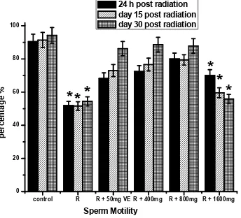
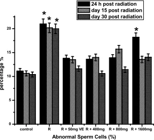
Fig. 3 shows the data on, sperm motility, sperm count, morphological abnormalities assay and live/dead ratio. There was a decrease in sperm motility, and count as shown in Figs 3a, b. Compared with controls, live/dead ratio decreased by 34, 36, and 38%, respectively, at 24 hours, days 15 and 30 post irradiation as shown in Fig. 3d and the decreases in sperm motility were 40, 41 and 43%, respectively as shown in Fig. 3a, while decrease in sperm count was 40, 41 and 39 at 24 hours, days 15 and 30 respectively as shown in Fig. 3b. The percentage of morphologically abnormal sperm increased by 47, 45 and 44 respectively when compared with control as shown in Fig. 3c. The altered sperm parameters were prevented by TOE pretreatment at doses of 400 and 800 mg/kg of b.wt at various points.
control R R + 50mg VE R + 400mg R + 800mg R + 1600mg 0
20 40 60 80 100
*
*
*
*
pe
rc
en
ta
ge
%
Sperm Motility
24 h post radiation day 15 post radiation day 30 post radiation
*
*
Fig. 3a. Effects ofTelfairia occidentalis(TOE) on gamma radiation induced alteration in sperm motility. R (Radiation only- 2 Gy), R + 50mg VE (Radiation +Vitamin E), R + 400mg (Radiation + 400mg dose of TOE), R + 800mg (Radiation + 800mg dose of TOE),
R + 1600mg (Radiation + 1600mg dose of TOE),*p<0.05
control R R + 50mg VE R + 400mg R + 800mg R + 1600mg
0 20 40 60 80 100
*
*
pe
rc
en
ta
ge
%
sperm count 24 h post radiation day 15 post radiation day 30 post radiation
*
Fig. 3b. Effects ofTelfairia occidentalis(TOE) on gamma radiation induced alteration in sperm count. R (Radiation only -2 Gy), R + 50mg VE (Radiation +Vitamin E), R + 400mg (Radiation + 400mg dose of TOE), R + 800mg (Radiation + 800mg dose of TOE),
control R R + 50mg VE R + 400mg R + 800mg R + 1600mg 0
5 10 15 20
* *
*
pe
rc
en
ta
ge
%
Abnormal Sperm Cells (%) 24 h post radiation day 15 post radiation day 30 post radiation *
Fig. 3c. Effects ofTelfairia occidentalis(TOE) on gamma radiation induced alteration in sperm morphology. R (Radiation only -2 Gy), R + 50mg VE (Radiation +Vitamin E), R + 400mg (Radiation + 400mg dose of TOE), R + 800mg (Radiation + 800mg dose of TOE), R + 1600mg
(Radiation + 1600mg dose of TOE),*p<0.05
control R R + 50mg VE R + 400mg R + 800mg R + 1600mg
0 20 40 60 80 100
*
*
*
*
*
pe
rc
en
ta
ge
%
Live / Dead ratio 24 h post radiation day 15 post radiation day 30 post radiation
*
Fig. 3d. Effects ofTelfairia occidentalis(TOE) on gamma radiation induced alteration in sperm live-dead ratio. R (Radiation only -2 Gy), R + 50mg VE (Radiation +Vitamin E), R + 400mg (Radiation + 400mg dose of TOE), R + 800mg (Radiation + 800mg dose of TOE), R + 1600mg
(Radiation + 1600mg dose of TOE),*p<0.05
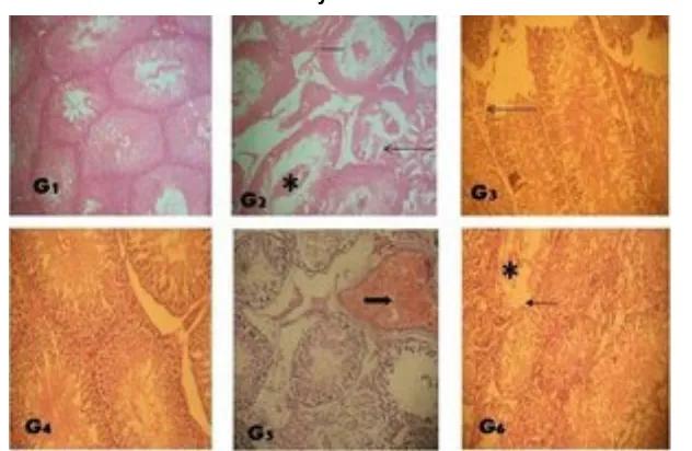
3.3 Histopathology
Fig. 4A. Amelioration of radiation-induced histological changes in testes of Wistar rats after 24 hours post-treatment byT. occidentalisextract. G 1– (Control): , G 2 – (Radiation only): reduced
height of germinal epithelium of seminiferous tubule (bar), defoliated germinal cells in the lumen of the tubules (asterick), G 3 - (vitamin E): no visible lesion, G 4 – (R+400mg): no visible
lesion, G 5 – (R+800mg): mild germinal epithelial erosion (thin arrow), G 6 – (R+1600mg): reduced height of seminiferous tubular germinal epithelium (bar). Magnification: × 400; Stain;
Haematoxylin and Eosin
Fig. 4B. Amelioration of radiation-induced histological changes in testes of Wistar rats at day 15 post-treatment byT. occidentalis extract. G 1 – (Control): shows normal architecture of testis
, G 2 – (Radiation only): foci of germinal erosion (thin arrow), sloughing with clumps of germinal cells in the lumen of the tubules (asterick) and reduced height of germinal epithelium
(bar) G 3 - (vitamin E): mild seminiferous tubular germinal epithelia erosion (thin arrow), G 4 – (R+400mg): no visible lesion, G 5 – (R+800mg): mild subcapsular congestion (dark thick arrow), G 6 – (R+1600mg): severe seminiferous tubular germinal epithelial erosion (thin arrow) with few
Fig. 4C. Amelioration of radiation-induced histological changes in testes of Wistar rats at Day 30 post-treatment byT. occidentalisextract. G 1 – (Control): shows normal architecture of testis
, G 2 – (Radiation only): severe germinal epithelial erosion (thin arrow) in some tubules and moderate subcapsular congestion (dark thick arrow), G 3 - (vitamin E): no visible lesion , G 4 – (R+400mg): no visible lesion, G 5 – (R+800mg): mild interstitial oedema (white thick arrow), G 6
– (R+1600mg): mild seminferous tubular germinal epithelium erosion (thin arrow). Magnification: × 400; Stain; Haematoxylin and Eosin
4. DISCUSSION
Exposure to ionizing radiation severely increases oxidative load on the testicular system. Reactive oxygen species (ROS) such as superoxide radicals, hydroxyl radicals, iron oxygen complexes, hydrogen peroxide and lipid peroxides are generated due to the ionization reactions mediated by gamma radiations [6]. When ROS generation is immense, as it is during gamma irradiation exposure, the cytotoxic effect is not merely local but may result in intracellular and extracellular transmission. This increases the interaction of the radicals with phospholipid structures, induce peroxidation and increase free radical induced DNA damage [10].
The significant increase in testicular LPO and concomitant accumulation of H2O2in irradiated
group as compared to control was a consequence of increased oxidative stress and cellular damage. However, decreases in levels of LPO and H2O2 generation was observed in
animals pretreated with TOE, prior to gamma irradiation exposure suggesting that TOE at doses of 400 and 800 mg/kg of b.wt strengthened the antioxidant defense system and prepared the animals to withstand the damaging effects of irradiation. Antioxidant enzymes i.e., SOD, GSH-Px and GST activity in testes showed significant decrease in gamma irradiated group probably due to the excessive generation of free radicals and increased oxidative stress. The superoxide anions are generally dismutated by SOD to form H2O2,
earlier studies, in which gamma irradiation exposure to animals showed marked increase in LPO and inhibition of SOD, GSH-Px and GST activity [10,27].
GST is directly responsible for the elimination of electrophilic oxidants at the expense of GSH [17]. The decrease in GST activity observed in testes of irradiated rats may be the result of the decrease in the availability of substrate (GSH) and also alterations in its protein structure under oxidative conditions [28]. Interestingly, pretreatment with VE 50 mg/kg of b.wt and TOE 400 and 800 mg/kg of b.wt showed radioprotection by increasing antioxidant capacity of the testicular micro environment. However higher dose of TOE (1600mg/kg of b.wt) tends to exacerbate the radiation induced testicular toxicity.
Exposure to gamma radiation markedly decreased epididymal sperm count, sperm motility and live/dead ratio, with a visible increase in sperm abnormalities in rats. Therefore, the observed decrease in testicular and epididymal sperm numbers may indicate that radiation affects the early stages of spermiogenesis. Abnormalities in sperm morphology observed in this study may be the consequence of increased lipid peroxidation of polyunsaturated fatty acids in the sperm plasma membrane. Impaired sperm motility may result in infertility caused by failure of the sperm to reach the site of fertilization as well as their ability to penetrate zonal pellucida [28]. It is commendable to note that pretreatment of TOE with its rich antioxidant properties prior to irradiation was able to improve sperm quality of rats. Histopathological studies showed that exposure to gamma radiation produced significant testicular damage in rats. The histological studies also suggest that pretreatment with TOE prior to gamma irradiation reduced the extent of testicular radiation damage and accelerated the recovery process at the cellular level. These effects may be attributed to the inhibition of oxidative stress and radioprotective function against gamma irradiation exposure by TOE.
TOE pretreatment protected or reduced the severity of irradiation induced testicular toxicity. Results from this experiment showed that the pretreatment with TOE (400 and 800 mg/kg of b.wt.) significantly increased activities of SOD, GSH-Px, GST, GSH content and reduced testicular damage.
5. CONCLUSION
Telfairia occidentalis has potent antioxidative potential and radioprotective effect against Gamma irradiation induced oxidative assault.
CONSENT
Not applicable.
ETHICAL APPROVAL
All authors hereby declare that "Principles of laboratory animal care" (NIH publication No. 85-23, revised 1985) were followed, as well as specific national laws where applicable. All experiments have been examined and approved by the appropriate ethics committee.
COMPETING INTERESTS
REFERENCES
1. Fabricant DS, Farnsworth NR. The value of plants used in traditional medicine for drug discovery. Environ Health Perspect.2001;109(1):69–75.
2. Singh RP, Banerjee S, Rao AR. Effect of Aegle marmelos on biotransformation enzyme systems and protection against free-radical-mediated damage in mice. J Pharm Pharmacol. 2000;52:991–1000.
3. Das UN. A radical approach to cancer. Med Sci Monit. 2002;8:79–82.
4. Badifu GI, Akpapunan, MA, Mgbemere VM. The fate of betacarotene in processed leaves of fluted pumpkin, a popular vegetable in Nigerian diet plant foods. Human Nutrition. 1995;48:141-147.
5. Alada AR. The haematological effects of Telfairia Occidentalis diet preparation. African Journal of Biomedical Research. 2000;3:185-186.
6. Mantena SK, Unnikrishnan MK, Joshi R. In vivo radioprotection by 5-aminosalicylic acid. Mutation Research. 2008;650:63-79.
7. Halliwell B, Gutteridge JMC. Free radical in Biology and Medicine, 2nd ed., Clarendon Press, Oxford; 1989.
8. Valko M, Rhodes CJ, Moncol J. Free radicals, metals and antioxidants in oxidative stress induced cancer. Chemico-Biological Interaction. 2006;160:41-40.
9. Farombi EO, Adedara IA, Ebokaiwe AP, Teberen R, Ehwerhemuepha T. Nigerian
bonny light crude oil disrupts antioxidant systems in the testes and sperm of rats. Archives of Environmental Contamination and Toxicology. 2010;59:166–174.
10. Deepti D, Amit KD, Harsha L, Bhalla Deepak B. Protective Effects of Terminalia
chebulain modulating Oxidative Damages against gamma irradiation induced lethality
In Rats. International Journal of Biological & Pharmaceutical Research. 2012;3:734-742.
11. Weiss JF, Kumar KS. Antioxidant mechanisms in radiation injury and radioprotection. In: Chow C, Cellular antioxidant defence mechanism, FL: CRC Press, Boca Raton. 1998;163-189.
12. Maneesh M, Jayalekshmi H. Role of reactive oxygen species and antioxidants on pathophysiology of male reproduction. Indian Journal of Clinical Biochemistry. 2006;21:80–89.
13. Chen H, Liu J, Luo L, Baig MU, Kim JM, Zirkin BR. Vitamin E, aging and Leydig cell steroidogenesis. Experimental Gerontology. 2005;40:728–736.
14. Hales DB, Allen JA, Shankara T. Mitochondrial function in Leydig cell steroidogenesis. Annals of the New York Academy of Sciences. 2005;1061:120–134.
15. Agarwal A, Gupta S, Sikka S. The role of free radicals and antioxidants in reproduction. Current Opinion in Obstetrics and Gynecology. 2006;18:325–332. 16. Misra HP, Fridovich I. The role of superoxide anion in the autooxidation of epinephrine
and a simple assay for superoxide dismutase. Journal of Biological Chemistry. 1972;247:3170–3175.
17. Habig WH, Pabst MJ, Jakoby WB. Glutathione S-transferase. The first enzymatic step in mercapturic acid formation. Journal of Biological Chemistry. 1974;249:7130–7139. 18. Rotruck JT, Pope AL, Ganther HE, Swanson AB, Hafeman DG, Hoekstra WG.
Biochemical role as a component of glutathione peroxidase. Science. 1973;179:588-90.
19. Jollow DJ, Mitchell JR, Zamppaglione Z, Gillette JR. Bromobenzene induced liver necrosis. Protective role of glutathione and evidence for 3, 4-bromobenzene oxide as the hepatotoxic metabolites. Pharmacology. 1974;11:151-157.
21. Farombi EO, Tahnteng JG, Agboola AO, Nwankwo JO, Emerole GO. Chemoprevention of 2-acetylaminofluorene induced hepatotoxicity and lipid peroxidation in rats by kolaviron a Garcinia kola seed extract. Food and Chemical Toxicology. 2000;38:535-41.
22. Zemjanis R. Diagnostic and Therapeutic Techniques in Animal Reproduction (2ndedn.)
Williams and Wilkins. 1970;139-153.
23. Pant N, Srivastava SP. Testicular and spermatotoxic effects of quinalphos in rats. Journal of Applied Toxicology. 2003;23:271–274
24. Wells ME, Awa OA. New technique for assessing acrosomal characteristics of spermatozoa. Journal of Dairy Science. 1970;53:227
25. Fridovich I. The biology of oxygen radicals. Science. 1978;201:875-880.
26. Kono Y, Fridovich I. Superoxide radical inhibits catalase. Journal of Biological Chemistry. 1982;257:5751-54.
27. Devipriya N, Sudheer AR, Srinivasan M. Quercetin ameliorates gamma radiation-induced DNA damage and biochemical changes in human peripheral blood lymphocytes. Mutation Research. 2008;654:1-7.
28. Farombi EO, Adedara IA, Ebokaiwe AP, Roy Teberen, Theresa Ehwerhemuepha. Nigerian Bonny Light Crude Oil Disrupts Antioxidant Systems in Testes and Sperms of Rats. Arch Environ Contam Toxicology. 2010;59:166-174.
© 2014 Adejuwon et al.; This is an Open Access article distributed under the terms of the Creative Commons Attribution License (http://creativecommons.org/licenses/by/3.0), which permits unrestricted use, distribution, and reproduction in any medium, provided the original work is properly cited.
Peer-review history:
The peer review history for this paper can be accessed here: