Electronic Theses and Dissertations Theses, Dissertations, and Major Papers
1-1-2006
Muscle contributions to knee joint stability: Effects of ACL injury
Muscle contributions to knee joint stability: Effects of ACL injury
and knee brace use.
and knee brace use.
Aaron Derouin University of Windsor
Follow this and additional works at: https://scholar.uwindsor.ca/etd
Recommended Citation Recommended Citation
Derouin, Aaron, "Muscle contributions to knee joint stability: Effects of ACL injury and knee brace use." (2006). Electronic Theses and Dissertations. 6943.
https://scholar.uwindsor.ca/etd/6943
by
Aaron Derouin
A Thesis
Submitted to the Faculty of Graduate Studies and Research through Kinesiology
in Partial Fulfillment of the Requirements for the Degree of Master of Human Kinetics at the
University of Windsor
Windsor, Ontario, Canada
2006
Published Heritage Branch
Direction du
Patrimoine de I'edition 395 W ellington Street
Ottawa ON K1A 0N4 Canada
395, rue W ellington Ottawa ON K1A 0N4 Canada
Your file Votre reference ISBN: 978-0-494-34902-1 Our file Notre reference ISBN: 978-0-494-34902-1
NOTICE:
The author has granted a non exclusive license allowing Library and Archives Canada to reproduce, publish, archive, preserve, conserve, communicate to the public by
telecommunication or on the Internet, loan, distribute and sell theses
worldwide, for commercial or non commercial purposes, in microform, paper, electronic and/or any other formats.
AVIS:
L'auteur a accorde une licence non exclusive permettant a la Bibliotheque et Archives Canada de reproduire, publier, archiver,
sauvegarder, conserver, transmettre au public par telecommunication ou par I'lnternet, preter, distribuer et vendre des theses partout dans le monde, a des fins commerciales ou autres, sur support microforme, papier, electronique et/ou autres formats.
The author retains copyright ownership and moral rights in this thesis. Neither the thesis
nor substantial extracts from it may be printed or otherwise reproduced without the author's permission.
L'auteur conserve la propriete du droit d'auteur et des droits moraux qui protege cette these. Ni la these ni des extraits substantiels de celle-ci ne doivent etre imprimes ou autrement reproduits sans son autorisation.
In compliance with the Canadian Privacy Act some supporting forms may have been removed from this thesis.
While these forms may be included in the document page count,
their removal does not represent any loss of content from the
Conformement a la loi canadienne sur la protection de la vie privee, quelques formulaires secondaires ont ete enleves de cette these.
MUSCLE CONTRIBUTIONS TO KNEE JOINT STABILITY: EFFECTS OF ACL INJURY AND KNEE BRACE USE
Aaron Joseph Derouin University o f Windsor, 2006
The purpose of this study was to develop an EMG-based biomechanical model of the
lower extremity for the purpose of evaluating the rotational joint stiffness of ACL-
deficient (n=9) and reconstructed (n=8) knees, with and without a functional knee brace.
Kinematic, EMG, and kinetic data were measured, while subjects performed stable and
unstable quasi-static squats, and were incorporated into biomechanical and anatomical
models. Individual and total muscle contributions to knee joint rotational stiffness were
calculated about the flexion-extension axis, using the method developed by Potvin and
Brown (2005) for four independent variables: Squat task; Leg condition (uninjured,
unbraced, braced); Knee angle (5-10°, 20-25°, 40-45°); Injury Status. Subjects had
significantly higher (p<0.05) total knee joint stiffness values while wearing the brace
compared to the control leg. The difference in knee joint stiffness values in the unstable
trials compared to stable trials was significantly greater at 5-10° versus 20-25° and 40-
The completion of this project would not have been possible without the valuable contributions and input from several significant individuals. I would like to first
acknowledge God, for blessing me with the opportunity and curiosity to undertake such a complex study and the ability, determination, and perseverance to finish.
Jim, I would first of all like to thank you for taking me on as a student. You allowed me to take on a project that I thought was a little different from the research you typically do and allowed me to satisfy some of my curiosities about stability, knee injury and knee braces. I have gained practical and hands on experience to compliment the excellent theoretical knowledge I learned from your graduate course in instrumentation and modelling. Most o f all, I am truly thankful for the all the opportunities you have provided to me, your generosity and your friendship.
Next, I would like to thank my committee members Dr. Andrews and Dr.
Altenhof for your involvement in this project. Your constructive feedback has improved my critical thinking skills and ultimately improved the quality of this document.
I would also like to acknowledge Dr.G. Jasey and Dave Stoute for their assistance in recruiting subjects. Additionally, Joel Cort, Christy Calder and Don Clark have
provided valuable assistance in the areas of programming, data collection, circuit control and electronics. Derek Kozier o f Clinical Orthotic Consultants of Windsor, also, provided equipment and materials to complete the data collection portion o f the study.
Innovation Sports graciously donated eight FLEX functional knee braces to fit on the subjects in this study. I would particularly like to thank Brett Guerin, the Marketing Director for his support and assistance.
Finally, I would like to thank my mother and John for your continued support and encouragement as I continue to fulfill my dreams. Mom, thanks so much for your
what lies within us.”
Ralph Waldo Emerson
ABSTRACT... iii
ACKNOWLEDGEMENTS...iv
LIST OF TABLES... ix
LIST OF FIGURES...x
LIST OF ABBREVIATIONS... xv
CHAPTER 1: INTRODUCTION...1
1.1 Background...1
1.2 Purpose... 8
1.3 Hypotheses...9
1.3.1a Overall Knee Joint Rotational Stiffness... 9
1.3.1b Overall Knee Joint Rotational Stiffness... 9
1.3.2 Individual Muscle Contributions to Knee Joint Rotational Stiffness... 10
CHAPTER 2: REVIEW OF LITERATURE... 12
2.1 Anatomy of the K nee...12
2.1.1 Passive Tissues... 12
2.1.2 Neuroanatomy and Mechanoreceptors o f the ACL and other joint structures...22
2.1.3 Muscles o f the K nee...24
2.2 Normal Knee Biomechanics...28
2.2.1 Passive Knee K inem atics... 28
2.2.2 Muscular Contributions to Knee Kinem atics... 31
2.2.3 Neuromuscular Recruitment and Reflexes...33
2.3 ACL Injury... 36
2.3.1 Diagnosis and Classification...36
2.3.2 Treatment and Prognosis...38
2.3.3 ACL-Deficient Pathomechanics... 42
2.4 Functional Knee Braces... 47
2.4.1 Brace Design: Types, Features and Other Considerations...48
2.4.2 Functional Considerations... 52
2.5 Joint Stability... 57
2.5.1 Knee Joint Stability... 57
2.5.2 Joint Mechanical Stability... 61
2.5.3 Calculation of Joint Stability...64
2.6 Biomechanical Modelling o f the Knee... 67
CHAPTER 3: METHODOLOGY... 73
3.1 Study Design...73
3.2 Subjects... 74
3.3 Functional Knee Brace Fitting... 76
3.4 Experimental Protocol... 76
3.5 Instrumentation and Data Acquisition... 80
3.5.1 Electromyography... 81
3.5.2 Force Plate D a ta ... 83
3.5.3 Single Leg Squat Kinematics...83
3.5.4 Subject D ata... 84
3.6 Data Analysis... 84
3.6.1 Anthropometric D ata... 84
3.6.2 Muscle Orientations...85
3.6.3 EMG Processing and Initial Force Estimates... 86
3.6.4 Biomechanical Model...87
3.6.5 Calculation of Knee Joint Rotational Stiffness... 91
3.7 Statistical A nalysis...93
CHAPTER 4: RESULTS... 95
4.1 Independent Variables... 95
4.1.1 Subject D a ta...95
4.1.2 Knee Angle and Knee Moment Data... 96
4.2 Dependent Variables... 97
4.2.1 Rotational Stiffness Data - Overall and Individual Muscular Contributions... 97
4.2.2 Rotational Stiffness Inputs- EMG and Moment Arm D a ta ... 104
CHAPTER 5: DISCUSSION...109
5.1 Knee Angle Effects...110
5.2 Injury status (ACL deficient vs. Reconstructed) Effects... 113
5.3 Leg Condition (Braced, Unbraced, Control) Effects... 115
5.4 Squat Task (Stable versus Unstable) Effects...116
5.5 Individual Muscle Contributions to Overall Joint Stiffness... 118
5.5.1 Knee Extensor Com partment...118
5.7 Limitations...125
5.8 Future Directions...128
5.9 Conclusions...130
APPENDICES... 132
APPENDIX A ...133
SUBJECT CONSENT AND FUNCTIONAL STATUS FO RM S... 135
APPENDIX B ...150
Wobble Board Control Circuit Description... 150
KNEE BRACE SPECIFICATIONS... 153
APPENDIX C ...155
SUBJECT DEMOGRAPHIC AND FUNCTIONAL STATUS DATA... 155
APPENDIX D ...156
FUNCTIONAL MOMENT ARM AND EMG D ATA ... 156
REFERENCES... 158
(p<0.0001) both had significant main effects (n=17)... 96
Table 4.2. Average knee moment (flexion-extension) and standard deviations (n=17)...97
Table 4.3. Significant interaction and main effects (p < 0.05) presented for overall stiffness and
the individual muscular contributions to stiffness...100
Table 4.4. Post hoc results for the significant interaction and main effects for overall stiffness and
individual muscular contributions to knee joint stiffness...101
Table AC-1 A. Subject demographic and anthropometric data...155
Table AC-1B. Subject functional injury status scores...155
Table AD-1A. Functional moment arm and average surface EMG data presented for the knee
extensors. Maximum force capcity (Fmax) and optimal length (rlen) of each muscle is
shown. Significant interaction and main effects (p<0.05) are displayed for the EMG
(%MVC) of each muscle... 156
Table AD-1B. Functional moment arm and average surface EMG data presented for the knee
flexors. Maximum force capcity (Fmax) and optimal length (rlen) of each muscle is
shown. Significant interaction and main effects (p<0.05) are displayed for the EMG
LIST OF FIGURES
Figure 1. A) Sagittal view of bone anatomy of knee joint. B) Posterior view of ligament
anatomy of knee joint. The ACL is not shown... 13
Figure 2. A) Relative orientation and position of the ACL on the medial surface of the right
lateral femoral condyle (Girgis et al., 1975). B) Schematic representation of the four-
bar linkage. The instant center of rotation is defined at the intersection of AB and CD
(Imran and O'Connor, 1998)... 18
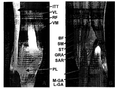
Figure 3. A) Anterior view of knee musculature: ITT- iliotibial tract, VL-vastus lateralis, RF-
rectus femoris, VM-vastus medialis, PL-patellar ligament. B) Posterior view: BF-
biceps femoris, SM-semimembranosus, ST-semitendonosus, GRA-gracilis,
SAR-sartorius, M-GA-medial gastrocnemius, L-GA-lateral gastrocnemius... 26
Figure 4. Average normalized antagonistic mean amplitude value versus joint angle for
hamstrings and quadriceps (Baratta et al., 1988)...34
Figure 5. Muscle moment arm variations with joint angle. Note the striking inverse pattern of
muscle moment arm and MAV between the hamstrings (4A & 5A) and quadriceps
(4B & 5B) (Baratta et al., 1988)... 34
Figure 6. A) Localized focal high pressure to the anterior aspect of the articular surface due to
the absence of hamstrings antagonistic activity. B) Evenly distributed low articular
pressure due to fully activated hamstrings co-contraction. (Baratta et al., 1988)... 35
Figure 7. The angle of the patellar tendon acquires a more perpendicular orientation (25° to 15°)
with quadriceps contraction in the ACL deficient knee.This effectively reduces the
quadriceps anterior shear component (Howell and Usafr, 1990)... 44
Figure 8. After sectioning of the ACL, the axis of tibial rotation is located about the MCL
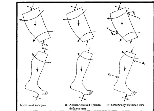
Figure 10. The forces acting on the weight-bearing lower extremity with the knee flexed for the
normal leg (A), the ACL deficient knee (B) and the orthotically stabilizes knee (C)
(Liggins and Bowker, 1991)... 51
Figure 11. A paradigm of functional stability, displaying the influence of mechanical instability
and proprioceptive deficits on functional stability following ligamentous injury
(Swanikef al., 1997)...58
Figure 12. Peak anterior tibial translation (ATT) is shown during isokinetic knee extension at 0,
30, 90, 180, and 300 deg/sec in the ACL deficient knee for different levels of
hamstring activation. The horizontal line at 7.5 mm represents peak ATT with out
hamstrings cocontraction and defines the limit of knee joint stability (Yanagawa et al.,
2002)... 59
Figure 13. Simplified ball analogy of stability during static equilibium. The ball in (c) returns to
its equilibrium position when perturbed and is stable, while both (a) and (b) are
unstable. (Crisco III and Panjabi, 1990)... 62
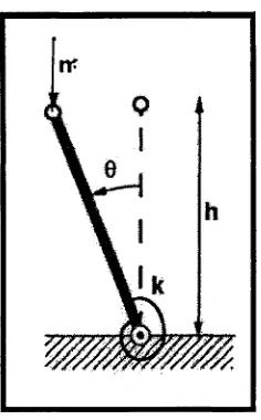
Figure 14. A single degree of freedom system to describe the components of stability: Mass (m),
height (h), and rotational stiffness (k). The mass applied to the system may cause a
change in the potential energy of the system by altering the height of system and
change the amount of energy stored in the spring, thus augmenting rotational stiffness.
(Crisco III and Panjabi, 1990)... 62
Figure 15. Dysfunction of the spinal stability system. Injury/disease may decrease the passive
and or active stability. The neural control unit enhances the stabilizing function of the
intact spinal components. This may lead to accelerated degeneration and may
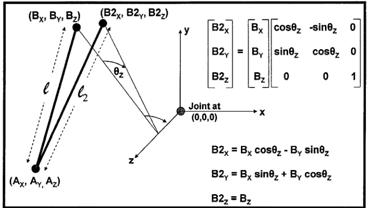
Figure 17. Stability may be calculated for any muscular system given the origin coordinates (Ax,
AY, Az) and insertion coordinates (Bx, By, Bz) relative to the axis of rotation of the
joint of interest. The length of the muscle may be determined from this coordinate
data. New insertion coordinates (B2X, B2y, B2Z) may be determined using a
transformation matrix and the new muscle length (£) and change in muscle length (At)
may then be calculated (Potvin and Brown, 2005)... 67
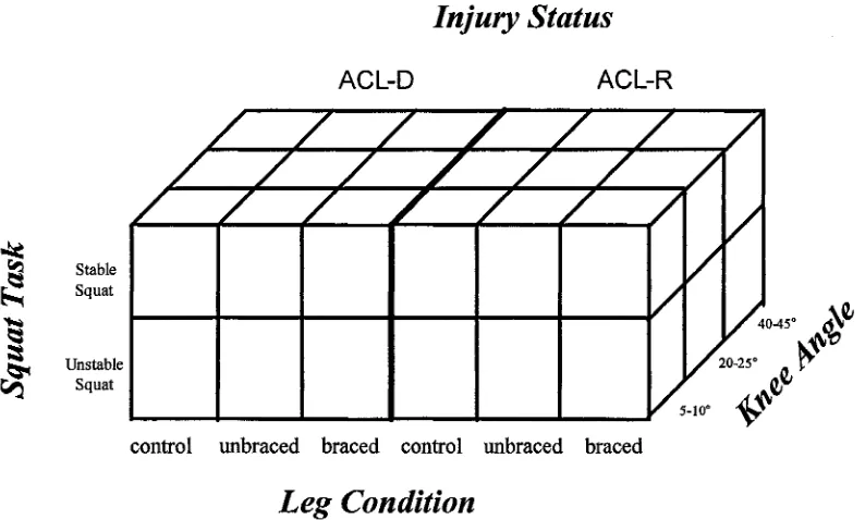
Figure 18. The study design is represented a s a 2 x 3 x 3 x 2 matrix of the independent variables
for ACL-Deficient and Reconstructed groups. For within variables, there were two
Squat Tasks, three Leg Conditions and three Knee Angles. The between variable of
Injury Status had two levels...74
Figure 19. FLEX Off the Shelf Functional Knee Brace from Innovation Sports...76
Figure 20. A) stable squat task, B) unstable squat task without brace, C) unstable squat with
brace, D) schematic diagram of the unstable platform dimensions. In A) the LED is
shown in the camera’s field of view, indicating that EMG and force plate data
collection has commenced. The anatomical markers, the pelvic fin with three
markers, and the two cluster sets of thigh and lower leg markers are shown... 80
Figure 21. Flowchart of the data analysis and model progression. Kinematic and kinetic data
were calculated to ensure static equilibrium was achieved before JSR was calculated.
model in a neutral posture C) the model is shown in a squat posture with the knee and
hip flexed to 90 degrees. Note the change in orientation of the patellar ligament (PL)
from the neutral posture to the squat posture (90 degrees of hip and knee flexion). ...89
Figure 23. Trend of injury status on overall JSr (p= 0.3881). ACL Deficient (n=9) and ACL
Reconstructed (n=8)... 97
Figure 24. Trend of injury status on individual muscular contributions to overall stiffness
(p>0.05). ACL Deficient (n=9) and ACL Reconstructed (n=8). Vastus lateralis
(p=0.2183) and semimembranosus (p=0.1271) displayed the most pronounced
trends...98
Figure 25. Main effect of leg condition on total knee joint stiffness (n=102). Standard deviation
bars are displayed. Post hoc results are presented... 99
Figure 26. Interaction effect between squat task (stable vs. unstable) and knee angle (n=51).
Standard deviation bars are displayed. Post hoc results are presented... 99
Figure 27. The effect of leg condition on individual muscle contributions to overall knee joint
stiffness (n=102)... 102
Figure 28. Interaction effect of knee angle and squat task on the absolute individual muscle
contributions to knee joint stiffness (n = 51)...103
Figure 29. The effect of knee angle on the individual muscle contributions to knee joint stiffness,
presented as a percentage of overall stiffness (n=102)... 104
Figure 30. Significant main effect of leg condition on average EMG of the individual muscles
that were used as inputs to calculate knee stiffness (n=102)...104
Figure 31. Interaction effect of knee angle and squat task on the average EMG of individual
Figure 32. A) Mean moment arm data showing effect of knee angle on moment arm and r2 value
used to calculate knee stiffness (n=102) B) Effect of leg condition on moment arm and
r2 value used to calculate knee stiffness (n=102)...107
Figure 33. Mean effect of leg condition, squat task, and knee angle on coactivation of the
semimembranosus relative to the vastus lateralis (n=17)... .107
Figure 34. Custom fabricated Fitter-Like wobble board. The platform of the wobble board
measured 22 x 20 cm, designed larger than the surface area of the force plate. A Vi”
rubberized mat (17%” x 19 Vi” cm) was mounted onto the force plate to minimize slip
from shear forces created while subjects performed unstable squat trials. Additionally,
self-adhesive sand paper was secured onto the rockered surface of the wobble board to
further minimize slip due to shear forces... 151
Figure 35. Solenoid engagement with wobble board. Pin is retained in the bracket by the spring.
Activation of solenoid disengages pin from the hole in the bracket, which is secured to
the board... 151
Figure 36. Wobble board control module. The Allen Bradley timer was set to one second
delay... 152
LIST OF ABBREVIATIONS
AAOS: American Academy of Orthopaedic Surgeons
ACL: Anterior Cruciate Ligament
AIIS: Anterior Inferior Illiac Spine
AMB: Anterior Medial Band
ANOVA: Analysis of Variance
ATT: Anterior Tibial Translation
BF: Biceps Femoris
BF-L: Biceps Femoris Long Head
BF-S: Biceps Femoris Short Head
BMI: Body Mass Index
CCI: Co-contraction Index
CMRR: Common Mode Rejection Ratio
COM: Center o f Mass
COP: Center o f Pressure
EMG: Electromyography
FKB: Functional Knee Brace
GA: Gastrocnemius
GA-L: Gastrocnemius Lateralis
GA-M: Gastrocnemius Medialis
GR: Gracilis
ITT: Illiotibial Tract
JSr: Joint Rotational Stiffness
k: Stiffness
KOS: Knee Outcome Survey
LCL: Lateral Collateral Ligament
MCL: Medial Collateral Ligament
Mr: Net Knee Reaction Moment
MRI: Magnetic Resonance Imaging
MVC: Maximum Voluntary Contraction
OA: Osteoarthritis
PCL: Posterior Cruciate Ligament
PF: Patellofemoral
PL: Patellar Ligament
PLB: Posterior Lateral Band
QF: Quadriceps Femoris
RF: Rectus Femoris
rz: Functional Moment Arm about Z-axis
SA: Sartorius
sEMG: Surface Electromyography
SM: Semimembranosus
TFL: Tensor Fascia Latae
V : Potential Energy
VI: Vastus Intermedius
VL: Vastus Lateralis
CHAPTER 1
INTRODUCTION
1.1 BACKGROUND
Disruption to the anterior cruciate ligament (ACL) is the most frequently
occurring ligament injury of the knee (Feagin and Curl, 1976) and is one of the most
dehabilitating injuries of the lower extremity. “The increasing popularity of physical
fitness, combined with activities that place the cruciate ligament at risk, seems to be
causing an almost exponential increase in the number of serious knee disruptions”(Ryder
et al., 1997; Johnson, 1988). In 1982, knee injuries in the United States were defined as a
national health problem (Feagin and Lambert, 1985), and today the incidence of serious
knee injuries continues to escalate. The estimated annual cost of surgical treatment is
about $1.5 billion, alone, without consideration of the cost of the initial evaluation and
treatment of those injured, the nonsurgical care of remaining patients, or the future of
medical treatment for those who develop post-traumatic arthritis (Kao et al., 1995).
Disruption to the ACL has become so commonplace that the seriousness of the injury is
often overlooked. In fact, the careers of thousands of athletes have been hampered or
completely terminated as a result of ACL disruption. The vulnerability of the knee to
injury may be accounted for by the fact that “no other joint in the leg depends less for its
stability on the shapes of its constituent articulating surfaces” (Laurence and Strachan,
1970) and that the knee joint is between the two longest bones in the body.
Each year in the United States, it is estimated that there are approximately
an analysis of athletic population segments, such as football, skiing and soccer, the
incidence of ACL injury is even higher (Smith et al., 1993).
Functionally, the ACL is the primary restraint to anterior translation of the tibia,
as it provides an average of 86 per cent of the total resisting force to anterior drawer
(Butler et al., 1980). The intersection of ACL together with the posterior cruciate
ligament defines the axis of rotation of the knee joint. Furthermore, the changing position
and relative orientation of the cruciate ligaments throughout the range o f knee motion is
responsible for the rolling and gliding functions o f the knee. “The ACL, in particular,
serves a highly specialized role in guiding knee motion, one that is vital to joint stability
and the maintenance o f normal knee function and overall kinematics” (Smith et al.,
1993). In addition to its structural stabilizing role, the ACL is thought to provide a
stabilizing function through a neurological feedback mechanism (Goldstein and Bosco,
2001). Mechanoreceptors, such as Golgi tendon-like organs, Pancinian and Ruffini
corpuscles, have been identified within the ACL and other joint structures. These
receptors are thought to be responsible for this feedback mechanism. It is postulated that
these receptors, when stimulated, provide afferent input along with information supplied
by muscle spindles and joint Golgi tendon organs (GTO’s), to the alpha and gamma
motor pathways to modulate muscle stiffness.
A loss of integrity to the ACL causes significant alterations to knee joint
kinematics and the recruitment patterns of muscles that surround the knee. The primary
concerns following injury to this ligament are: functional impairment and the
development of symptomatic knee instability (Evans and Stannsih, 2001). Instability
instability defined as giving way during activity (Lysholm and Gillquist, 1982). Without
appropriate treatment, a complete rupture of the ACL may result in progressive knee
instability, which in turn leads to recurrent intra-articular damage and eventual
osteoarthritis (OA) (Daniel et al., 1994). Furthermore, it is difficult to select patients for a
specific treatment protocol because the relationship between quantitatively measured
laxity, such as that determined by a knee joint arthrometer, is not at all related to the
functional status of the patient. This inability to predict the functional outcome of ACL
deficient patients is further compounded by the fact that not every patient displays
symptoms of instability and that not all knees with ongoing ACL insufficiency progress
to developing osteoarthritis (Evans and Stannsih, 2001).
Typically, the focus of treatment for the ACL deficient patient is the restoration of
normal knee joint mechanics in an effort to relieve pain, reduce instability, and minimize
the risk of post-traumatic osteoarthritis (MacWilliams et al., 1999). The treatment options
available to the ACL injured individual are surgical reconstruction and non-operative
treatment. Reconstruction is generally advocated for the young active individual, whereas
non-operative management is recommended to patients with low athletic demands or
sedentary occupations (Segawa et al., 2001). Recently, the term non-operative
management has been clarified and described as a progressive exercise based program
which may include the following components: isometric, isotonic, isokinetic, closed
chain exercises, and functional exercises involving perturbation training and sport-
specific agility exercises. An additional treatment option prescribed to both surgically and
non-surgically managed ACL-deficient patients is the use of functional knee braces. In a
prescription practices, only 13% and 3% of physicians never prescribed functional braces
to ACL-reconstructed and ACL-deficient patients, respectively.
Despite the overwhelming recommendations of functional knee brace use by
physicians that manage ACL injured patients, there exists significant controversy in the
literature regarding the effectiveness of these braces in stabilizing the knee. Cawley et al.
(1991) reviewed the subjective response data from several studies and found that 90% of
subjects reported fewer episodes of giving way (instability) and were functionally
improved with brace use. Subjective response, although a valuable tool to the researcher,
must always be correlated to biomechanical measures (Cawley et al., 1991). The ability
of functional knee braces to passively restrain anterior tibial translation in cadaveric
specimens has been demonstrated by several authors to occur at relatively low loads
(140 N) (Beynnon et al., 2004), well below estimated physiological loads of 400N
(Noyes et al., 1980). Despite these consistent findings, the inability o f functional knee
braces to control tibial displacement under high physiological loads has yet to be
validated in vivo. Loading o f the knee in vivo with physiological loads is a significant
ethical constraint that has yet to be resolved.
Recently, electromyography (EMG) has been used to evaluate the effectiveness of
knee braces in modifying muscle activity in favourable manner to stabilize the ACL-
deficient knee. The effects of functional knee bracing have been demonstrated by a
limited number of studies to exert its influence on the neuromuscular system through
various mechanisms. Compensatory recruitment of the hamstrings and quadriceps is
required following ACL injury to maintain stability of the knee joint. Although, several
2003; Wojtys et al., 1996b) have established that bracing the ACL deficient patient alters
the EMG activity o f knee musculature, no conclusive evidence exists that these changes
actually enhance joint stability. Unfortunately, these modifications to muscular activity
are speculatively postulated as favourable or unfavourable based on qualitative data.
Favourable responses to knee bracing, as described by Smith et al. (2003), were
considered as: earlier activation (shorter onset latency) of either the hamstrings or
gastrocnemius muscles groups relative to the quadriceps muscle groups. The results of
this study, along with the findings of some other investigations (Wojtys and Huston,
1994; Wojtys et al., 1996a), suggest that inter-individual differences exist in muscle
reflex patterns and that there is a lack of a universal recruitment pattern in response to
anterior tibiofemoral shear. To date, no known study has quantitatively assessed how
functional knee bracing alters mechanical knee joint stability in ACL-deficient patients.
Doorenbosch and Harlaar (2003) quantified co-activation of the hamstrings and
quadriceps using a co-contraction index and found that this index was significantly higher
in ACL-deficient subjects compared to normal subjects performing isokinetic
contractions about the knee. It was suggested that quantifying co-contraction is a useful
parameter to evaluate clinical interventions and rehabilitation processes. The usefulness
of this approach in assessing joint stability is limited by one central assumption.
Rotational equilibrium of the joint is achieved when flexor and extensor torques are
applied simultaneously and if the flexing and extending moments of the muscle forces are
equal. However, this does not necessarily mean that the components of the flexor and
extensor forces parallel to the tibial plateau will also be equal and opposite (O'Connor,
absence of the ACL, the collateral ligaments and joint capsule must resist anterior tibial
displacement that occurs when the shear force generated by the quadriceps is greater than
that of the hamstrings. O’Connor (1993) identified a critical flexion angle of 22 degrees,
where these respective parallel components exactly balance each other and no ligament
forces are required. Although, static equilibrium is one of the required conditions to
calculate joint stability, it does not guarantee that a particular musculoskeletal system is
stable from a mechanical perspective. No known study to date has calculated the
mechanical stability o f the healthy or ACL-deficient knee.
In 1989, Bergmark’s quantitative assessment o f lumbar spine stability was the
first study to fully examine and define the mechanical stability of a muscular system. He
affirmed that when the Potential Energy (V) of an entire system is at a relative minimum
then the muscular system can be described as stable. Thus, a system in a stable state of
equilibrium must always be capable of returning to its original state of equilibrium in
response to a perturbation around this state. Two conditions must be satisfied in order for
a system to be classified as stable. Initially, the system must be in a state of rotational or
mechanical equilibrium, that is the first derivative of V (net moment) with respect to
angle (0) must be zero. Next, the second derivative of V must be positive definite (greater
than zero) indicating that V is at some minimum.
Muscular contributions, to the potential energy of a system during a perturbation,
are dependent on how this perturbation changes the length of the muscle and the
orientation of the muscles relative to the joint being examined. The ability of a muscle to
store or release elastic energy is related to its stiffness, which in turn depends on the
a very significant role in stabilizing the muscles surrounding a joint and/or system of
joints.
A novel approach to calculating stability about any specified joint has recently
been proposed by (Potvin and Brown, 2005). This method is suggested to be
mathematically less complex than the approach used by Bergmark and subsequent
researchers (Cholewicki and McGill, 1996; Crisco and Panjabi, 1991; Granta and Wilson,
2001). The method used by Bergmark is thought to be limited to only few researchers, as
the mathematical analysis required to determine stability limits its application.
Additionally, this new approach allows the determination of stability for a multi-muscle
system, overcoming the shortcomings of the method used by Cholewicki et al. (1999) and
Granata and Orishimo (2001), which is limited to one single-equivalent flexor and one
single upright single-equivalent extensor muscle. According to Potvin and Brown
(2005), the stability of any specified joint may be assessed with an EMG-driven
biomechanical model given the coordinates of the individual muscles that cross the joint,
the pre-activation level of the muscle prior to being perturbed, the muscles functional
moment arm, the length of the muscle, the stiffness of the muscle and the potential
energy of the system being examined.
To date, the efficacy of functional knee braces in stabilizing the ACL deficient
knee is equivocal. Unfortunately, the scientific validation of functional knee braces has
not kept pace with the proliferation of knee brace companies and their innovative designs.
The evaluation o f the effectiveness of braces using knee arthrometers on cadaveric
specimens has proven to be redundant, as the loads employed and the testing mode do not
braces are designed for athletically active individuals wishing to return to their pre
morbidity status and, consequently, should be evaluated under conditions that duplicate
their normal environment. Up to now, relatively few studies have attempted to elucidate
the effects of functional knee braces on muscle control in the ACL-deficient knee.
Numerous researchers (Aciemo et al., 1995; Branch et al., 1989; Ramsey et al., 2003;
Smith et al., 2003; Wojtys et al., 1996b) have all documented notable changes to EMG
amplitudes and patterns of ACL-deficient patients with brace use. However, the stability
that functional knee braces potentially provide, thus far, has only been qualitatively
described. The new approach to calculating joint stability proposed by Potvin and Brown
(2005) will enable a quantitative evaluation of the stabilizing potential of functional knee
braces.
1.2 PURPOSE
The goal o f this study was to develop an EMG-based biomechanical model of the
lower extremity for the purpose o f evaluating the rotational joint stiffness (JSr) of the
ACL injured knee with and without a functional knee brace. JSrwas evaluated for two
single-leg squat tasks, a stable isometric squat and an unstable isometric squat, performed
on a custom fabricated fitter-like wobble board. The individual contributions, and total
contribution, of leg muscles to knee JSrwere assessed at three knee joint angle ranges (5-
10,20-25, and 40-45 degrees) that the subject was instructed to maintain over the course
1.3 HYPOTHESES
1.3.1a Overall Knee Joint Rotational Stiffness
Total muscle contributions to knee flexion-extension JSr, will show a statistically
significant main effect (p<0.05) between leg conditions. Post hoc analysis will reveal that
the brace involved leg condition will have significantly lower total knee JSr compared to
the unbraced involved leg.
In two studies, a statistically significant decrease in hamstrings torque or muscle
activity has been demonstrated while wearing a functional knee brace (Aciemo et al.,
1995; Wojtys et al., 1996b). It is hypothesized that the proprioceptive feedback that the
brace provides will optimize the gain on neuromotor control of the hamstrings and will
decrease its activity. Preliminary biomechanical modelling has shown that particular knee
flexors, such as biceps femoris long and short head, are destabilizing and have negative
stability values when the model is positioned in a neutral posture (neutral hip extension
and full knee extension). The brace may provide the nervous system with sufficient
proprioceptive information to decrease the gain on the hamstrings throughout a range of
joint angles that are destabilizing.
1.3. lb Overall Knee Joint Rotational Stiffness
Total muscle contributions to knee flexion-extension JSr will show a statistically
significant interaction (p<0.05) between leg condition and squat task. Further Post hoc
analysis will reveal that the highest total JSr will be displayed fo r the unbraced involved
leg performing the unstable single leg squat, compared to any other leg x squat task
Typically, squat exercises performed on a wobble board provide a greater
challenge to the neuromuscular system, and result in increased co-activation of the
flexors and extensors of the knee, compared to similar stable squat exercises. Kean et al.
(2006) found that a fixed foot balance training program on a wobble board significantly
increased rectus femoris muscle activation during the landing phase of a jumping test,
compared to functionally directed training (supervised jump training). Additionally, static
balance improved 33% more with the fixed foot protocol compared to functionally
directed training. In a different study, both conservatively managed ACL deficient and
surgically treated subjects had significantly higher hamstrings co-activation patterns
compared to that o f the normal-control subjects (Grabiner and Weiker, 1993). It is
hypothesized that the proprioceptive feedback that the brace provides will optimize the
gain on neuromotor control of the hamstrings and will decrease its activity. The brace
may provide the nervous system with sufficient proprioceptive information to decrease
the gain on the hamstrings throughout a range of joint angles that are destabilizing. It is
expected that the unstable squat task will elicit greater reliance on the proprioceptive
information provided by the brace, as it has a greater requirement for mechanical
stability.
1.3.2 Individual Muscle Contributions to Knee Joint Rotational Stiffness
The vastus lateralis will exhibit the greatest contribution to knee flexion-extension JSr o f
all the extensor muscles, while the semimembranosus will display the highest JSr o f all
the knee flexors. The ratio o f vastus lateralis and semimembranosus EMG will be used to
between co-contraction and rotational jo in t stiffness. This will be calculated by dividing
the percent maximum voluntary contraction (%MVC) fo r the semimembranosus by the
%MVC fo r o f the vastus lateralis.
In the literature, it is widely regarded that co-activation of the hamstrings and
quadriceps is the primary mechanism that is used by the neuromuscular system to
enhance stability about the knee joint in ACL injured patients. According to Delp et al.
(1990), the vastus lateralis and the semimembranosus have the greatest maximal force
capacity of the muscles in the extensor and flexor compartments, respectively.
Additionally, preliminary biomechanical modelling (Derouin and Potvin, 2005) has
shown that the vastus lateralis and semimembranosus.have the greatest potential to
CHAPTER 2
REVIEW OF LITERATURE
In order to understand the destabilizing effects of injury to the anterior cruciate ligament
it is first necessary to describe the anatomy of the largest and the most complex joint in
the body, the knee (Tortora and Grabowski, 1996)
2.1 ANATOMY OF THE KNEE
2.1.1 Passive Tissues
The knee is composed of 3 bones, 3 joints, 5 primary ligaments and several other
accessory structures. The distal end of the femur, proximal portion of the tibia and the
patella make up the knee. The inherent complexity of the knee may be attributed to the
uniquely shaped articular surfaces of the femoral condlyes, the tibial plateau and to the
configuration of the cruciate ligaments.
2.1.1.1 Bone Anatomy
The femur is the longest, strongest bone in the body and its distal end is
broadened for articulation with the tibia (Moore, 1992). The articular surfaces of the
condyles are pulley shaped and are convex in both the sagittial and frontal planes
(Kapandji, 1987) (Figure 1A). The articular surface of the medial femoral condyle is
somewhat larger than that of the lateral condyle (Brantigan and Voshell, 1941). The neck
o f the pulley is represented by the central groove on the patellar surface anteriorly and by
superior/anterior surfaces of femoral condyles related to the patella and is primarily
responsible for limiting hyperextension of the knee (Brantigan and Voshell, 1941).
In an examination o f 100 cadaveric knees, Brantigan and Voshell (1941) found
that the knee will go into greater than 90 degrees o f hyperextension after cutting away the
femoral condyles and leaving the ligaments intact.
Intercondylar ' notch
„ Lateral Meniscus Medial ^
Meniscus
Figure 1. A) Sagittal view of bone anatomy of knee joint. B) Posterior view of ligament anatomy of knee joint. The ACL is not shown
The tibia is the second largest bone of the skeleton and is situated on the
anteromedial side of the leg (Moore, 1992). The proximal surface is expansive compared
to the rest of the tibia, due to its articulation with the large condyles of the femur. The
tibial surfaces are reciprocally curved to the femoral condyles and consist of two curved
and concave parallel gutters that are separated by the intercondylar eminence (Kapandji,
1987). More precisely, the medial condyle of the tibia is biconcave in both the sagittal
and frontal plane, while the lateral condyle is concave in the frontal plane and convex in
the sagittal plane. The medial femoral condyle is relatively stable inside the concave
medial tibial condyle, whereas the lateral femoral condyle is unstable as it travels on the
on the integrity of the anterior cruciate ligament. The intercondylar eminence is oriented
in an anterior-posterior direction and fits into the intercondylar notch between the femoral
condyles. The planed shape of the intercondylar eminence acts as a pivot, allowing axial
rotation of the tibia through a vertical axis that passes through the medial intercondylar
spine (Kapandji, 1987). An additional noteworthy feature of the tibia is the prominent
tibial tuberosity, located anteriorly, where the patellar ligament inserts (Moore, 1992).
The patella, or kneecap, is a triangular shaped sesamoid bone embedded in the
quadriceps femoris tendon with its apex directed inferiorly (Moore, 1992). The posterior
surface of the patella, which articulates with the femur, is comprised of two biconcave
facets. The lateral and medial facets are separated by the median vertical ridge.
The 3 joints that comprise the knee are: 1) the patellofemoral joint between the
inner surface of the patella and the patellar surface of the femur; 2) the lateral
tibiofemoral joint between the lateral condyle of the femur, lateral meniscus, and lateral
condyle of the tibia; and 3) the medial tibiofemoral joint between the medial condyle of
the femur, medial meniscus, and medial condyle o f the tibia (Tortora and Grabowski,
1996). Tortora and Grabowski (1996) describe the patellofemoral joint as a gliding joint.
Generally, the tibiofemoral joint may be considered a modified hinge joint, referring to
the ability o f the tibiofemoral joint to produce moderate amounts of transverse rotational
motion, small amounts of varus-valgus or frontal plane motion, in addition to the flexion-
extension motion in the sagittal plane.
2.1.1.2 Ligament Anatomy
Ligaments are relatively inelastic and highly adapted tissues comprised of short
which connect bone to bone. Ligaments are obligated to function in a state of tension,
with its functional position considered as the position in which a vast majority of its
fibres are taut (Fuss, 1989). This state of ligament tension is dependent on whether or not
the distance between the origin and insertion of the ligament fibres remains constant or
variable as the knee joint moves throughout its normal range of motion (Fuss, 1989). The
variability of this distance is dependent both on the osseous attachment of the ligaments
and the surface anatomy of the tibiofemoral articular surfaces. They function at lower
applied loads to guide joint motion, at higher loads to limit joint motion and to assist
other joint structures in the protection of periarticular soft tissues during both normal and
pathological knee motions (Smith et al., 1993).
The 5 primary structural ligaments in the knee are the two collateral ligaments
(medial and lateral), the two cruciate ligaments (anterior and posterior) and the patellar
ligament/tendon (Figure IB). These ligaments along with the joint capsule and the
menisci (articular cartilage) are responsible for stabilizing the knee during movement.
According to Ryder et al. (1997), the cruciate ligaments form the central pivot and serve
as the key components to stability o f the femoral-tibial articulation. Several other
accessory structures not as significant to stability of the knee include: the medial and
lateral patellar retinaculae, the infrapatellar fat pad, the oblique and arcuate popiteal
ligaments, the transverse ligaments of the menisci, and the bursae.
Collateral Ligaments
The collateral ligaments are mostly responsible for providing medial and lateral
translational and rotational stability to the knee. The tibial (medial) collateral ligament
extends from the medial condyle of the femur to the medial condyle of the tibia (Tortora
and Grabowski, 1996). The MCL functions primarily as a passive medial joint stabilizer,
as it is taut throughout flexion. It also aids in restraining hyperextension and provides the
main valgus stability in these positions (Pope, 1994).
The fibular (lateral) collateral ligament (LCL) is positioned on the lateral aspect
of the tibiofemoral joint and originates on the lateral condyle of the femur and inserts
onto the lateral side of the head of the fibula. The LCL is a rounded strong ligament that
is tense in knee extension but relaxed in knee flexion. Its primary role is to prevent
abnormal (varus) lateral motion when the knee is extended.
Patellar Ligament
The patellar ligament or tendon is a connective tissue structure that connects the
patella to the tibia. Pope (1994) classified this structure as either a ligament or a tendon
on the characterization of the patella, as either a sesamoid bone or as a separate bone.
Normally, the patellar ligament is the same length as the patella.
The Cruciate Ligaments
The cruciate ligaments are intracapsular and extrasynovial structures that traverse
the knee joint, attaching to the tibia and the femur (Tortora and Grabowski, 1996). The
cruciates were originally named due to their crossed arrangement (Goldblatt and
Richmond, 2003). The posterior cruciate ligament (PCL) originates on the anterior aspect
of the medial surface o f the medial condyle of the femur and courses in a lateral,
posterior, and inferior direction to insert onto a depression located between the medial
and lateral tibial plateaus. The primary function of the PCL is to resist posterior
movement and external rotation (Girgis et al., 1975). According to Gollehon et al.
(1987), the PCL is the only ligament in isolation that provides primary restraint to the
posterior translation at all angles o f flexion. The PCL provides no resistance to anterior
drawer (Butler et a l, 1980), but does provide a check against extreme hyperextension
only after the ACL has been severed (Girgis et al., 1975).
The anterior cruciate ligament originates on the posterior aspect of the medial
aspect of the lateral femoral condyle and runs medially, anteriorly and inferiorly to insert
onto an area anterior to the intercondylar eminence of the tibia (Tortora and Grabowski,
1996). The ACL functions primarily to resist anterior movement of the tibia on the femur
and control internal rotation, but also acts in conjunction with the PCL to resist varus-
valgus movement.
Normal functioning of the knee is very dependent on the anatomy of the ACL,
specifically, the position, shape and dimensions of its areas of attachment (Fuss, 1989).
Its spatial orientation within the joint can be directly related to its function as a constraint
of joint motion. Dienst et al. (2002) precisely defined the location of the origin of the
ACL as slightly inferior to the most superoposterior quadrant o f the intercondylar fossa.
The orientation and shape of this area has been described, by several authors, as a
segment/arc of a circle or an ellipse (Amockzky, 1983; Ellison and Berg, 1985; Fuss,
1989; Girgis et al., 1975; Goldblatt and Richmond, 2003; Moore, 1992), and may be seen
in Figure 2A.
The ACL consist of fibres of varying lengths that attach to the tibia and femur,
not as a singular unit, but as a collection of individual fascicles that spread out over a
having 2 or 3 distinct divisions called fascicles, while as other anatomical studies have
found the ACL to contain no distinct divisions (Butler et al., 1980; Odensten and
Gillquist, 1985). The two fascicular bundles have been described as the anterior medial
band (AMB) and the posterolateral band (PLB) (Amockzky, 1983; Dienst et a l, 2002;
Ellison and Berg, 1985; Furman et al., 1976; Girgis et a l , 1975; Smith et a l, 1993), in
reference to their relative tibial attachments. An additional fascicular bundle called the
intermediate band has been identified by the work of Amis and Dawkins (1991) and is
described by Kapandji (1987). Unfortunately, no consensus exists regarding the
fascicular anatomy of the ACL. However, a two bundle description has been accepted for
understanding the function of the ACL (Dienst et al., 2002) and will be utilized
throughout the remainder of this document.
F e m u r
Proximat
A.CL
PCL
- }$ * - Posterior
Tibia
Figure 2. A) Relative orientation and position of the ACL on the medial surface of the right lateral femoral condyle (Girgis et al, 1975). B) Schematic representation of the four-bar linkage. The instant center of rotation is defined at the intersection of AB and CD (Imran and O'Connor,
1998).
According to Dienst et al. (2002), the fascicles of the anteromedial band originate
band originate at the distal aspect of the femoral attachment and insert at the
posterolateral aspect of the tibial attachment. The PLB represents the largest portion of
the ligament, with a smaller number of fascicles making up the AMB (Smith et al.,
1993). As well, Girgis et al. (1975) found the AMB to be relatively shorter compared to
the PLB. The relative position of the AMB and the PLB attachment sites is critical in
their respective functions during flexion and extension of the knee joint. Several
investigators have reported a functional difference between these two bundles, noting that
the AMB is tight in flexion, whereas the PLB is tight in extension (Amockzky, 1983;
Dienst et al., 2002; Ellison and Berg, 1985; Furman et a l, 1976; Girgis et al., 1975;
Smith et a l, 1993). The functional significance of the relative attachment of the two
bundles during knee flexion is very well described by Girgis et al. (1975).
Since the femur flexes 120° - 130°, the vertical attachment of the anterior cruciate ligament becomes horizontal. This change brings the bulk of the attachment of the anterior cruciate ligament closer so that it becomes loose. Only the fascicles from the maximum convexity of the femoral attachment which form a thin anterior medial band become taut. The reason that these fascicles become taut during flexion is that the maximum convexity of the femoral attachment moves inferiorly and posteriorly rather than anteriorly as does the bulk of the anterior cruciate, and thus this part of the ligament becomes taut.
Smith et al. (1993) described this functional relationship as a continuum, with
different fascicles being taught at different angles of knee flexion as the knee moves
throughout its full range of motion. The reciprocal relationship of the anteromedial and
the posterolateral bundles, is described by Ellison and Berg (1985), “as a four bar linkage
within the anatomy of this single ligament and provides stability throughout the entire arc
Isometric fibres of both the posterior and anterior cruciate ligaments act together
as guiding bundles to control the rolling and gliding motion of the tibiofemoral joint
(Fuss, 1989) and prevent unphysiologic or excessive motion (Ellison and Berg, 1985).
The kinematics o f the tibiofemoral joint are uniquely complex and are a result of the
interaction of the shape o f the femoral condyles and the guiding and restraining motion
provided by the cruciate ligaments. Ellison and Berg (1985) stated that “kinematic
guidance is therefore determined by the relative tension exhibited in the various
collagenous fascicles of a given ligament”. During flexion of the knee, the ratio of
rolling to gliding movement changes and as result alters the position of the axis of
rotation of the knee in the sagittal plane (Ellison and Berg, 1985). Muller (1983) revealed
that the variable axis of rotation of the knee can be simulated from crossed four bar link,
where as Fuss (1989) described the four bars as the femur, tibia and the two guiding
bundles of each respective cruciate ligament (Figure 2B). The distal attachments of the
ACL and PCL are fixed and the distance between the two ends does not change in flexion
or extension. According to Dienst et al.(2002), the gliding intersection of the crossed bars
(i.e. the cruciate ligaments) represents the instant center o f rotation of the knee joint. The
relationship of the cruciate ligaments is nearly perpendicular when comparing the tibial
attachment of the ACL to the line of attachment of the PCL on the posterior aspect of the
tibia. In the sagittal plane both ligaments are separate from each other and also cross each
other in the frontal plane.
2.1.1.3 Accessory Structures
The articular capsule o f the knee is a fibrous sleeve that covers the distal end of
along with the medial and lateral menisci partitions the synovial cavity into almost
separate and distinct medial and lateral halves. According to Tortora and Grabowski
(1996), the capsule is not complete or independent but is continuous with the muscle
tendons and their expansions and is closely associated with the cruciate ligaments. The
capsule consists of two layers: an outer layer is made up of dense, irregular connective
tissue and an inner layer called the synovial membrane. This membrane secretes synovial
fluid, which lubricates and reduces friction in the knee joint (Tortora and Grabowski,
1996). The articular disc or menisci of the knee are pads of fibrocartilage interposed
between the joint surfaces of the femur and tibia. The relative size and thickness of lateral
meniscus is greater than that of its medial counterpart. This is due to the greater lack of
congmency o f articular surfaces between the lateral femoral and tibial femoral condyles
compared to the medial side. The lateral joint space is an average of 2.95 mm larger than
its medial counterpart. As described earlier, the medial meniscus is relatively immobile
compared to its lateral counterpart, as it is bound to the medial tibial plateau (Mackenzie
and Dixon, 2001) and has fibres blended with the MCL and joint capsule. This relative
immobility of the medial meniscus makes it more liable for damage during various forms
of trauma (Mackenzie and Dixon, 2001). The lateral meniscus is required to be relatively
mobile as it moves with the lateral femoral condyle during the screw home mechanism as
the knee approaches full extension. The menisci function to correct the lack of
congruency between the tibiofemoral joint surfaces and maintain joint stability.
Additional functions of the menisci include: transmission of compressive loads, shock
absorption, stress reduction, joint lubrication, and nutrient distribution (Allen et a l,
2.1.2 Neuroanatomv and Mechanoreceptors of the ACL and other joint structures
The ACL, typically, is thought of as a purely mechanical structure. However, its
role in knee joint proprioception, kinaesthesia and recruitment of agonistic muscles
during knee destabilizing activities has been investigated by a plethora o f researchers.
Early histological studies (Boyd, 1954; Freeman and Wyke, 1967; Halata, 1977) of
various knee joint structures, primarily on cats, found the presence of several types of
mechanoreceptors in the ACL and other surrounding structures such as the joint capsule,
PCL, collateral ligaments and the menisci. Ruffini and pacinian corpuscles, golgi-tendon
like organs, and free nerve endings have been reported in these structures. Later
histological studies on humans (Haus and Halata, 1990; Schultz et a l, 1984; Schutte et
al., 1987; Zimny et a l , 1986; Zimny and Wink, 1991) confirmed the sensory and
proprioceptive potential of the ACL and other connective tissues o f the knee, reporting
the presence of these same mechanoreceptors, as in cat preparations. Understanding the
proprioceptive and reflexive potential of the ACL and the various other joint structures is
of a paramount importance to gaining insight into the mechanisms of mechanical joint
stability. For example, injury to the ACL may disrupt its afferent feedback potential,
which will require neural adaptation and increased reliance on the mechanoreceptors in
the remaining intact structures. Ultimately, this will affect the level of muscular
recruitment and the stiffness of the muscles supporting the knee, directly influencing the
joint’s rotational stability.
Mechanoreceptors play a fundamental role in the organization of the central
nervous system, functioning as transducers, converting physical energy expressed as
described by Kennedy et al. (1982) as a structure that has the ability “to initiate a reflex
which protects the joint by muscular splinting in situations of abnormal stress”. The 3
mechanoreceptors described thus far, each adapt to mechanical stimuli at different
thresholds and are categorized as either slow or rapidly adapting. Functionally, Ruffini
corpuscles and Golgi tendon organs are slowly adapting, but the Ruffini corpuscles have
a low threshold while the GTO’s have a high threshold to physical strain (Freeman and
Wyke, 1967; Zimny e ta l., 1986).
These 3 mechanoreceptors when stimulated provide afferent feedback to
divergent neurons and intemeurons in the spinal cord to regulate the control of movement
along with input from supraspinal centers. The afferent signals o f these mechanoreceptors
along with feedback from muscle spindles and musculotendinous GTO’s provide input to
the alpha and gamma motor neuron pathways to regulate muscle stiffness. The alpha
motor neurons innervate extrafusal muscle fibres, while the gamma motor neurons supply
the smaller diameter intrafusal muscle fibres. Activation of gamma motor neurons results
in shortening of the intrafusal fibres and a concomitant elevation of muscle spindle
sensitivity. Kandel et al. (2000) describe the synergistic activation of the gamma motor
neurons in a nearly parallel fashion with the alpha motor neurons as alpha-gamma
coactivation. In cat preparations, spindle responsiveness when muscles are stretched are
preset at a fairly steady level, but vary according to the specific task or context (i.e. is a
perturbation expected or unexpected??; is the task complexity high or low??) Kandel et
al. (2000). Significant controversy exists regarding the exact mechanisms in which
afferent feedback from ligament and joint mechanoreceptors influence the
of reflexive (i.e. monosynaptic or polysynaptic input o f joint receptors) muscular
activation, strain or electrical stimulation to the ACL results in the antagonistic activation
of the hamstrings and the inhibition of the quadriceps (Solomonow et al., 1987; Tsuda et
al., 2001). Injury to the ACL may dramatically alter the level of muscle spindle activity,
which ultimately affects knee joint stability by augmenting muscle stiffness, which serves
as an integral role in rotational joint stability. This neuromuscular control mechanism acts
in conjuction with the ACL’s passive stabilizing role, as the joint approaches the end
range of knee extension.
2.1.3 Muscles of the Knee
Activation of the muscles that cross the knee produces movement at the joint and
function synergistically along with ligamentous restraints to stabilize the knee,
throughout its full range of motion. Therefore, it is necessary to describe in detail the
muscles examined in this thesis. The following section has been adapted from two
primary sources (Tortora and Grabowski, 1996; Moore, 1992). Additional resources,
when required to add detail or highlight supplementary information not provided by the
above two sources, will be cited accordingly.
2.1.3.1 Anterior (Extensor) Compartment
The anterior compartment of the thigh is comprised of the quadriceps femoris, the
sartorius, and the tensor fasciae latae (Figure 3A).
Quadriceps Femoris
The quadriceps femoris (QF) is a composite muscle comprised of four distinct
intermedius. The QF heads have a common insertion onto the patella and their overall
function is to extend the leg on the thigh or extend the knee joint.
The rectus femoris (RF) originates on the anterior inferior iliac spine (AIIS) and
also functions as a hip flexor. Its fibres course straight down the thigh. The vastus
lateralis (VL) originates on the greater trochanter and lateral lip of the linea aspera of the
femur. Its fibres are obliquely oriented in a lateral to medial direction. The vastus
medialis (VM) originates on the intertrochanteric line and medial lip of the linea aspera
of the femur. Its fibres are also obliquely oriented, but in medial to lateral direction. The
vastus intermedius (VI) originates on the anterior and lateral surfaces of the body of the
femur. It lies between VL and VM and is deep to RF. The VI and RF are both bipennate
muscles and each of their respective tendons run straight down.
Sartorius
The sartorius (SA) attaches proximally onto the anterior superior iliac spine
(ASIS) and the superior part of the notch inferior to it. It inserts onto the superior aspect
of the medial surface o f the tibia. The SA is responsible for flexing the lower leg on the
thigh (knee flexion) and additionally acts at the hip to flex and laterally rotate the thigh.
Its fibres are oriented in an oblique fashion, running in a lateral to medial direction. The
SA is most superficial muscle in the anterior compartment.
Tensor Fasciae Latae
The tensor fasciae latae (TFL) originates on the ASIS and anterior aspect of the
external lip of the iliac crest. Its fibres attach distally by way of the iliotibial tract (ITT) to
the lateral condyle of the tibia. It acts across two joints. Contraction of the TFL enables
gluteus maximus muscle to maintain the knee in an extended position. At the hip, the
TFL is responsible for abduction, medial rotation, and flexion o f the thigh.
2,1.3.2 Posterior (Flexor) Compartment
The posterior compartment, the hamstrings, is a collective name for three distinct
muscles: the biceps femoris, the semitendinosus, and the semimembranosus (Figure 3B).
Overall the hamstrings act to flex the knee and extend the hip. All of these muscles have
a common origin on the ischial tuberosity, with the exception of the short head of the
biceps femoris.
M-GA
L-GA
The biceps femoris (BF) is composed of two divisions, a short and long head that
have a common insertion onto the head of the fibula and the lateral condyle of the tibia.
The biceps femoris short head (BF-S) originates on the lateral lip of the linea aspera,
while the biceps femoris long head (BF-L) originates on the ischial tuberosity. The BF-S
only acts at the knee, flexing the leg on the thigh. Both BF muscle groups are oriented in
a medial to lateral direction. Due to its orientation, the BF also is responsible for laterally
rotating the leg.
The semitendinosus (ST) inserts on the medial surface o f the superior aspect of
the tibia. Its fibres course in a slight lateral to medial direction and account for its ability
to medially rotate the knee when both the leg and thigh are flexed.
The semimembranosus (SM) inserts onto the posterior aspect of the medial
condyle of the tibia. Its fibres course in a lateral to medial direction, similar to the ST,
and also functions to medially rotate the leg on the thigh.
Gracilis
The gracilis (GR) is part of the medial (adductor) compartment of the thigh and
originates on the pubic symphysis and pubic arch. It inserts to the medial surface of the
body of the tibia. Its fibres run along the medial side of the thigh and knee and it
functions as a knee flexor, medial rotator and as a hip adductor.
Gastrocnemius
The gastrocnemius (GA) is the most superficial of all the posterior compartmental
muscles of the lower leg. It consists of two heads, crosses two joints, and has a common
insertion via the Achilles tendon onto the posterior surface of the calcaneus. The
superior to the medial condyle. The gastrocnemius lateral head (GA-L) attaches
proximally onto the lateral aspect of the lateral condyle of the femur. Collectively, both
heads function to plantarflex the ankle and flex the knee.
2.2 NORMAL KNEE BIOMECHANICS
2.2.1 Passive Knee Kinematics
The intricate three dimensional movement of the tibiofemoral joint is a result of
complex interactions between the changing orientation of the cruciate ligaments and the
changing radii of curvature of the femoral condyles that simultaneously occur during
flexion and extension motions. Understanding the joint kinematics of the knee is further
compounded when the motion of the patellofemoral joint is considered.
2.2.1.1 Tibiofemoral Kinematics
The role of the cruciate ligaments and the geometry of the articular surfaces of the
tibiofemoral joint in determining its movements are inseparable and do not act in
isolation (Jakob and Staubli, 1990). “The shapes of the articular surfaces must fulfill the
requirement that they move in contact with one another while maintaining the neutral
fibers of the cruciate ligaments at constant length (Daniel et al., 1990).
The cruciate ligaments assist in guiding the knee through 6 potential degrees of
freedom: 3° of translation (anterior-posterior, medial-lateral, and proximal-distal) and 3°
of rotation (flexion-extension, external-internal rotation, and abduction). According to
Butler et al. (1980), the ACL provides an average of 86% of the total resisting force to