Feature Extraction by Gabor Filter and
Classification of Skin Lesion using Support
Vector Machine
Jisha Gopinath
1, Maya John
21
PG Student, Dept. of CSE, Sree Buddha College of Engineering for Women, Elavumthitta, Pathanamthitta, Kerala, India
2
Assistant Professor, Dept. of CSE, Sree Buddha College of Engineering for Women, Elavumthitta,Pathanamthitta, Kerala, India
ABSTRACT: Melanoma is a type of malignant pigmented skin lesion, and currently is amongthe most dangerous existing cancers. However, differentiating malignant and benign cases is a hard task even for experienced specialists, and a computeraideddiagnosis system can be an useful tool. Usually, the system starts by preprocessingthe image, i.e. removing undesired artifacts such as hair, freckles or shading effects. Next, the system performs a segmentation step to identify the lesion boundaries. Finally, based on the image area identified as lesion, several features are computed and a classification is provided. Skin cancer is the most common type of cancer and represents 50 percentage of all new cancers detected each year. The deadliest form of skin cancer is melanoma and its incidence has been rising at a rate of 3 percentage per year. Due to the costs for dermatologists to monitor every patient, there is a need for an computerized system to evaluate a patients risk of melanoma using images of their skin lesions captured using a standard digital camera. In the proposed system, gabor filter is used for extracting features from the input medical image. Features of an image with skin lesion and an image having no skin lesion are extracted. Support vector machine and artificial neural network techniques are used for classification purpose.
KEYWORDS: Classification; feature extraction; gabor filter; support vector machine; neural network I. INTRODUCTION
Object classification is an important task within the field of computer vision. Image classification[2] refers to the labelling of images into one of a number of predefined categories. Classification includes image sensors, image pre-processing, object detection, object segmentation, feature extraction and object classification. Classification between the objects is easy task for humans but it has proved to be a complex problem for machines. The raise of high-capacity computers, the availability of high quality and low-priced video cameras, and the increasing need for automatic video analysis has generated an interest in object classification algorithms. A simple classification system consists of a camera fixed high above the interested zone, where images are captured and consequently processed. Classification includes image sensors, image pre-processing, object detection, object segmentation, feature extraction and object classification. Classification system consists of database that contains predefined patterns that compares with detected object to classify in to proper category. Image classification is an important and challenging task in various application domains, including biomedical imaging, biometry, videosurveillance, vehicle navigation, industrial visual inspection, robot navigation, and remote sensing.
process. The goal of this research is to increase diagnosticaccuracy and optimize decision time allowing a detailed analysis of a largenumber of images in the shortest time.
Melanoma[4] is a type of malignant pigmented skin lesion, and currently is among the most dangerous existing cancers. However, differentiating malignant and benign cases is a hard task even for experienced specialists, anda computer-aided diagnosis system can be an useful tool. Usually, the systemstarts by pre-processing the image, i.e. removing undesired artifacts such as hair, freckles or shading effects. Next, the system performs a segmentation stepto identify the lesion boundaries. Finally, based on the image area identifiedas lesion, several features are computed and a classification is provided.
II. RELATED WORK
P.Jayapal et al.[5] proposed a method which uses hybrid spatial features representation and radial basis type network classifier to classify melanoma skin lesion. There are five different skin lesions commonly grouped as Actinic Keratosis(AK), Basal Cell Carcinoma(BCC), Melanocytic Nevus(MN), Squamous Cell Carcinoma(SCC), Seborrhoeic Keratosis(SK). To classify the queried images automatically and to decide the stages of abnormality, the automatic classifier PNN with RBF will be used. This approach is based on learning with some training samples of each stage. Here, the color features from HSV space and discriminate texture features such as gradient, contrast, kurtosis and skewness are extracted. The lesion diagnostic system involves two stages of process such as training and classification. An artificial neural network radial basis types is used as classifier. The accuracy of this neural scheme is high among five common classes of skin lesions. This will give the most extensive result on non-melanoma skin cancer classification from color images acquired by a standard camera. Final experimental result shows that the texture descriptors and classifier yields the better classification accuracy in all skin lesion stages.
Berrar [6] presented a gene expression profiling by microarray technology which has been successfully applied to classification and diagnostic prediction of cancers. Various machine learning and data mining methods are currentlyused for classifying gene expression data. However, these methods have notbeen developed to address the specific requirements of gene microarray analysis.First, microarray data is characterized by a high-dimensional feature spaceoften exceeding the sample space dimensionality by a factor of 100 or more. In addition, microarray data exhibit a high degree of noise. Most of the discussedmethods do not adequately address the problem of dimensionality and noise. Furthermore, although machine learning and data mining methods are basedon statistics, most such techniques do not address the biologists requirement for sound mathematical confidence measures. Finally, most machine learning and data mining classification methods fail to incorporate misclassification costs, i.e. they are indifferent to the costs associated with false positive and false negative classifications. This method presents a probabilistic neural network(PNN) model that addresses all these issues. The PNN model provides sound statistical confidences for its decisions, and it is able to model asymmetrical misclassification costs. Furthermore, they demonstrate the performance of the PNN for multiclass gene expression data sets and compare the performance of the proposed PNN model with two machine learning methods, adecision tree and a neural network. To assess and evaluate the performance of the classifiers, they use a lift-based scoring system that allows a fair comparison of different models. The PNN clearly outperformed the other models. Theresults demonstrate the successful application of the PNN model for multiclass cancer classification.
Sigurdsson[7] devised a Skin lesion classification based on in vitro Raman spectroscopy which is approached using a nonlinear neural network classifier. The classification framework is probabilistic and highly automated. The framework includes a feature extraction for Raman spectra and a fully adaptive androbust feed forward neural network classifier. Moreover, classification ruleslearned by the neural network may be extracted and evaluated for reproducibility,making it possible to explain the class assignment.
The objective function is optimized using the bacterialforaging algorithm which gives image specifc values to the parametersinvolved in the algorithm.
Ganzeli[9] proposed a system named as SKAN: Skin Scanner System for Skin Cancer Detection Using Adaptive Techniques combines computer engineeringconcepts with areas like dermatology and oncology. Its objective is todiscern images of skin cancer, specifically melanoma, from others that showonly common spots or other types of skin diseases, using image recognition.This work makes use of the ABCDE visual rule, which is often used by dermatologists for melanoma identification, to define which characteristics areanalyzed by the software. It then applies various algorithms and techniques, including an ellipse-fitting algorithm, to extract and measure these characteristicsand decide whether the spot is a melanoma or not.
Wei Xu[10] presented an image analysis of cancer cells which is important for cancer diagnosis and therapy, because it is recognized as the most efficient and effective way to observe its proliferation. For the purpose of adaptiveand accurate cancer cell image segmentation, a double threshold segmentationmethod is proposed. Based on a single gray value histogram of the RGBcolor space, a double threshold, the key parameters of threshold segmentation can be fixed by a fitted-curve of the RGB component histogram. With thepost-processing of mathematical morphology and division of whole image, thebetter segmentation result can be finally achieved. By the comparison withother advanced segmentation methods such as level set and active contour,the proposed double thresholding has been found as the simplest strategywith shortest processing time as well as highest accuracy. This method can be effectively used in the detection and recognition of cancer stem cells in images.
III.CLASSIFICATIONOFSKINLESION
Melanoma is the deadliest form of skin cancer. Incidence rates of melanoma have been increasing, but survival rates are high if detected early. Due to the costs for dermatologists to screen every patient, there is a need for an automated system to assess a patients risk of melanoma using images of their skin lesions captured using a standard digital camera. The main aim of this work is to perform classification of skin lesion so that melanoma can be detected at an early stage. The stages of the proposed system is shown below. Gabor filter is used for extracting features from the skin lesion. Usinggabor filter, first we have to extract features from images with skin lesion and images with no skin lesion. Next training is done on these images using Support Vector Machine (SVM). These trained images are used for classification purpose where the input image is compared with these trained images. After comparison, SVM classifies the input image as either skin lesion or normal image. Another classification technique, Artificial neural Network (ANN) is used for making a comparative study with SVM.
A. Overview of the Proposed System:
The proposed system consists of the following modules:
Feature Extraction By Gabor Filter.
SVM Training And Classification.
Classification Using ANN.
B. Feature Extraction by Gabor Filter:
A Gabor filter is a linear filter[11] used for edge detection in image processing which is named after Dennis Gabor. Gabor filter frequency and orientationrepresentations are similar to those of human visual system, for texture representation and discrimination it has been found to be remarkably appropriate.A sinusoidal plane wave has been modulating a 2D Gabor filter which is aGaussian kernel function in the spatial domain. From one parent wavelet all filters can be generated by dilation and rotation, thus the Gabor filters are self-similar.
complex sinusoidal[12]grating modulated by a 2-D Gaussian function. The parameters of the Gabor function are specified by the frequency, the orientation of the sinusoid, and the scale of the Gaussian function.
Figure 3.1: Proposed system Figure 3.2: Supervised and unsupervised classification A harmonic function multiplied by a Gaussian function gives Gabor filter's impulse response. The convolution of the Fourier transform of the Gaussianfunction and the Fourier transform of the harmonic function is the Fourier transform of a Gabor filter's impulse response because of Convolution theorem.Orthogonal directions are represented by an imaginary and a real component of the filter. The two components may be shaped into a complex number orused individually.
C. SVM training and classification:
Support Vector Machines(SVMs) have been extensively researched in the data mining and machine learning communities for the last decade and actively applied to applications in various domains. SVMs are typically used for learning classification, regression, or ranking functions, for which they are called classifying SVM, support vector regression (SVR), or ranking SVM (or RankSVM) respectively. Two special properties of SVMs are that SVMs achieve high generalization by maximizing the margin and support an efficient learning of nonlinear functions by kernel trick.
SVMs were initially developed for classification and have been extendedfor regression and preference (or rank) learning. The initial form of SVMs is a binary classifier[34] where the output of learned function is either positive or
keyproperties margin maximization and kernel trick. The classification problemcan be restricted to consideration of the two-class problem without loss ofgenerality. In this problem the goal is to separate the two classes by a function which is induced from available examples. The goal is to produce a classifier that will work well on unseen examples, i.e. it generalises well. Consider the example in Figure 3.3. Here there are many possible linear classifiersthat can separate the data, but there is only one that maximises the margin (maximises the distance between it and the nearest data point of each class). This linear classifier is termed the optimal separating hyperplane. Intuitively, we would expect this boundary to generalise well as opposed to the other possible boundaries.
Figure 3.3: Optimal separating hyperplane Figure 3.4: Support vectors closest to the separating hyperplane
D. Classification using artificial neural networks:
A technical neural network consists of simple processing units, the neurons, and directed, weighted connection between those neurons. Here, the strengthof a connection (or the connecting weight) between two neurons i and j is referred to as w(i, j). So the weights can be implemented in a square weight matrix W or, optionally, in a weight vector W with the row number of thematrix indicating where the connection begins, and the column number of thematrix indicating, which neuron is the target. Indeed, in this case the numeric 0 marks a non-existing connection. Neural networks have emerged as an important tool for the classification. The recent research activities in neural classification have established that neural networks are a promising alternative to different conventional classification
methods. The advantage of neural networks resides in the following theoretical aspects. First, neural networks are data driven self-adaptive methods in whichthey can adjust themselves to the data, without any explicit specification offunctional or distributional form with the underlying model. Second, neuralnetworks are universal functional approximations which can approximate anyfunction with arbitrary accuracy. Since any classification procedure finds afunctional relationship between the group membership with the attributes of the object, accurate identification of this underlying function is very important.Third, neural networks are nonlinear models, which makes them flexible
Neural networks play an important role in classifications by using its supervised and unsupervised techniques. Self-organizing maps (SOM) of neuralnetworks are useful in cluster based classification of medical images. Thismethodology can be used in categorization and in computer-aided diagnostic decision making. ANN is a parallel distributed processor that has a naturaltendency for storing experiential knowledge. A key benefit of neural networks
is that a model of the system can be built from the available data. Image classification using neural networks is done by texture feature extraction andthen applying the back propagation algorithm.
IV.SIMULATIONRESULTS
Experimental results of the proposed technique for feature extraction using gabor filter and classification of skin lesion using support vector machine, arediscussed in this section. This method is implemented using MATLAB. Gabor filter is used for feature extraction. Classification of images is done usingSVM and a comparative study is performed using artificial neural network todetermine the performance of the system.
A. Results:
The proposed medical image classification technique has been applied against different images with skin lesion and images with no skin lesion. The system have been tested over various skin lesion images and normal images. The tested images are shown from figure 4.1 to figure 4.6. Features are extracted using gabor filter and classified using SVM and compared it with artificial neural network classification technique.
B. Discussion:
These results prove that the proposed technique provide better classification results and the original images can be classified accurately so that melanomacan be detected at an early stage. Support vector machine provides better classification results than the artificial neural network technique. Feature extractionby gabor filter is more accurate since its frequency response is similar to that of the human visual system.
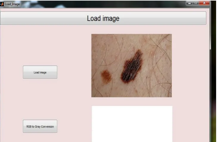
Figure 5.2: RGB to gray-scale conversion. The input image loaded will be an RGB image. The RGB image should be converted to gray-scale image.
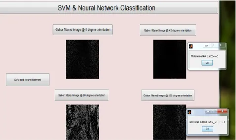
Figure 5.4: Loading input image. The input image is not a skin lesion.
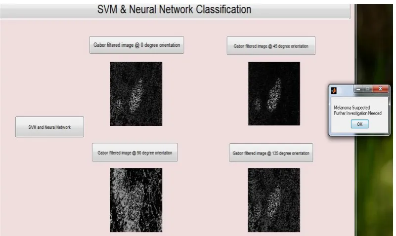
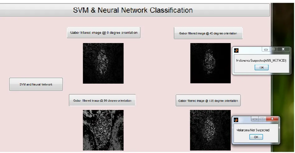
Figure 5.6: Melanoma suspected using SVM and not suspected using neural network. The input image is a skin lesion. SVM produces better results than neural networks.
V. CONCLUSION
Melanoma is the deadliest form of skin cancer. Incidence rates of melanoma have been increasing, but survival rates are high if detected early. An automatedsystem to assess a patients risk of melanoma using images of their skinlesions captured using a standard digital camera is developed. Classificationof skin lesion is done so that melanoma can be detected at an early stage. Gabor filter is used for extracting features from the skin lesion. Using gabor filter, features from images with skin lesion and images with no skin lesionare extracted. Training is done on these images using support vector machine.These trained images are used for classification purpose where the input imageis compared with these trained images. SVM classifies the input image aseither skin lesion or normal image. Another classification technique, artificialneural network is used for making a comparative study with SVM. It is found that SVM produces better results when compared with ANN. SVM is ableto suspect skin lesion whereas ANNis not able to suspect skin lesion.
REFERENCES
1. Jeffrey Glaister, David A. Clausi, ‘Segmentation of Skin Lesions From Digital Images Using Joint Statistical Texture Distinctiveness’,
IEEE Transactionson Biomedical Engineering, Vol. 61, No. 4, April 2014.
2. PoojaKamavisdar, SonamSaluja, Sonu Agrawal, "A Survey on Image Classification Approaches and Techniques," International Journal of AdvancedResearch in Computer and Communication Engineering Vol. 2, Issue1, January 2013.
3. D. Lu, Q. Weng, "A Survey of ImageClassication Methods and Techniques for Improving Classification Performance," International Journal ofRemote Sensing Vol. 28, No. 5, 10 March 2007, 823-870.
4. R. W. Demetrius, H. W. Randle, "High-risk Nonmelanoma Skin Cancers,"Dermatol. Surg. 24, 1272-1292, 1998.
5. P.Jayapal, R.Manikandan, M.Ramanan, R.S. ShiyamSundar, T.S. UdhayaSuriya, "Skin Lesion Classication Using Hybrid Spatial Features andRadial Basis network," International Journal of Innovative Research in Science,Engineering and Technology Vol. 3, Issue 3, March 2014.
6. Daniel P. Berrar, "Multiclass Cancer Classification Using Gene Expression Profiling and Probabilistic Neural Networks," Pacific Symposium onBiocomputing Vol. 8, 2003.
8. Vamsi K. Madasu, Brian C. Lovell, "Blotch Detection in Pigmented SkinLesions using Fuzzy Co-Clustering and Texture Segmentation," IEEE Conferenceon Digital Image Computing: Techniques and Applications, 2009.
9. H. S. Ganzeli, "SKAN: Skin Scanner- System for Skin Cancer DetectionUsing Adaptive Techniques," , IEEE Latin America Transactions,
Vol. 9,No. 2, April 2011.
10. JinWeiXu, "A double thresholding method for cancer stem cell detection,"7th International Symposium on Image and Signal Processing
and Analysis(ISPA 2011) September.
11. T. S. Lee, "Image representation using 2D Gabor wavelets," IEEE Trans.Pattern Analysis and Machine Intelligence, 18(10), 1996.
12. L. Shen, L. Bai, "A review of Gabor wavelets for face recognition," PatternAnalysis Application 9: 273-292, 2006.
13. 14.
BIOGRAPHY