ABSTRACT
BASNAYAKE, VERONICA ROSHANI. Identification and characterization of the Red clover necrotic mosaic virus origin of assembly sequence. (Under the direction of Dr. Steven A. Lommel).
Red clover necrotic mosaic virus (RCNMV) is a single-stranded
positive-sense RNA plant virus of the Dianthovirus genus, family Tombusviridae. Each
RCNMV virion contains 180 subunits of the 37 kDa capsid protein (CP) forming a non-enveloped, isometric particle of T=3 quasi symmetry, 30-35 nm in diameter. The RCNMV genome consists of two RNAs, RNA-1 and RNA-2. RNA-1 codes for three proteins: i) p88, the RNA-dependent RNA polymerase, ii) p27, the replicase related protein and iii) the CP. RNA-2 is monocistronic and codes for the movement protein (MP).
Currently, the RNA complement within RCNMV virions has not been
observations suggest that the RCNMV virion population consists of two distinct types of particles having similar densities.
Based on the above RNA complement findings, I proceeded to delineate the specific viral sequences that determine the RNA content of an RCNMV virion. During virion assembly, the RCNMV CP must be able to distinguish and package both RNA-1 and RNA-2 while excluding heterologous host RNAs present in the cell. Viral CP subunits recognize specific cognate genomic sequences and/or structures that are unique to each viral genome. These are designated as origin of assembly
sequences or packaging signals. To elucidate the assembly mechanism of RCNMV, I searched for the presence of distinct packaging signals on each of the genomic RNAs. Various constructs of RNA-1 and RNA-2 were tested for their assembly efficiencies in vivo using a plant assay system. While it has been previously
demonstrated that RNA-1: RNA-2 base pairing directs the synthesis of the CP subgenomic RNA (sgRNA) from RNA-1 via the trans-activating (TA) element on
virus vector directed the co-packaging of RCNMV RNA-1 into virions proving it to be
essential for RCNMV assembly. Deletion mutagenesis also revealed that RCNMV has an RNA packaging size requirement for production of stable virions.
IDENTIFICATION AND CHARACTERIZATION OF THE RED CLOVER NECROTIC MOSAIC VIRUS ORIGIN OF ASSEMBLY SEQUENCE
by
VERONICA ROSHANI BASNAYAKE
A dissertation submitted to the Graduate Faculty of North Carolina State University in partial fulfillment of the requirements for the Degree of Doctor of Philosophy
DEPARTMENT OF PLANT PATHOLOGY
Raleigh 2005
APPROVED BY
Steven A. Lommel James W. Moyer
Chair of Advisory Committee Member of Advisory Committee
Cynthia L. Hemenway Eric L. Davis
BIOGRAPHY
ACKNOWLEDGEMENTS
The years I spent at graduate school at NC State would not have been the great experience it was if not for a lot of people. I would like to express my gratitude to them.
I am truly grateful to Dr. Steven A. Lommel, my major thesis advisor, for providing me with the opportunity to work in his lab as well as for his guidance and encouragement. I also wish to thank the members of my advisory committee, Drs. Cynthia Hemenway, James W. Moyer and Eric L. Davis, for their support.
I have to thank all the members of the Lommel lab for sharing a memorable time with me. Most importantly I thank Dr. Tim L. Sit who has, literally, shown me the way. He helped me in innumerable ways. Tim, I will never forget the times we
laughed or danced with music blaring in the background, while burning the midnight oil.
Valerie Knowlton, for looking at countless numbers of grids under the electron microscope. Drs. Tim Petty and A.L.N Rao for helpful discussions. Drs. David
Ritchie and Pete Lindgren, for helping to make life at NC State less stressful. I greatly appreciate the office staff of the Department of Plant Pathology for all their dedication and help. I am also thankful to the Plant Pathology students and faculty for making life at NC State memorable.
My wonderful friends for their love, support and friendship. My heartfelt thanks to you as I don’t think I could have done it without you. I would be remiss if I do not mention my undergraduate advisors who were the foundation for my scientific career. My childhood friend Subashini Nagendran, for always being there.
TABLE OF CONTENTS
list of tables ...vii
list of figures...viii
list of abbreviations ...x
Chapter 1 Red Clover Necrotic Mosaic Virus: A Review ...1
Introduction ...2
Symptomatology ...4
Cytopathology...4
Transmission ...5
Control ...6
Physical, chemical and biochemical properties...6
1. Purification of RCNMV ...6
2. Electron microscopy...7
3. Virion properties...7
Serology...8
Genome properties ...9
RCNMV RNA-1...9
RCNMV RNA-2...10
Ribosomal frameshifting ...12
Replication ...13
Trans-activation ...15
Movement ...17
The research project...23
References ...33
Chapter 2 Virus Packaging Signals: A Review...44
Introduction ...45
Animal virus OASs...49
Retroviruses (family Retroviridae)...49
Human immunodeficiency virus -1...54
Alphaviruses (family Togaviridae)...59
Mouse hepatitis virus (family Coronaviridae) ...61
Hepadnaviruses (family Hepadnaviridae) ...63
Adenoviruses (family Adenoviridae) ...64
Flock house virus (family Nodaviridae)...65
Plant Virus OASs ...66
Tobacco mosaic virus...66
Papaya mosaic virus (family Potexviridae) ...69
Tobacco vein mottling virus (family Potyviridae)...69
Peanut clump virus ...70
Turnip crinkle virus (family Tombusviridae) ...71
Cowpea chlorotic mottle virus (family Bromoviridae)...73
Brome mosaic virus (family Bromoviridae) ...74
Summary ...75
References ...90
Chapter 3 The Packaging Scheme of Red Clover Necrotic Mosaic Virus Genomic RNAs...105
Introduction ...106
Materials and methods...110
Results...113
An RNA-1: RNA-2 heterodimer is produced upon UV irradiation of RCNMV virions...113
An RNA-1: RNA-2 heterodimer is also formed upon heat treatment of virions...115
Formation of the RNA-1: RNA-2 heterodimer is temperature dependent ...116
The RNA-1: RNA-2 heterodimer is formed within virions by means of a non-covalent interaction...116
RCNMV RNA does not form dimer complexes upon heating in vitro...117
Discussion ...118
Acknowledgements...124
References ...133
Chapter 4 The Red Clover Necrotic Mosaic Virus Origin of Assembly: A Third Function for the RNA-2 Trans-Activator...137
Introduction ...139
Materials and methods...144
Results...149
RCNMV sgRNA is not packaged into RCNMV virions ...149
RNA-1 does not package into virions in the absence of RNA-2 ...150
A 209 nt region of RCNMV RNA-2 directs encapsidation of heterologous TBSV RNA in the presence of RCNMV CP...151
Refining the minimal OAS on RCNMV RNA-2 by deletion mutagenesis...152
TA loop mutation does not disrupt OAS function...153
The RCNMV TA element functions as an OAS in a heterologous context ...154
Encapsidation of heterologous RNAs defines an upper packaging size limit ...154
Acknowledgements...165
References ...180
Chapter 5 Red Clover Necrotic Mosaic Virus Particles Consist of Both Native and Expanded Forms...186
Introduction ...188
Materials and methods...192
Results...195
Optiprep™ and CsCl density gradient separation yields swollen and native forms of RCNMV virions...195
Differential density of the RCNMV virions is not due to a modification of CP ...196
Virion RNA composition of the swollen and native RCNMV virions ...197
Discussion ...197
LIST OF TABLES
Chapter 4
Table 1. Sequences of the oligonucleotides used for the synthesis of
LIST OF FIGURES Chapter 1
Figure 1. Symptoms caused by RCNMV on Nicotiana benthamiana...27
Figure 2. The electron microscopic image of RCNMV virions...28
Figure 3. The genome organization of RCNMV...29
Figure 4. The 3' termini of RCNMV RNA-1 and RNA-2 ...30
Figure 5. Trans-activation and CP sgRNA synthesis...31
Figure 6. The structure of the TBSV particle architecture...32
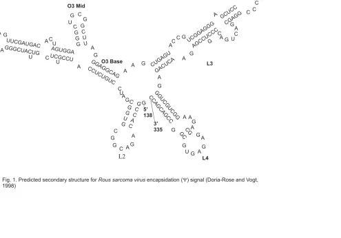
Chapter 2 Figure 1. Predicted secondary structure for Rous sarcoma virus encapsidation (Ψ) signal...77
Figure 2. The secondary structure model of the Avian sarcoma leukosis virus packaging region...78
Figure 3. Secondary structure of the Moloney murine leukemia virus encapsidation signal (Ψ)...79
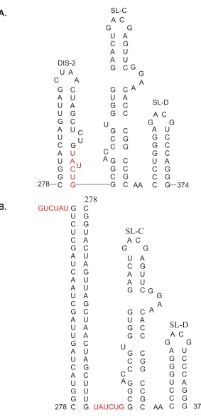
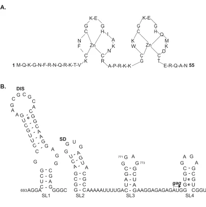
Figure 4. HIV-1 packaging signal (Ψ) ...80
Figure 5. Alternative foldings of the HIV-1 leader ...81
Figure 6. Secondary structure of the Bovine leukemia virus discontinuous encapsidation signal ...82
Figure 7. Predicted secondary structure of the Mouse hepatitis virus 69-nucleotide packaging signal...83
Figure 8. The predicted secondary structure of Hepatitis B virus encapsidation (ε) signal ...84
Figure 9. Secondary structures of duck and heron hepatitis B virus encapsidation signals...85
Figure 10. Predicted structure of the packaging signal of Flock house virus...86
Figure 11. The Tobacco mosaic virus origin of assembly...87
Figure 12. Secondary structure of the Turnip crinkle virus essential element for packaging ...88
Figure 13. The Turnip yellow mosaic virus 5' leader region with two hairpins containing protonatable internal loops ...89
Chapter 3 Figure 1. Red clover necrotic mosaic virus genome and various possibilities for packaging the genomic RNAs...125
Figure 2. UV crosslinking of RCNMV virions ...126
and unheated RCNMV virions ...127
Figure 4. Denaturing gel electrophoresis and northern analysis of heat treated and UV-crosslinked RCNMV virion RNA ...128
Figure 5. Temperature range for RCNMV RNA complex formation and effects of heating on virion morphology ...129
Figure 6 RNase T1 treatment of heated and unheated virions ...130
Figure 7. Heating of RCNMV transcripts and virion purified RNA...131
Figure 8. A comparison of the TA, TABS and TABSM sequences ...132
Chapter 4 Figure 1. Red clover necrotic mosaic virus genome organization...167
Figure 2. RCNMV CP sgRNA is not packaged into virions...168
Figure 3. RCNMV RNA-1 does not contain a discrete OAS ...169
Figure 4. RCNMV virion assembly directed by the TBSV sgRNA containing a RCNMV RNA-2 OAR fragment ...170
Figure 5. Virion formation of the RNA-2 deletion mutants ...171
Figure 6. The role of TA terminal loop in assembly ...175
Figure 7. The RCNMV RNA-2 TA is the OAS...176
Figure 8. RNA size requirement for RCNMV packaging...177
Figure 9. An RCNMV assembly model ...179
Chapter 5 Figure 1. Separation of swollen and native RCNMV virions ...202
Figure 2. CP of swollen virions do not appear to be modified...203
LIST OF ABBREVIATIONS
ARM arginine-rich motif ASLV Avian sarcoma leukosis virus
BLV Bovine leukemia virus
BMH branched multiple hairpins BMV Brome mosaic virus
BYDV Barley yellow dwarf virus
C hepadnavirus core protein CA retrovirus capsid protein CP capsid protein
CCMV Cowpea chlorotic mottle virus
CRSV Carnation ringspot virus
DHBV Duck hepatitis B virus
DI-RNA defective interfering RNA DIS dimerization initiation site d.p.i. days post-inoculation
ε hepadnavirus encapsidation signal ELISA enzyme-linked immunosorbent assay EM electron microscope
ER endoplasmic reticulum
env retrovirus envelope gene
FHV Flock house virus
FNSV Furcrea necrotic streak virus
Gag retrovirus Gag polyprotein GFP green fluorescent protein HAdV Human adenovirus
HBV Human hepatitis B virus
HHBV Heron hepatitis B virus
HIV-1 Human immunodeficiency virus –1 kb kilobases
kDa kilodalton
lac Z beta galactosidase gene
LDI long distance interaction MA matrix protein
MHV Mouse hepatitis virus
MoMuLV Moloney murine leukemia virus MP movement protein
mw molecular weight NC nucleocapsid protein OAS origin of assembly ORF open reading frame
P hepadnavirus reverse transcriptase PapMV Papaya mosaic virus
PBS primer binding site
pgRNA pregenomic RNA PLRV Potato leaf roll virus
RCNMV Red clover necrotic mosaic virus
RdRp RNA-dependent RNA polymerase RpRSV Raspberry ringspot virus
RRV Ross river virus
RYMV Rice yellow mottle virus
S Svedberg unit
SCNMV Sweet clover necrotic mosaic virus
SCPMV Southern bean mosaic virus cowpea strain SEL size exclusion limit
SFV Semliki forest virus
sgRNA subgenomic RNA snRNA small nuclear RNA SNV Spleen necrosis virus
TA trans-activator
TABS trans-activator binding site TBSV Tomato bushy stunt virus
TCV Turnip crinkle virus
TGB triple gene block movement proteins TLS tRNA-like structure
TMV Tobacco mosaic virus
TVMV Tobacco vein mottling virus
TYMV Turnip yellow mosaic virus
UV ultra violet wt wild type
Chapter 1
Red Clover Necrotic Mosaic Virus: A Review
Introduction
Red clover necrotic mosaic virus (RCNMV) was first described by Musil and
Matisova (1967) in Czechoslovakia based on symptoms in red clover (Trifolium
pratense) and alfalfa (Medicago sativa). The virus was isolated and distinguished
from other clover infecting viruses by host range, physical properties, electron
microscopy and serological studies (Musil, 1969 a and b). RCNMV is a
positive-sense single-stranded RNA plant virus with isometric particles 30-35 nm in diameter.
Different isolates and strains have been reported from northern Europe (primarily
England, Czechoslovakia and Sweden), Canada, Australia and New Zealand.
RCNMV naturally occurs in red clover, sweet clover, white clover and alfalfa in field
crops or in pastures (Hiruki, 1987). The virus is easily mechanically transmissible to
a wide range of herbaceous hosts although the natural host range is limited. They
can systemically infect a number of species in the host families Solanaceae,
Leguminosae, Cucurbitaceae and Asteraceae, and others locally. Nicotiana
clevelandii, N. benthamiana and Phaseolus vulgaris are suitable propagation
species while Chenopodium quinoa is a good local lesion host. There are no known
vectors for RCNMV. RCNMV is transmitted through soil (Hiruki, 1987, Hollings and
Stone, 1977), but not seed-borne (Hollings and Stone, 1977).
Taxonomically RCNMV resides in the family Tombusviridae, which is a
diverse family of icosahedral viruses with single-stranded, positive-sense RNA
Necrovirus, Dianthovirus, Machlomovirus, Panicovirus, Avenavirus and Aureovirus.
The members of the family share two important features: the RNA-dependent RNA
polymerase is phylogenetically conserved in all members and the viruses are
primarily transmitted in soil. All members of Tombusviridae contain a single genomic
RNA, except dianthoviruses. Dianthoviruses are distinguished from the other
members of the family due to the segmented nature of their genome. Members of
Tombusviridae comprise of Capsid proteins (CP) with two distinct phylogenetic
origins. One group is formed with CP with a molecular weight of 37-48 kDa
containing protruding domains and the other lacking protruding domains with a
molecular weight of 27-29 kDa (Lommel et al., 2000). The members of the genus
Dianthovirus, created in 1981 by the ICTV (Matthews, 1982), contain two
non-homologous single-stranded positive-sense RNAs. RCNMV produces T=3
icosahedrons composed of 180 units of a 37 kDa CP. The dianthovirus CP is
grouped within the CP with protruding domains (Aureusvirus, Avenavirus,
Carmovirus, Dianthovirus, and Tombusvirus) giving rise to virions with a granular
outline. The phylogenetic group lacking the protruding domain (Machlomovirus,
Panicovirus, and Necrovirus) produces virions with a smooth outline. There are three
known members of the genus: Carnation ringspot virus (CRSV), the type member,
RCNMV and Sweet clover necrotic mosaic virus (SCNMV). Furcrea necrotic streak
virus (FNSV), which causes a necrotic streak of fique (Furcrea macrophylla and F.
cabuya) is serologically related to RCNMV and could be a fourth possible species
(Morales et al., 1992). Dianthoviruses do not cause severe crop losses. Their
is tropical in range. CRSV spread is worldwide whereas RCNMV is commonly found
in temperate Europe, Canada, Australia and New Zealand. SCNMV has only been
found in Alberta, Canada.
Symptomatology
Symptoms on infected red clover include necrotic lesions, severe leaf mottle,
necrosis of the veins, distortion of leaves and moderate to severe stunting (Fig. 1).
The symptoms are more pronounced in the winter, but masked in hot weather,
particularly in newly infected plants. Inoculated red clover or Nicotiana species
primarily show local necrotic ringspot lesions. Although a wide variation in
symptomatology has been reported for RCNMV, Okuno et al. (1983) observed that
in controlled growth chambers with temperature maintained at 17 -26°C plants
showed reproducible symptoms. The virus is present in leaves, stems and roots and
has been observed in soil surrounding the rootstock.
Cytopathology
Virions have been observed scattered in the cytoplasm and vacuole of N.
clevelandii leaf mesophyll cells (Francki et al., 1985). Large aggregates of virions as
well as tubular inclusions occur in CRSV-infected sweet william and cowpea
(Weintraub et al., 1975). RCNMV or SCNMV do not frequently produce inclusions,
however, amoeboid inclusion bodies have been observed in less than 5% of the
been associated with microtubules in cells infected with both CRSV and SCNMV,
but not observed with RCNMV (Francki et al., 1985).
Transmission
Contact transmission and soil-borne transmission without the aid of vectors
seem to be the natural modes of transmission for RCNMV. Dianthovirus virions are
readily released from root cells into soil water where they may remain infective for
months (Hollings and Stone, 1977; Kegler and Kegler, 1981; Hiruki, 1986). The
remarkably stable nature of the particles enables the viruses to remain infective for
long time periods. RCNMV is more stable than other dianthoviruses and is known to
be more resistant to swelling and proteolysis of the virion (Gould et al., 1981;
Hamilton and Tremaine, 1996). Although dianthoviruses have been reported to be
transmitted by longidorid nematodes they have not been confirmed (Fritzsche, 1968;
Fritzsche and Schmelzer, 1967; Kleinhempel et al., 1980). Transmission of RCNMV
was enhanced 2- to 20-fold in the presence of the Chytridiomycete, Olpidium
brassicae suggesting that the fungal vector might be involved in its transmission
(Gerhardson and Insunza, 1979). MacFarlane (1982) reported that O. bornovanus
(Sahtiyanci), which is more prevalent in Britain, transmitted RCNMV to clover.
Although fungal and/or nematode vectors have been thus far suggested as modes of
dianthovirus transmission, it is likely that the contamination of soil water with
infective virions may have been responsible for the infections. Further work is
vectors predispose plant roots to infection by creating wounds for initial virus
infection.
Control
In vegetatively propagated carnation and fruit trees (in the case of CRSV)
control of virus has been achieved by thermotherapy (Brierley, 1964) or meristem-tip
culture (Stone, 1968; Kowalska, 1974). Usage of such propagation techniques
coupled with constant monitoring with enzyme linked immunosorbent assay (ELISA)
should eradicate virus infections (Lommel et al., 1983). Control of RCNMV could
pose greater problems due to its broader host range however dianthovirus plant
diseases have not so far posed economical concerns.
Physical, chemical and biochemical properties
1. Purification of RCNMV
High viral concentrations are achieved in RCNMV infected plants. Local
lesions on N. benthamiana leaves appear 2-3 days after inoculation of the virus and
systemic symptoms appear 4-5 days post inoculation. Virus titer is largely dependent
on environmental conditions and plants grown at 18-20°C and 16 hours/day
illumination at a light intensity of ~5000 lux consistently produce high virus
concentrations, approximately 100 µg of virus per gram of leaf tissue (Hollings and
7.4 (Gould et al., 1981) and is also successfully achieved by 0.2 M Tris-acetate
buffer, pH 5.2. RCNMV virions appear to be most stable at pH 6.8.
2. Electron microscopy
Virion morphology of the dianthoviruses is very similar to other members of
Tombusviridae family with similar phylogenetically conserved CP having a protruding
domain. RCNMV virions exhibit a spherical morphology with a granular surface (Fig.
2). Diameter for RCNMV ranges from 27-35 nm (Hiruki et al., 1984b; Hollings and
Stone, 1965; Bowen and Plumb, 1979; Kuhne and Eisbein, 1983).
3. Virion properties
RCNMV virions sediment as a single component with a sedimentation
coefficient (S20,w) at about 130-135 S at, pH 5.0 (Hollings and Stone, 1970, 1977).
Virus concentration, age of the virus preparation, virus purification methods and the
pH of the virus preparation are known to affect the sedimentation coefficient (Hiruki,
1987). RCNMV has a buoyant density of approximately 1.363 g/cm3 in cesium
chloride. The Swedish strain of RCNMV has an extra component at 1.356 g/cm3
(Hollings and Stone, 1977). RCNMV genome is encapsidated by 180 subunits of 37
kDa capsid protein to form a virion of T=3 symmetry (Lommel, 1983; Morris-Krsinich
et al., 1983). The capsid protein subunit consists of approximately 339 amino acids
and the amino acid composition of different RCNMV strains is basically similar
(Hamilton and Tremaine, 1996). The RNA complement within the virion was
has thus far not been determined. CRSV particles swell slowly when pH is changed
from 5.0 to 7.5 (Tremaine and Ronald, 1976). Treatment with EDTA accelerates this
swelling but Mg2+ or Ca2+ ions reverse it (Tremaine et al., 1976). However, RCNMV
which is more stable than CRSV (Hamilton and Tremaine, 1996) does not seem to
swell at pH 7.0 but proteolysis of virions samples has been observed in storage.
RCNMV-Can strain when treated with pH 7.5 for 1 hour exhibited swelling in 20% of
particles. Half of the virions were dissociated when treated with 1% SDS. RCNMV
completely dissociated when pH was elevated to 8.25. Capsid protein subunits
reassembled in the presence of the nucleating agent sodium dextran sulfate to form
T=1 and T=3 virions, which could be readily distinguished by electron microscopy
(J.H. Tremaine and W.P. Ronald, unpublished results, Hamilton and Tremaine,
1996).
Serology
Serological studies of RCNMV initially distinguished three serotypes, A
(RCNMV-TpM34) and B (RCNMV-TpM48) from Czechoslovakia (Musil, 1969b), and
C (RCNMV-Sw) from Sweden (Gerhardson and Lindsten, 1973). Serological
analysis by intragel cross-absorption experiments revealed that the Canadian strain
(RCNMV-Can) belongs to serotype B and Australian (RCNMV-Aus) and English
(RCNMV-Eng) strains belong to serotype D (Rao et al., 1987). RCNMV-Aus and
RCNMV-Eng were serologically indistinguishable and did not react with the antisera
of any of the three serotypes of the virus. There was no cross-reaction between
antibody sandwich ELISA, however, indirect ELISA gave weak cross-reactions with
them (Van Regenmortel and Burckard, 1980). The isoelectric points of RCNMV
strains are in the range of pH 4.75-5.1 (Pappu and Hiruki, 1989).
Genome properties
The bipartite nature of RCNMV (Fig. 3) was first described by Ragetli and
Elder (1977) who observed two distinct RNA species in density gradient
centrifugation of the products of sodium dodecyl sulfate (SDS)-denatured virus
particles. Although this observation was attributed to clover primary leaf necrosis
virus, it is now considered to be the RCNMV-Ca strain based on host range,
serology, nucleic acid and capsid mobilities (Rao and Hiruki, 1985; Hamilton and
Tremaine, 1996). The RNA1 of the dianthoviruses are similar in size (CRSV type
-3756 bases, RCNMV-Aus- 3889 and SCNMV-59 alfalfa isolate- 3876). The RNA-2
molecules however, differ slightly in size (CRSV-1394 bases, RCNMV-Aus- 1448
and SCNMV-59-1449). The complete sequence of RCNMV RNA-1 and RNA-2 has
been published (Xiong and Lommel, 1989; Lommel et al., 1988). Both RNA-1 and
RNA-2 are required for infectivity.
RCNMV RNA-1
RNA-1 consists of three open reading frames (ORF) (Fig. 3). The 5’ terminal
ORF encodes a 27 kDa polypeptide (p27) and an 88 kDa (p88) polypeptide, which is
translated as an extension of p27 due to ribosomal frameshift near the stop site of
(Xiong and Lommel, 1989). The p88 polypeptide is the viral RNA-dependent RNA
polymerase (RdRp) and p27 is a replicase related protein. The 3’ terminal CP gene
is expressed in vivo from a subgenomic RNA. The genomic RNA-1 has a 5’
122-nucleotide leader followed by the p27 ORF initiating at 122-nucleotide 123 and
terminating at an amber terminator at 831. The polypeptide p88 extends the p27 and
terminates at 2423. The 3’ terminal CP ORF initiates at 2427 and terminates at
3444. There is a 445-nucleotide 3’ terminal non-coding region.
RCNMV RNA-2
The RNA-2 molecule is monocistronic and codes for a 35 kDa movement
protein (MP; Lommel et al., 1988). The first initiation codon of the ORF of MP is at
nucleotide 80 and ends at nucleotide 1030 (Fig 3.B). The C-terminal region of MP is
required for the movement function. The MP C-terminus shares some identity with
bromoviruses, Brome mosaic virus and Cowpea chlorotic mottle viruses, at the
sequence level (Lommel et al., 1988; Allison et al., 1989). Computer translation of
the RNA-2 revealed the presence of another ORF downstream of the MP ORF, with
the start position at 1224 and ending at 1358. This has the probability to encode a
4.8 kDa polypeptide, although none has been observed in vitro. Subgenomic RNA
corresponding to it has not been detected either (Lommel et al., 1988) and deleting
this ORF results in no change in phenotype.
Folding of the RNA-1 5’ leader sequence does not reveal significant
secondary structure formation and this is typical for plant viruses. The 5’ leaders of
promoter. The 5’ RNA-2 leader sequence is 79 nucleotides in length and is in the
range observed for other spherical RNA plant viruses (Davies and Hull, 1982).
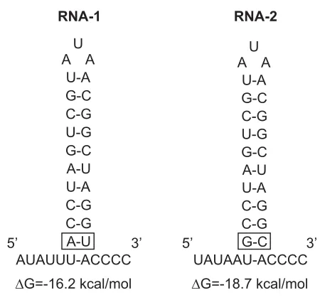
RNA-2 has high adenine content (44% of the nucleotides). The 3’ termini of both RNA-1
and RNA-2 on the other hand contain two highly homologous domains (Fig. 4). The
first homologous domain extends from RNA-1 position 3817 to 3852 and RNA-2
from 1315 to 1346 nucleotides. The second homologous region on RNA-1 is from
3863 to 3889 and on RNA-2 from 1422 to 1448. The 27 nucleotide terminal
homologous sequence forms a highly stable stem loop. The 3’ non-coding regions of
viral RNAs naturally serve as replicase recognition sites with complex secondary
structures or sequences (Dreher et al., 1984; Bujarski et al., 1986). Turner and Buck
(1999) showed that the 5’ leader and most of the 3’ untranslated region of RNA-2
were required for replication and that the 3’ proximal stem loop was indispensable
for replication. Most recently, an element on RNA-2 was identified as an essential
cis-replicating RNA element of RNA-2 (Tatsuta et al., 2005). The 3’ termini for both
RNA-1 and RNA-2 are longer than the typical 150- to 250-base non-coding regions
for other viruses (Davies and Hull, 1982). There is the possibility that there are extra
ORFs in these regions for which the corresponding polypeptides have not been
identified.
RCNMV genomic RNAs lack a 5’ cap structure (m7GpppN; Mizumoto et al.,
2003). The eukaryotic cap structure together with the 3’ poly (A) tail act as binding
sites for the translational machinery for initiation of translation. In the absence of
these standard translation elements secondary sequences functions as translational
both lack a 5’ cap and a poly (A) tail, however, the genomic RNA consists of
translation enhancer sequences in the 3’ untranslated region that confer
cap-independent translation (Wang et al., 1997; Wu and White, 1999). Since RCNMV
RNAs are uncapped and without 3’ polyadenylation, it was assumed that they
contained translation enhancer sequences within their genomes. Recently RCNMV
RNA-1 was demonstrated to contain a translation element in the 3’ untranslated
region (nts 3596 to 3732 (Mizumoto et al., 2003). A stem loop within this translation
element that is conserved among the dianthoviruses appears to be the main
component of the cap-independent translation. This stem loop was shown to be
nearly identical to the translation enhancers identified from BYDV and Tobacco
necrosis virus.
Ribosomal frameshifting
Ribosomal frameshifting is an approach for regulating protein expression at
the translational level that is utilized by eukaryotic organisms. This phenomenon was
first observed in Rous sarcoma virus synthesis of viral reverse transcriptase (Jacks
and Varmus, 1985; Jacks et al., 1988). A similar mechanism operates in the
synthesis of RNA polymerases of BYDV (Veidt et al., 1988) and Potato leafroll
luteovirus (Prufer et al., 1992) as well as coronaviruses (Brierley et al., 1989) and
other retroviruses (Varmus, 1988). A slippage model of -1 ribosomal frameshifting
was proposed as a specific heptanucleotide where the ribosome slippage occurs
(Jacks et al., 1998). The shifty heptanucleotides conform to a generalized sequence
of X XXY YYZ (X=A,G, or U; Y=A or U; Z=A,C, or U). After slippage into the –1
frameshift event and to ensure continuity of translation. An RNA stem loop structure
immediately downstream of this shifty heptanucleotide is also believed to be
facilitating the frameshift event (Rice et al., 1985; Brierley et al., 1989; Prufer et al.,
1992). The shifty heptanucleotide causes tRNA slippage during translation and the
stable secondary stem loop downstream is believed to stall or slow ribosome
migration increasing the probability of slippage leading to the -1 frameshift event
(Somogyi et al., 1993). The frameshift mechanism of the heptanucleotide RCNMV
was established by point and frameshift mutations in in vitro synthesis of 88 kDa
polymerase (Kim and Lommel, 1994; Xiong et al., 1993b). The shifty
heptanucleotide of RCNMV has similarities to those of BYDV, Rous sarcoma virus
and Mouse mammary tumor virus. The RCNMV shifty heptanucleotide was
functionally interchangeable with many plant shifty heptanucleotide signals but not
with those of mammalian signals. RCNMV was able to revert the shifty signal
mutants back to the wild type. Similar structures have been identified in RNA-1 of
CRSV and SCNMV that could be involved in a frameshift mechanism (Ryabov et al.,
1994). An RNA secondary structure, a stem loop element downstream of the shifty
heptanucleotide is implicated to assist frameshift event similar to others. Mutations
that destabilized this stem loop did not function in vitro, and they were unable to
replicate in vivo (Kim and Lommel, 1998).
Replication
RCNMV replicates rapidly in host plants kept at 22°C and the virus titer
ambient temperature in which they are grown. Since RCNMV produces a high virus
titer in Nicotiana species, it is well suited for assembly and replication studies.
RCNMV replication occurs in two stages: the RNA negative-strand is synthesized
from the parental positive-strand genomic RNA and, which in turn acts as a template
to synthesize progeny positive-strand RNA. Virus encoded RdRp catalyses the
synthesis of progeny RNA molecules from nucleoside triphosphate substrates.
Koonin (1991), proposed three phylogenetic supergroups of RdRp based on
sequence similarities of conserved amino acid motifs. Supergroup I include virus
genera, Picornavirus, Nodavirus, Comovirus, Nepovirus, Potyvirus, Bymovirus,
Sobemovirus and others. Members of Carmovirus, Necrovirus, Tombusvirus, and
Hepatitis C virus belong to supergroup II. Supergroup III consists of Tobamovirus,
Tobravirus, Hordeivirus, Alfamovirus, Bromovirus, Cucumovirus, Alphavirus, and
Potexvirus among others. The RCNMV 88 kDa polypeptide (p88) contains the GDD
(Glycine: Aspartic acid: Aspartic acid) motif which is conserved in all viral RNA
polymerases (Koonin, 1991). Based on the high degree of sequence similarity,
RCNMV RdRp is grouped in supergroup II together with other members of family
Tombusviridae and Luteoviridae (Koonin and Dolja, 1993; Koonin, 1991). RCNMV
RdRp is template specific and therefore only able to utilize RCNMV RNAs but not
RNAs of taxonomically different viruses (Bates et al., 1995). RCNMV 27 kDa
polypeptide (p27) was found to be a component of the viral RdRp purified from N.
clevelandii suggesting that it could be playing a role in replication such as template
Recently it was established that positive-strand RNA viruses replicate in
association with intracellular membranes (Lee et al., 2001; Miller et al., 2001;
Restrepo-Hartwig and Ahlquist, 1999). Viruses were shown to cause proliferation of
host membranes possibly to amplify sites available for replication. Positive-strand
RNA viruses utilize a variety of membranes as sites of replication. They range from
outer membranes of endoplasmic reticulum (ER), mixed compartments of secretory
pathway, endosomes, mitochondria and other organelles (Westaway et al., 1997;
Pedersen et al., 1999; Suhy et al., 2000; Froshauer et al., 1988; Hatta et al., 1973).
The type of membrane used is specific to the virus. In the case of BMV, modulating
the levels of replication factors changed the structure of membrane rearrangement
but still supported virus replication (Schwartz et al., 2004). To determine RCNMV
replication sites, Aequoria victoria green fluorescent protein (GFP) fusions were
made to both p27 and p88, and GFP was used to visualize the location of these
fusions in N. benthamiana protoplasts. Both the p27 and p88 proteins individually
localized to the ER suggesting it as the possible RCNMV replication site (Turner et
al., 2004). RCNMV infection caused invaginations of perinuclear ER and proliferation
of smooth ER as further evidence of ER being replication sites.
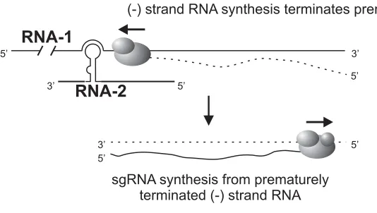
Trans-activation
Capsid protein is encoded by RNA-1 and CP subgenomic RNA (sgRNA) is
produced by a process of RNA-2 trans-activation. The accumulation of sgRNA
corresponding to CP in vivo was very low and infrequent (Zavriev et al., 1996). The
region that shows 13 of the 14 nucleotides identical to that of 5’ RNA-1 leader. This
subgenomic leader is likely to be the positive strand promoter. Sit et al., (1998) in an
elegant experiment demonstrated a novel RNA-mediated regulation of transcription
that resulted in the synthesis of CP sgRNA (Fig. 5). An RCNMV RNA-1 cDNA clone
that expressed the GFP in place of CP was engineered. Fragments of RNA-2 were
expressed from the TBSV replicon. A 34-nucleotide sequence in RNA-2 (756-789)
was demonstrated to be sufficient to produce GFP as well as synthesize sgRNA
corresponding to GFP. This 34-nucleotide region on RNA-2, denoted as the trans
-activator (TA), is predicted to form a simple stem loop. Sequence alignments
revealed that the 8-nucleotide terminal loop of this stem loop was complementary to
an 8-nucleotide sequence on RNA-1, the trans-activator binding site (TABS), located
within the CP subgenomic promoter. Mutations in the RNA-2 TA and RNA-1 TABS
revealed that the base pairing between these two elements was a prerequisite for
the trans-activation function. Based on these results a model for trans-activation in
RCNMV was proposed: RNA-1 progeny molecules are synthesized in the initial
phase of replication, later RNA-2 molecules are synthesized and the TA on RNA-2
base pairs with the TABS on RNA-1 thereby trans-activating CP sgRNA synthesis.
This mechanism could be mediated by viral or host proteins. This base pairing could
form a steric hindrance to the replicase from completing synthesis of full-length
RNA-1 negative strands. The truncated complementary strands that are transcribed
possibly serve as templates for the synthesis of positive-sense sgRNA. NMR and
structural computer modeling were used to investigate the TA-TABS interaction at
were observed to form two stacked helices with the stem of RNA-2 TA stem loop
forming one helix and the other formed by the intermolecular base pairing between
the TA loop and RNA-1 (Guenther, et al., 2004).
Movement
Virus movement in animals takes place by means of receptor-mediated
endocytosis or cell fusion. Plant viruses are unable to utilize this route due to the
presence of the rigid cell wall of plant cells. A plant virus when introduced in to a
plant cell replicates and then moves from the initially infected cell into neighboring
cells to cause a productive infection. These viruses circumvent the rigid cell wall by
moving through plasmodesmata, the intercellular connections connecting the cellular
contents of plant cells (Gibbs, 1976; Atabekov and Dorokov, 1984). The viruses
eventually reach the vascular transport system and then systemic spread occurs
through the phloem (Gilbertson and Lucas, 1996). Virus encoded non-structural
proteins termed movement proteins mediate cell-to-cell movement through
plasmodesmata (reviewed in Hull, 1989; Atabekov and Taliansky, 1990). Although
there is little similarity between various virus movement proteins, comparison of viral
MPs from distinct groups of plant viruses have identified conserved amino acid
motifs that may reflect functional ancestry (Melcher, 1990; Koonin et al., 1991).
Infectivity studies demonstrated that RCNMV RNA-1 and RNA-2 were
required for a systemic infection (Gould et al., 1981; Okuno et al., 1983) with RNA-2
required for the expansion of the local lesions (Osman et al., 1986) suggesting that
infection. In the absence of the movement protein the RNA-1 is unable to move from
inoculated cells to adjoining cells although it replicates efficiently.
Deletions less than 39 amino acids in the C-terminus of the RCNMV MP
exhibited wild type movement function whereas larger deletions failed to exhibit the
cell-to-cell movement function (Xiong et al., 1993a). A spontaneous mutant identified
from cowpea, had a single base deletion at nucleotide 790 which was responsible
for a truncated MP and failed to cause a systemic infection (Osman et al., 1991).
The exact mechanism of the movement function of the RCNMV MP is unknown but
believed to be similar to that of tobamoviruses. The tobamovirus (TMV) movement
protein accumulates in the plasmodesmata and modifies the aperture to allow
passage of genomic RNA (Lucas and Wolf, 1993). The TMV MP modifies the size
exclusion limit (SEL) of plasmodesmata from the 800 Da limit to more than 10 kDa in
MP transgenic plants (Wolf et al., 1989). Tobamovirus MP binds to single stranded
RNA in vitro, and it is believed that the MP-RNA in the form of a ribonucleoprotein
complex moves intercellularly in plants (Hull, 1989; Citovsky et al., 1992). This is
further supported by the observation that TMV CP is not required for cell-to-cell
movement (Culver and Dawson, 1989; Saito et al., 1990). RCNMV MP is similar to
TMV as it accumulates in the cell wall (Osman and Buck, 1991), binds to single
stranded RNA, increases the SEL of plasmodesmata (Osman et al., 1992;
Giesman-Cookmeyer and Lommel, 1993) and does not require CP for cell-to-cell movement
(Xiong et al., 1993a). The TMV movement protein has two single strand
Gel shift and photochemical crosslinking studies identified a single-stranded
RNA-binding domain in the region between amino acids 181 and 225 in the RCNMV
MP (Osman et al., 1993). Alanine scanning mutagenesis of the RCNMV MP
identified three distinct functional domains (Giesman-Cookmeyer and Lommel,
1993): an RNA-binding domain, cooperative RNA-binding domain and, a domain
necessary for the cell-to-cell movement function. The RNA-binding domain resides
in the N-terminal half of the MP and mutations in the amino acids 122 and 128
reduced the level of RNA binding up to 10% of wild type. However, very little
RNA-binding activity is required for the cell-to-cell movement function (≤ 20% of wild type).
Alanine substitutions in amino acids 27-31, 122, 128 and 305 appear to disrupt the
cooperative RNA-binding domain, which is not necessary for movement. The third
domain (amino acid 278) which exhibits both binding and cooperative
RNA-binding is deficient in cell-to-cell movement suggesting its involvement in either
targeting the MP-RNA complex to plasmodesmata, modifying the plasmodesmata in
interaction with host factors or involved in proper folding of the ribonucleoprotein
complex. Among dianthovirus movement proteins there is high amino acid
conservation in the amino terminal region (Kendall and Lommel, 1992) and this
suggests that it maybe involved in the movement function. There is very little
homology between the movement proteins of RCNMV and TMV, but both have
highly ordered structures with repeating hydrophobic and hydrophilic domains typical
of membrane bound proteins (Xiong et al., 1993a). The function of the movement
proteins is believed to be dependent more on the three-dimensional structure than
cell-to-cell movement function to the heterologous RCNMV and vice versa
demonstrating their functional equivalence (Giesman-Cookmeyer et al., 1995b).
Transgenic N. benthamiana plants expressing either TMV or RCNMV MP
were able to complement the cellular movement function of the heterologous RNA.
Furthermore both were able to perform as helper viruses to provide cell-to-cell
spread of the movement-deficient heterologous virus. The MP furthermore may be
involved in host range determination. TMV upon infecting resistant plants, replicate
in initially infected cells but fail to invade adjacent cells suggesting that the
resistance is due to the inability of the virus to move (Sulzinski and Zaitlin, 1982).
There are many examples of plant viruses that replicate in protoplasts of
non-hosts (Ponz and Bruening, 1986) and the transport function of the virus is
considered to be a determinant in its host range. Although N. tabacum is a
non-systemic host for RCNMV, RNA-1 replicates in BY-2, a cell line derived from N.
tabacum (Ragetli and Elder, 1977; Paje-Manalo and Lommel, 1989). This suggests
that the resistance observed is due to the inability of RCNMV to move in N. tabacum
and suggests that MP functions as a host range determinant. Immunocytochemical
studies on TMV movement protein have shown its localization to the plasmodesmata
both in infected plants and in MP expressing transgenic plants (Tomenius et al.,
1987; Atkins et al., 1991). Movement proteins of other plant viruses have also been
localized to the cell wall and plasmodesmata (Albrecht et al., 1988;
Godefroy-Colburn et al., 1986; Lehto et al., 1990; Linstead et al., 1988; Moser et al., 1988).
The RCNMV MP was detected in the cell wall fraction of infected N. clevelandii leaf
recently demonstrated to be the cell wall, presumably the plasmodesmata, and an
intimate correlation between localization and cell-to-cell movement was identified
(Tremblay et al., 2005). Similar to TMV, it has been shown that RCNMV MP was
capable of modifying plasmodesmata to enable movement of macromolecules, as
well as trafficking RCNMV RNA (Fujiwara et al., 1993). Although MP binds to both
single stranded RNA and single stranded DNA, it was observed that it selectively
transported single stranded RNA but not DNA in vivo. The RCNMV MP does not
unfold the RNA in the MP-RNA nucleoprotein complex. There is further evidence of
plant viral MP-GFP fusions being co-localized to the cytoskeleton (Heinlein et al.,
1995; McLean et al., 1995) and ER-derived membranes (Mas and Beachy, 1999).
Since these are the components of the intra- and intercellular transport machinery it
is likely that the plant viruses have evolved means to subvert the intercellular
communication network to facilitate the spread of infection.
Virion formation is believed to be the essential requirement for translocation
of many small RNA spherical and rod-shaped viral genera such as Tobamovirus,
Potyvirus, Furovirus, Sobemovirus, Cucumovirus and Bromovirus (Seron and
Haenni, 1996). Long distance transport is believed to take place through the
vascular system. Vascular transport is believed to occur in the form of virions in
order to protect the viral genomic RNA from nuclease activity in the vasculature
(Saito et al., 1990). There is also the possibility that the CP could be interacting with
a host factor enabling vascular transport. TMV has been demonstrated to require
CP, possibly in the form of virions for long distance transport (Saito et al., 1990).
and N. benthamiana plants, but with some delay compared to wild-type infection
(Scholthof et al., 1993). However, systemic infection near wild type occurred only
when the mutants were able to package into virions (Qu and Morris, 2002). TCV, a
member of the Tombusviridae family, also requires virus assembly for vascular
movement (Cohen et al., 2000; Heaton et al., 1991). RCNMV moves cell-to-cell in
the absence of CP while CP is required for long distance transport depending on the
host genotype and temperature. These capsid protein mutants were unable to infect
either N. benthamiana or N. clevelandii systemically at 25°C although systemic
spread was observed at 15°C (Xiong et al., 1993a). This has also been observed
with Brome mosaic virus (BMV; Ding et al., 1999). Systemic infection due to
cell-to-cell spread through the stem is observed 2-4 weeks post inoculation.
RCNMV RNA-1 together with RNA-2 is required for CP accumulation and
virion formation, and capsid protein in the form of virions is required in the
long-distance movement of RCNMV (Vaewhongs and Lommel, 1995). Although
plasmodesmata SEL expanded when TMV MP was expressed in transgenic tobacco
plants, it had no effect on the SEL of plasmodesmata connecting bundle sheath and
phloem parenchyma cells (Ding et al., 1992). Similarly Cowpea chlorotic mottle virus
(CCMV), when infecting a non-systemic soybean host, was not observed in
companion cell-sieve element (CC-SE) although it was detected in the vascular
parenchyma (Goodrick et al., 1991). Certain RCNMV movement protein mutants that
facilitated cell-to-cell spread were unable to cause a systemic infection and they
were confined to the bundle sheath cells and/or phloem parenchyma (Wang et al.,
distinct from the long distance function of the movement protein. RCNMV, when
infecting N. tabacum, a non-systemic host, causes a local infection but was
restricted to bundle sheath cells and phloem parenchyma with low levels detected in
the companion cells, but none detected in the sieve element. The barrier to long
distance transport may reside within the phloem parenchyma-companion cell
interface and plasmodesmata in this location may require both viral movement
protein as well as host factors to enable movement to the vascular transport system.
The research project
RCNMV is a simple single-strand positive-sense RNA virus with icosahedral
virions. Since infectious transcripts are available and RCNMV readily infects
Nicotiana sp. it is an ideal virus model to be studied. However, it is also a complex
system where its small genome codes for all the requirements of its life cycle.
Therefore exploration of the RCNMV molecular processes will likely provide novel
and remarkable insights into plant virus biology.
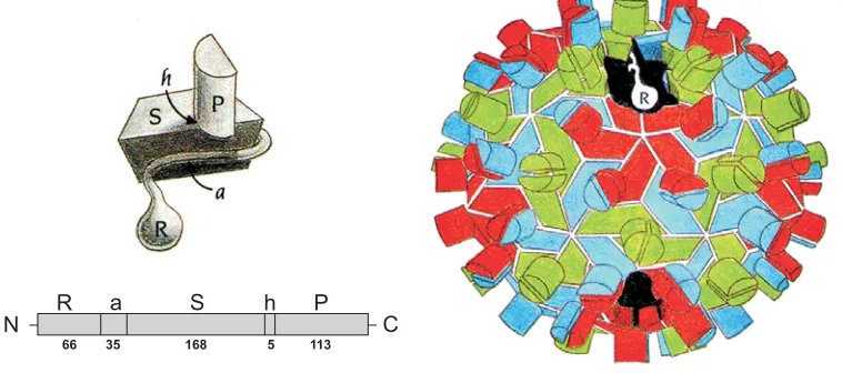
Morphologically RCNMV is most similar to TBSV on the basis of size (34.2
nm), round virion outline and granular surface morphology (Hatta and Francki,
1984). Based on the primary amino acid sequence similarity to the TBSV capsid
protein, RCNMV CP is divided into four domains (Fig. 6). The N-terminus of the CP
is a flexible region composed of two domains: the RNA-binding (R) domain and the
arm (a) region downstream. The arm connects the R domain to the Shell (S)
domain. The R domain contains many basic amino acids and is involved in binding
domain forms the shell of the virion and it consists of two sets of four-stranded
anti-parallel β sheet structures in a jellyroll conformation. There are clusters of negatively
charged residues with an invariant aspartic acid residue within the S domain that
may constitute a Ca2+ binding site which maybe involved in viral assembly and/or
structural stability. The C-terminal protruding (P) domain forms surface projections
that give the virion a granular appearance (Giesman-Cookmeyer et al., 1995a). Each
projection is composed of two P domains. There is also a hinge (h) sequence that
links S and P domains, which provides CP the flexibility to change conformations.
The S and P domains of the capsid protein have a high sequence similarity with that
of Tombusvirus and a moderate sequence similarity with the Carmovirus genus
(Rochon et al., 1991).
There is very little known of RCNMV assembly and the packaging
complement within RCNMV virions is unknown. If RCNMV produces only a single
type of virions it must package both RNA molecules into a single virion. If multiple
types of virions are produced, the two RNAs could be packaged separately, or in
different combinations. Packaging of RNA-1 in one virion and three RNA-2
molecules in another virion has been suggested. This however, does not explain the
single component observed by buoyant density centrifugation of RCNMV virions in
cesium chloride. Encapsidation of both RNA segments into the same virion, as is the
case with Nodaviruses, insect-infecting bipartite RNA virus (Schneemann and
Marshall, 1998), is not possible due to the ratio of RNA-1: RNA-2 which is
approximately 1:3 (S.A. Lommel, unpublished data). Turnip crinkle virus which is
trimer of CP dimers with the origin of assembly sequence on the genomic RNA. The
origin of assembly of TCV was determined to be a distinct bulged hairpin loop in the
CP ORF (Qu and Morris, 1997). Since RCNMV genome consists of two RNAs, each
molecule may have its own packaging signal or the origin of assembly could be the
secondary structure formed by the intermolecular base-pairing of RNA-1 and RNA-2.
RCNMV RNA-1 and RNA-2 of RCNMV are required for a systemic infection
and CP in the form of virions is required for vascular transport. Since formation of
virions is an integral part in the virus life cycle it is important to determine the
processes involved in producing virions in order to characterize the infectious
process of RCNMV. Formation of virions ultimately affects the virus life cycle and
subsequently the disease process. Disruption of virion formation will lead to the
control of the virus. With that goal in mind, this research was designed to identify the
genomic signals required for assembly of virions. These signals, referred to as
packaging signals or origin of assembly sequences (OAS), have been and are being
studied for a number of viruses including animal viruses such as HIV-1. To date TCV
is the only icosahedral plant RNA virus whose packaging signal has been completely
characterized (Qu and Morris, 1997). Furthermore although virions are
morphologically similar, TCV is consists of a monopartite genome whereas RCNMV
contains a bipartite genome. The packaging signal of Flock house virus, an
insect-infecting RNA virus with a bipartite genome, has been characterized. Although it
contains two RNAs that are packaged together, the packaging signal has been
identified only on RNA2 (Zhong et al., 1992; Newman and Brown, 1973; Krishna and
delineated to a sequence that folds into a stem loop structure. It is important to
identify the origin of assembly of RCNMV and to determine common packaging
aspects between these viruses. This will provide us with insights as to the forces and
mechanisms directing virus assembly. Furthermore, determining the specific
packaging signal on RCNMV would enable us to utilize this knowledge in developing
techniques such as viral packaging systems that are presently pursued for
Fig. 1. Symptoms caused by RCNMV onNicotiana benthamiana.On left, necrotic ringspot lesions (local lesions) produced on the inoculated leaf. On right, systemic symptoms include characteristic mottling, cupping and leaf distortion.
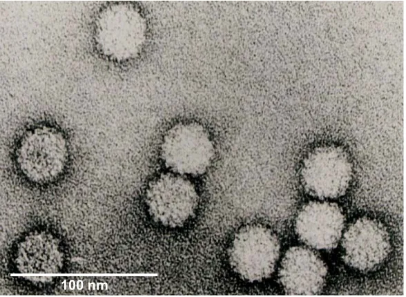
Fig. 2. The electron microscopic image of RCNMV virions. Note the spherical outline and the granular surface of the virions, a characteristic ofTombusviridaevirus CP with protruding domains.
100nm
Fig. 3. The genome organization RCNMV. A schematic representation of RNA-1 and RNA-2. The encoded proteins and the ORFs are shown. ORF nucleotide locations of start and stop positions are indicated below. The -1 frame shift position is shown and the arrow depicts the CP subgenomic RNA promoter.
RNA-1 CP
-1 FS
p88-RdRp p27
RNA-2
3889 nt
1448 nt MP
CP
CP sgRNA 1525 nt
2427
831 2423
3444 123
RNA-1
RNA-1
CCUAGUCGUAUAACGGCUAGGUACCCC CCUAGUCGUAUAACGGCUAGGCACCCC RNA-2
RNA-2
5’ 5’
3’
3’ 3’
A-U
A-U U-A
U-A
AUAUUU-ACCCC U
A A
G-C G-C C-G
C-G C-G U-G
A-U U-A
U-A
UAUAAU-ACCCC U
A A
G-C
G-C G-C C-G
C-G C-G U-G
DG=-16.2 kcal/mol DG=-18.7 kcal/mol
(-) strand RNA synthesis terminates prematurely
sg
terminated
RNA synthesis from prematurely (-) strand RNA
RNA-2 RNA-1
5’
5’
5’
3’
3’
3’ 5’
5’
Fig. 6. The structure of the TBSV particle architecture. RCNMV particle structure and the CP domains are similar to the TBSV. Each CP subunit folds into three domains (R,S,P) with a 35 residue connecting arm (a) between R and S and a hinge (h) between S and P. The number of amino acid residues in each structural module is indicated. The CP subunits pack into the virus particle in one of the three conformations, A (red), B (blue) and C (green). The P domains project out of the surface of the particle (from, Harrison, et al., 1978).
N R a S h P C
References
Albrecht, H., Geldreich, A., Menissier de Murcia, J., Kirchherr, D., Mesnard, J.-M., Lebeurier, G. 1988. Cauliflower mosaic virus gene 1 product detected in a well wall-enriched fraction. Virology 163, 503-508.
Allison, R.F., Janda, M., Ahlquist, P. 1989. Sequence of cowpea chlorotic mottle virus RNAs 2 and 3 and evidence of a recombination event during bromovirus evolution. Virology 172, 321-330.
Atabekov, J.G., Dorokov, Y.L. 1984. Plant virus-specific transport function and resistance of plants to viruses. Adv. Virus Res. 29, 313-364.
Atabekov, J.G., Taliansky, M.E. 1990. Expression of a plant virus-coded transport function by different viral genomes. Adv. Virus Res. 38, 201-248.
Atkins, D., Hull, R., Wells, B., Roberts, K., Moore, P., Beachy, R.N. 1991. The tobacco mosaic virus 30K movement protein in transgenic tobacco plants is localized to plasmodesmata. J. Gen. Virol. 72, 209-211.
Bates, H.J., Farjah, M., Osman, T.A.M., Buck, K.W. 1995. Isolation and characterization of an RNA-dependent RNA polymerase from Nicotiana clevelandii plants infected
with red clover necrotic mosaic dianthovirus. J. Gen. Virol. 76, 1483-1491.
Bos, L., Maat, D.Z. 1965. A distinctive strain of the red clover mottle virus in the Netherlands. Neth. J. Plant Pathol. 71, 8-13.
Bowen, R., Plumb, R.T. 1979. The occurrence and effects of red clover necrotic mosaic virus in red clover (Trifolium pratense). Ann. Appl. Biol. 91, 227-236.
Brierley, I. Digard, P., Inglis, S.C. 1989. Characterization of an efficient coronavirus ribosomal frameshifting signal: requirement for an RNA pseudoknot. Cell 57, 537-547.
Brierley, P. 1964. Heat cure of carnation viruses. Plant Dis. Rep. 48, 143.
Bujarski, J.J., Ahlquist, P., Hall, T.C., Dreher, T.W., Kaesberg, P. 1986. Modulation of replication, aminoacylation and adenylation in vitro and infectivity in vivo of BMV
RNAs containing deletions within the multifunctional 3' end. EMBO J. 5, 1769-1774.
Cohen, Y., Gisel, A., Zambryski, P.C. 2000. Cell-to-cell and systemic movement of recombinant green fluorescent protein-tagged turnip crinkle viruses. Virology 273, 258-266.
Culver, J.N., Dawson, W.O. 1989. Tobacco mosaic virus coat protein: An elicitor for the hypersensitive reaction but not required for the development of mosaic symptoms in
Nicotiana sylvestris. Virology 173, 755-758.
Davies, J.W., Hull, R. 1982. Genome expression of plant positive-strand RNA viruses. J. Gen. Virol. 61, 1-14.
Ding, B., Haudenshield, J.S., Hull, R.J., Wolf, S., Beachy, R.N., Lucas, W.J. 1992. Secondary plasmodesmata are specific sites of localization of the tobacco mosaic virus movement protein in transgenic tobacco plants. Plant Cell 4, 915-928.
Ding, X.S., Flasinki, S., Nelson, R.S. 1999. Infection of barley by brome mosaic virus is restricted predominantly to cells in and associated with veins through a temperature-dependent mechanism. Mol. Plant-Microbe Interact. 12, 615-623.
Dreher, T.W., Bujarski, J.J., Hall, T.C. 1984. Mutant viral RNAs synthesized in vitro show altered aminoacylation and replicase template activities. Nature 311, 171-175.
Francki, R.I.B., Milne, R.G., Hatta, T. 1985. In: Atlas of Plant Viruses Vol. 2, CRC Press, Boca Raon, Florida. pp. 47-52.
Fritzsche, R. 1968. Okologie und Vektoreignung von Longidorus macrosoma Hooper. Biol. Zentralbl. 87, 139-146.
Fritzsche, R., Schmelzer, K. 1967. Ubertragbarkeit des Nelkenringflecken Virus durch Nematoden. Naturwissenschaften 54, 498-499.
Froshauer, S., Kartenbeck, J., Helenius, A. 1988. Alphavirus RNA replicase is located on the cytoplasmic surface of endosomes and lysosomes. J. Cell Biol. 107, 2075-2086.
Fujiwara, T., Giesman-Cookmeyer, D., Ding, B., Lommel, S.A., Lucas, W.J. 1993. Cell-to-cell trafficking of macromolecules through plasmodesmata potentiated by the Red clover necrotic mosaic virus movement protein. Plant Cell 5, 1783-1794.
Fulton, R.W. 1948. Hosts of the tobacco streak virus. Phytopathology 38, 421-428.
Gerhardson, B., Insunza, V. 1979. Soil transmission of red clover necrotic mosaic virus. Phytopathol. Z. 94, 67-71.
Gibbs, A. 1976. Viruses and plasmodesmata. In: Gunning, B.E.S., Robards, A.W. (Eds.), Intercellular Communication in Plants. Studies on Plasmodesmata. Springer Verlag, Berlin. pp. 149-164.
Giesman-Cookmeyer, D., Lommel, S.A. 1993. Alanine scanning mutagenesis of a plant virus movement protein identifies three functional domains. Plant Cell 5, 973-982.
Giesman-Cookmeyer, D., Kim, K.-H., Lommel, S.A. 1995a. Dianthoviruses. in: Pathogenesis and Host-Specificity in Plant Diseases: Histopathological,
Biochemical, Genetic and Molecular Basis. In: Singh, R.P., Singh, U.S., Kohmoto, K. (Eds.), Viruses and Viroids, vol. 3. Pergamon Press, Oxford, UK. pp. 157-176.
Giesman-Cookmeyer, D., Silver, S., Vaewhongs, A.A., Lommel, S.A., Deom, C.M. 1995b. Tobamovirus and dianthovirus movement proteins are functionally homologous. Virology 213, 38-45.
Godefroy-Colburn, T., Gagey, M.-J., Berna, A., Stussi-Garaud, C. 1986. A
non-structural protein of alfalfa mosaic virus in the walls of infected tobacco cells. J. Gen. Virol. 67, 2233-2239.
Goodrick, B.J., Kuhn, C.W., Hussey, R.S. 1991. Restricted systemic movement of cowpea chlorotic mottle virus in soybean with nonnecrotic resistance.
Phytopathology 81, 1426-1431.
Gould, A.R., Francki, R.I.B., Hatta, T., Hollings, M. 1981. The bipartite genome of red clover necrotic mosaic virus. Virology 108, 499-506.
Guenther, R.H., Sit, T.L., Gracz, H.S., Dolan, M.A., Townsend, H.L., Liu, G., Newman, W.H., Agris, P.F., Lommel, S.A. 2004. Structural characterization of an
intermolecular RNA-RNA interaction involved in the transcription regulation element of a bipartite plant virus. Nucl. Acids Res. 32, 2819-2828.
Hamilton, R.I., Tremaine, J.H. 1996. Dianthoviruses: Properties, Molecular Biology, Ecology, and Control (Chapter 10). In: Harrison, B.D., Murant, A.F. (Eds.), The Plant Viruses. Vol. 5:Polyhedral Virions and Bipartite RNA Genomes. Plenum press, New York, NY. pp. 251-282.
Harrison, B.D. 1958. Raspberry yellow dwarf, a soil-borne virus. Ann. Appl. Biol. 46, 221-229.
Hatta, T., Bullivant, S., Matthews, R.E. 1973. Fine structure of vesicles induced in chloroplasts of Chinese cabbage leaves by infection with turnip yellow mosaic virus. J. Gen. Virol. 20, 37-50.
Hatta, T., Francki, R.I.B. 1984. Differences in the morphology of isometric particles of some plant viruses stained with uranyl acetate as an aid to their identification. J. Virol. Methods 9, 237-247.
Heaton, L.A., Lee, T.C., Morris, T.J. 1991. Point mutations in the turnip crinkle virus capsid protein affect the symptoms expressed by Nicotiana benthamiana. Virology
183, 143-150.
Heinlein, M., Epel, B.L., Padgett, H.S., Beachy, R.N. 1995. Interaction of tobamovirus movement proteins with the plant cytoskeleton. Science 270:1983-1985.
Hiruki, C. 1986. Incidence and geographic distribution of sweet clover necrotic mosaic virus in Alberta. Plant Dis. 70, 1129-1131.
Hiruki, C. 1987. The dianthoviruses: A distinct group of isometric plant viruses with a bipartite genome. Adv. Virus Res. 33, 257-299.
Hiruki, C., Rao, A.L.N., Furuya, Y., Figueiredo, G. 1984a. Serological studies of
dianthoviruses using monoclonal and polyclonal antibodies. J. Gen. Virol. 65, 2273-2275.
Hiruki, C., Rao, D.V., Chen, M.H., Okuno, T., Figueiredo, G. 1984b. Characterization of sweet clover necrotic mosaic virus. Phytopathology 74, 482-486.
Hollings, M. Stone, O.M. 1977. Red clover necrotic mosaic virus. CMI/AAB Descript. Plant Viruses, No. 181.
Hollings, M., Stone, O.M. 1970. Carnation ringspot virus. CMI/AAB Descript. Plant Viruses, No. 20.
Hollings, M., Stone, O.M. 1965. Investigation of carnation viruses. II. Carnation ringspot. Ann. Appl. Biol. 56, 73-86.
Hull, R. 1989. The movement of viruses in plants. Annu. Rev. Phytopathol. 27, 213-240.
Jacks, T., Madhani, H.D., Masiarz, F.R., Varmus, H.E. 1988. Signals for ribosomal frameshifting in the Rous sarcoma virus gag-pol region. Cell 55, 447-458.
Jacks, T., Varmus, H.E. 1985. Expression of the Rous sarcoma virus pol gene by
Kegler, G., Kegler, H. 1981. Beitrage zur Kenntnis der vektorlosen Obertragung pflanzenpathogener Viren. Arch. Phytopathol. Pflanzenschutz 17, 307-323.
Kendall, T.L., Lommel, S.A. 1992. Nucleotide sequence of carnation ringspot dianthovirus RNA-2. J. Gen. Virol. 73, 2479-2482.
Kim, K.H., Lommel, S.A. 1998. Sequence element required for efficient –1 ribosomal frameshifting in red clover necrotic mosaic dianthovirus. Virology 250, 50-59.
Kim, K.H., Lommel, S.A. 1994. Identification and analysis of the site of –1 ribosomal frameshifting in red clover necrotic mosaic virus. Virology 200, 574-582.
Kleinhempel, H., Gruber, G., Kegler, H. 1980. Investigations on carnation ringspot virus in fruit trees. Acta Phytopathol. Acad. Sci. Hung. 15, 107-111.
Koonin, E.V. 1991. The phylogeny of RNA-dependent RNA polymerases of positive-stranded RNA virses. J. Gen. Virol. 72, 2197-2206.
Koonin, E.V., Dolja, V.V. 1993. Evolution and taxonomy of positive-strand RNA viruses: Implications of comparative analysis of amino acid sequences. Criti. Rev. Biochem. Mol. Biol. 28, 375-430.
Koonin, E.V., Mushegian, A.R., Ryabov, E.V., Dolja, V.V. 1991. Diverse groups of plant RNA and DNA viruses share related movement proteins that may possess
chaperone-like activity. J. Gen. Virol. 72, 2895-2903.
Kowalska, A. 1974. Freeing carnation plants from viruses by meristem-tip culture. Phytopathol. Z. 79, 301.
Krishna N.K., Schneemann, A.1999. Formation of an RNA heterodimer upon heating of nodavirus particles. J. Virol. 73, 1699-1703.
Kuhne, T., Eisbein, K. 1983. Untersuchungen zur Stabilitat des Nelkenringflecken-Virus. Acta Phytopathol. Acad. Sci. Hung. 18, 101-110.
Lapchic, L.G., Kuznetzova, L.L. Melniczenko, V.S., Vocelko, S.K., Sjedin, A.A. 1975. Red clover mottle virus in Ukraine. Phytopathol. Z. 82, 339-346.
Lehto, K., Bubrick, P., Dawson, W.O. 1990. Time course of TMV 30 K protein accumulation in intact leaves. Virology 174, 290-293.