INVESTIGATION
Comparing Enhancer Action in
Cis
and in
Trans
Jack R. Bateman,1Justine E. Johnson, and Melissa N. Locke Biology Department, Bowdoin College, Brunswick, Maine 04011
ABSTRACTStudies from diverse systems have shown that distinct interchromosomal interactions are a central component of nuclear organization. In some cases, these interactions allow an enhancer to act intrans, modulating the expression of a gene encoded on a separate chromosome held in close proximity. Despite recent advances in uncovering such phenomena, our understanding of how a regulatory element acts on another chromosome remains incomplete. Here, we describe a transgenic approach to better understand enhancer action in trans in Drosophila melanogaster. Using phiC31-based recombinase-mediated cassette exchange (RMCE), we placed transgenes carrying combinations of the simple enhancer GMR, a minimal promoter, and different fluorescent reporters at equivalent positions on homologous chromosomes so that they would pair via the endogenous somatic pairing machinery of
Dro-sophila. Our data demonstrate that the enhancer GMR is capable of activating a promoter intransand does so in a variegated pattern, suggesting stochastic interactions between the enhancer and the promoter when they are carried on separate chromosomes. Fur-thermore, we quantitatively assessed the impact of two concurrent promoter targets incisand intransto GMR, demonstrating that each promoter is capable of competing for the enhancer’s activity, with the presence of one negatively affecting expression from the other. Finally, the single-cell resolution afforded by our approach allowed us to show that promoters incisand intransto GMR can both be activated in the same nucleus, implying that a single enhancer can share its activity between multiple promoter targets carried on separate chromosomes.
I
N an oversimplified view of the genome, each chromosome could be considered a linear arrangement of genic units, with each gene controlled solely by nearby cis-acting regu-latory sequences. However, extensive analyses based on DNAfluorescencein situhybridization (DNA FISH) and chromatin conformation capture (3C) have shown that the three-dimensional organization of the nucleus involves physical in-teractions between distant genomic regions, suggesting that gene regulation is more complex than this simplified model (reviewed by Gondor and Ohlsson 2009; Naumova and Dekker 2010). Further complicating our view, long-distance interactions are not limited to genomic regions on the same chromosomes (in cis), but are also observed between spe-cific loci on separate chromosomes (intrans) (reviewed by Williams et al.2010). Through genome-wide 3C-based ap-proaches, extensive interchromosomal interactions have been identified in yeast (Duanet al.2010),Drosophila(Sexton et al.2012), mice (Zhanget al.2012), and humans
(Lieberman-Aidenet al.2009) and are thus a component of nuclear organi-zation in diverse organisms.
Long-distance intra- and interchromosomal interactions can result from colocalization of unlinked genomic regions to a common nuclear compartment, including loci undergo-ing active transcription or that bind insulator or Polycomb Group proteins (reviewed by Bantignies and Cavalli 2011; Bulger and Groudine 2011; Dean 2011). In some cases, long-distance interactions could reflect communication between regulatory sequences that will directly affect gene expression, as is the case when a distant enhancer loops toward a pro-moter to activate transcription (reviewed by Bartkuhn and Renkawitz 2008; Bulger and Groudine 2011). Interchromo-somal interactions that directly affect gene expression have been demonstrated in several developmental contexts, in-cluding X-inactivation (Bacheret al.2006; Xuet al.2006), Ifng expression in naive T helper cells (Spilianakis et al. 2005), and the establishment of replication timing of imprinted loci (Sandhu et al. 2009) in mice, and IFN-b expression in response to virus infection in humans (Apostolou and Thanos 2008). In addition, physical interactions between chromosomes have been postulated to explain epigenetic phe-nomena such as repeat-induced point mutation inNeurospora (reviewed by Galagan and Selker 2004) and paramutation in Copyright © 2012 by the Genetics Society of America
doi: 10.1534/genetics.112.140954
Manuscript received April 3, 2012; accepted for publication May 22, 2012 Supporting information is available online at http://www.genetics.org/content/ suppl/2012/05/29/genetics.112.140954.DC1.
1Corresponding author: Biology Department, Bowdoin College, 6500 College Station,
maize (Chandler and Stam 2004). However, our ability to identify and catalog interchromosomal interactions has grown more quickly than our ability to determine their poten-tial impact on gene expression. Thus, a central question remains: Is it common for sequences that regulate gene expres-sion to communicate between chromosomes when they are physically juxtaposed?
Drosophilaprovides an excellent model system to better understand the potential impact of interchromosomal inter-actions on gene regulation. InDrosophila, homologous chro-mosomes are stably paired from end to end in virtually all cells of the organism (reviewed by McKee 2004). The po-tential for communication between Drosophila homologs was first postulated .100 years ago by Nettie Stevens (Stevens 1908) and was demonstrated genetically in 1954 by Ed Lewis, who coined the term transvection to describe the phenomenon of pairing-dependent gene regulation (Lewis 1954). Transvection has since been shown to influence many genes in Drosophila and can lead to either upregulation or silencing of gene expression by several mechanisms (reviewed by Duncan 2002; Kennison and Southworth 2002).
In one form of transvection, an enhancer on one homolog acts in transto activate a promoter on a paired homolog, leading to upregulation of a linked gene. Enhancer action in transis often uncovered as an unexpected case of intragenic complementation involving specific types of mutant alleles. For example, one mutant allele may carry a nonfunctional enhancer while another carries a compromised promoter; acting alone, neither allele can support gene expression, but when paired, the remaining functional enhancer and promoter interact in transto activate transcription (Figure 1A). For many genes, enhancer action intransis thought to produce reduced levels of transcription relative to activation incis, as evidenced by the partial complementation of mu-tant phenotypes that result from trans-action (e.g., Lewis 1954; Geyer et al.1990; Leiserson et al. 1994). However, precise quantification of specific enhancer–promoter inter-actions in transhas been difficult due to the complexity of regulatory sequences surrounding most genes.
Despite decades of genetic studies of transvection, several questions regarding enhancer action intransremain. First, is the capacity to act intransa common property of enhancers? Only a handful of the thousands of enhancers encoded in the Drosophila genome have been demonstrated to act in trans, which could reflect that this is a rare property, but could also be a consequence of the specific types of alleles required to easily observe trans activity. Second, are specific sequences required to promote enhancer action intrans? Some examples of transvection at the Abdominal-B locus require a 10-kb sequence dubbed the transvection-mediating region (tmr) (Hopmannet al.1995), and transgenic studies have shown that thetmrcan positively affect pairing (Ronshaugen and Levine 2004). Similarly, long-distance interactions between enhancers and promoters inciscan be facilitated by promoter tethering sequences (Zhou and Levine 1999; Calhoun et al. 2002; Calhoun and Levine 2003; Lin 2003; Akbariet al.2008;
Fujioka et al. 2009; Kwon et al. 2009). It is as yet unclear whether similar sequences are required for any enhancer to act in trans or, alternatively, whether transvection between regulatory elements is a natural consequence once chromo-somes are physically paired. Finally, in light of the extensive pairing in the Drosophila genome, how does an enhancer choose a target promoter when simultaneously presented with promoters incisand intrans? For several genes, the presence of a promoter incisreduces the strength of enhancer action in trans(Martinez-Labordaet al.1992; Casareset al.1997; Gohl et al.2008), suggesting that promoters incisand intrans com-pete for an enhancer’s activity. Because these observations have been largely based on mutant phenotypes in whole animals, the cellular basis for this competition is not yet clear. Does an en-hancer from each cell choose either the promoter incisor the promoter intrans? Or can a single enhancer in one cell activate both targets?
Here we describe a transgenic approach to the study of enhancer action intrans. Notably, the use of transgenic report-ers greatly simplifies the manipulation of regulatory sequences via the use of straightforwardin vitrocloning techniques, permitting the construction of various“alleles”with which to better understand transvection. Our approach was in-formed by a prior experiment that used transposons carrying
yellow alleles, including flanking regulatory sequences, that were designed to mimic mutations known to support en-hancer action in trans at the natural yellow locus. That study showed theDrosophilagenome to be generally per-missive to transvection, demonstrating the feasibility of a transgenic approach to the analysis of enhancer action intrans (Chenet al.2002). In the present study, our goal was to construct transgenes based onfluorescent reporters such as GFP and mCherry to facilitate quantification of gene expression and to provide cellular resolution of trans-gene activation. Using the eye-specific enhancer GMR, we
first established that simple constructs containing different combinations of enhancer, promoter, and transcriptional unit can support enhancer action intrans, suggesting that special sequences are not required to mediate this form of transvection. Furthermore, we showed that GMR action in transoccurs in a variegated pattern in cells of the eye disc. Variegation was not seen when GMR acts incis, even when the distance between the enhancer and the promoter was increased, suggesting that stochastic interactions between the enhancer and the promoter are specific to enhancer action in trans. Finally, we quantitatively assessed the im-pact of promoter competition on GMR function, supporting a model wherein promoters in cis and in trans will both compete for an enhancer’s activity.
Materials and Methods
Fly stocks and husbandry
target line 38F is an insertion of the same target cassette at polytene band 38F. All insertions are intergenic. Flies were maintained at 25on standardDrosophilacornmeal, yeast, sugar, and agar medium with p-hydroxybenzoic acid methyl ester as a mold inhibitor (Morriset al.1998).
Plasmid construction
The RMCE donor vector piB-GFP was previously described (Bateman et al. 2006). To create GMR-GFP carrying the Completegfp construct, the GMR enhancer was amplified
by PCR from the plasmid pGMR (a gift from J. Settleman), using primers 5xGMR1 and 5xGMR2 carrying additional ClaI recognition sequences at their 59ends (note that two additional cloning steps below make use of the same primers that instead carry BamHI or AscI sites), and subcloned into pcr2.1 using a TOPO-TA cloning kit (Invitrogen, Carlsbad, CA). The enhancer was then digested from pcr2.1 and inserted upstream of the minimal hsp70 promoter in piB-GFP using ClaI.
To create GMR-lacZ carrying the CompletelacZconstruct,
wefirst digested piB-GFP withXbaI andHindIII to remove all promoter, GFP, and UTR sequences. We then isolated a 4.6-kb SpeI-HindIII fragment carrying the minimalhsp70 promoter, the b-galactosidase open reading frame, and the SV40 39-UTR from the plasmid pCaSpeR-hs43-lacZ and cloned it into the remaining piB-GFP backbone to create piB-lacZ (consisting of the lacZ transgeneflanked by phiC31 attB sequences to allow for insertion into RMCE sites). The GMR enhancer was then cloned into aBamHI site upstream of thehsp70minimal promoter.
To create the Promoterless construct, a 4.5-kbSalI frag-ment consisting of theb-galactosidease open reading frame and the SV40 39-UTR but omitting the minimalhsp70 pro-moter was isolated from pCaSpeR-hs43-lacZ. This fragment was cloned into theSalI-digested backbone of piB-GFP, pro-ducing piB-lacZ(P-), carrying a promoterless lacZ transgene
flanked by attB sites. Next, we introduced a minimalhsp70 promoterflanked by loxP sites that could be removedin vivo by treatment with the Cre recombinase. We amplified the hsp70 promoter from piB-GFP using loxTATA1 and lox-TATA2, subcloned this fragment into pcr2.1 using a TOPO-TA kit, and then liberated the loxP-promoter-loxP fragment from pcr2.1 usingHindIII and inserted it into a unique Hin-dIII site upstream of the lacZ gene in piB-lacZ(P-) to create piB-LTL-lacZ. Finally, the GMR enhancer was inserted into aBamHI site upstream of the loxP-promoter-loxP cassette to create GMR-LTL-lacZ. Once integrated into flies via RMCE (see below), the final Promoterless transgene was created by removing the promoter between the two loxP sites via crosses toflies carrying the Cre recombinase on the balancer CyO (Siegal and Hartl 1996). Promoter deletions were ver-ified by PCR using primers Pry1 and RNXG2.
To create GMR-mCherry carrying the CompletemC
con-struct, wefirst used gene splicing by overlap extension (SOE-ing) (Hortonet al.1990) to combine an mCherry ORF from pmCherry (Clonetech) with the minimal hsp70 promoter.
Briefly, thehsp70promoter was amplified from piB-GFP using cherrySOE1_for and cherrySOE2_3RC_rev primers, and the mCherry ORF was amplified using cherrySOE3_2RC_for and cherrySOE4_rev. The two products were combined in a PCR reaction containing cherrySOE1_for and cherrySOE4_rev, and the resulting product was subcloned into pcr2.1 via TOPO-TA cloning. Thehsp70-mCherry fragment was then digested from pcr2.1 using BamHI andXbaI and cloned intoBamHI/Xba I-digest piB-GFP(-S) (a piB-GFP derivative generated by religa-tion after XbaI/SpeI digestion, removing BamHI, SpeI, and XbaI sites from downstream of the 39-UTR) to create piB-mCherry, carrying thehsp70promoter, an mCherry ORF, and the SV40 39-UTRflanked by attB sites. Finally, the GMR en-hancer was inserted into the BamHI site upstream of the promoter.
To create GMR-l3-GFP carrying the Complete+3.0
con-struct, a 3.0-kb fragment that has previously been demon-strated to lack insulator activity (Chunget al.1993; Singer et al. 2009) was amplified from phage lambda DNA using primers lambda3kb_Bam_3 and lambda3kb_BamAsc_5, and the resulting fragment was subcloned into pcr2.1 via TOPO-TA cloning. Next, the GMR enhancerflanked withAscI sites was cloned into theAscI site at one end of the lambda fragment. Finally, the GMR-lambda fragment was liberated with BamHI and cloned into theBamHI site upstream of the pro-moter in piB-GFP(-S).
Site-specific transformation via RMCE
All constructs were integrated into RMCE target sites as previously described (Bateman and Wu 2008), using a ge-nomic source of the phiC31 integrase (Grothet al.2004; Bischof et al.2007). Following integration, the orientation of insertion was assessed using primers lac4 or 39Pend1, which are complementary to the 59and 39P-element ends
flanking the RMCE cassette, respectively, and RNXG9, which is complementary to the SV40 39-UTR. All constructs were analyzed in the same orientation with the enhancer or pro-moter near the 39P-element end, with the exception of the Complete+3.0construct, which generated only one insertion in
the opposite orientation.
Tissue preparation, immunostaining, and microscopy
primary antibody incubation, discs were rinsed and then washed 2 ·20 min in PBST, blocked in 4% NGS/PBST for 1 hr, and then incubated for 1 hr in secondary antibodies, goat anti-mouse Cy3 (Jackson ImmunoResearch) at 1:250 and goat anti-rabbit AlexaFluor-488 (Invitrogen) at 1:2000. Discs were then washed 4·20 min in PBST and mounted in Vectashield with DAPI or Fluoromount G with DAPI.
For low-magnification imaging, discs were visualized on an Olympus BX51, using a Media Cybernetics Evolution VF color camera. For higher-magnification imaging, samples were visualized using a Zeiss Axioplan 2 microscope with a 510 Meta confocal laser scanning system, with optical sections collected at 0.7-mm increments. At least 10 discs of each genotype were analyzed; all discs with the same genotype showed similar patterns of expression.
Quantification of variegation
To quantify the degree of variegation in GFPcis- andtrans -activation, confocal z-stacks of immunostained discs were
first max-projected into a single-plane image. Using ImageJ imaging software, a small elliptical region of interest (ROI) was placed over cell R4 of each ommatidial cluster of the
five posterior-most rows and the mean GFPfluorescence was measured for each R4 cell. Next, the mean backgroundfl uo-rescence, determined by averaging the mean fluorescence of 10 ROIs in nonfluorescent regions of the disc, was subtracted from each measurement. Finally, each background-corrected measurement was converted to a relative intensity by express-ing it as a percentage of the highest-intensity cell in thefield of view.
Reverse transcription and quantitative PCR
For each sample, eye-antennal discs were dissected from 10 wandering third-instar larvae (20 discs total) and frozen at
280. Tissue homogenization, genomic DNA elimination, and RNA purification were carried out using a QIAGEN RNeasy plus kit according to the manufacturer’s protocol. Briefly, discs were suspended in RLTplus buffer, passed through a 25-gauge needle eight times, and then spun through a qiash-redder spin column. The lysate was then spun through a gDNA eliminator spin column, and theflow-through was transferred to an RNeasy spin column for RNA purification. Generation of cDNA was performed using a QIAGEN QuantiTect Reverse Transcription Kit according to the manufacturer’s protocol, in-cluding pretreatment with gDNA Wipeout Buffer to eliminate any remaining traces of genomic DNA.
Quantitative PCR was carried out on a StepOne Real-Time PCR system (Applied Biosystems, Foster City, CA). For each reaction, cDNA was diluted 1:5 in water and combined with SYBR green PCR mastermix (Applied Biosystems) and primers. GFP cDNA was amplified using primers GFPRT_1 and GFPRT_2, and mCherry cDNA was assayed using primers cherryRT_1F and cherryRT_1R. Primers 58F and RP49-175R targeting the housekeeping rp49 cDNA (Moon et al. 2008) were used as an internal reference. Each reaction was performed in triplicate, and each genotype was assayed using
at least three independent cDNA samples from separate crosses. Relative levels of transcript were calculated via the
DDCtmethod, using StepOne software. Unpairedt-tests were performed using GraphPad Software’s onlinet-test calculator (www.graphpad.com).
Primer sequences
Primers used in this study are indicated below in 59to 39 orientation: 5XGMR1: GATCCCCCTAGAATCCCAAA 5XGMR2: TATACTCCGGCGCTCTTTTC loxTATA1: AAGCTTATAACTTCGTATAGCATACATTATACGA AGTTATAAGAGCGCCGGAGTATAAATAG loxTATA2: AAGCTTATAACTTCGTATAATGTATGCTATACGA AGTTATCTGCAGATTGTTTAGCTTGTTCAG RNXG2: CAGCTGCGCTTGTTTATTTG Pry1: CCTTAGCATGTCCGTGGGGTTTGAAT cherrySOE1_for: ATGTCGAGGTCGACGGTATC cherrySOE2_3RC_rev: ATGGTGGCGACCGGTACTCTTCTTT CTCGGTAACTTGTTGA cherrySOE3_2RC_for: TCAACAAGTTACCGAGAAAGAAGA GTACCGGTCGCCACCAT cherrySOE4_rev: CGGCGCTCAGTTGGAAT lac4: ACTGTGCGTTAGGTCCTGTTCATTGT 39Pend1: GTCGGCAAGAGACATCCACT RNXG9: GTGGTTTGTCCAAACTCATCAA GFPRT_1: ATTCTCGTGGAACTGGATGG GFPRT_2: AGCTTTCCAGTGGTGCAGAT cherryRT_1F: CCCGCCGACATCCCCGACTA cherryRT_1R: CTGGGTCACGGTCACCACGC RP49-58F: TACAGGCCCAAGATCGTGAAG RP49-175R: GACGCACTCTGTTGTCGATACC. Results
Our goal was to better understand enhancer action intrans and its relationship to expression in cisby comparing these modes of gene activation under controlled conditions. We rea-soned that a transgenic approach would be advantageous, per-mitting us to easily manipulate the identities of the transgene’s components, to vary simple parameters such as deletions and relative distances between regulatory regions, and to accu-rately quantify levels of gene expression. We chose to focus our analysis on the simple enhancer GMR, which consists of
five tandem binding sites for the transcription factor Glass and drives tissue-specific expression of reporter constructs in the photoreceptors, or R cells, posterior to the morphogenetic fur-row of the developing eye (Moses and Rubin 1991).
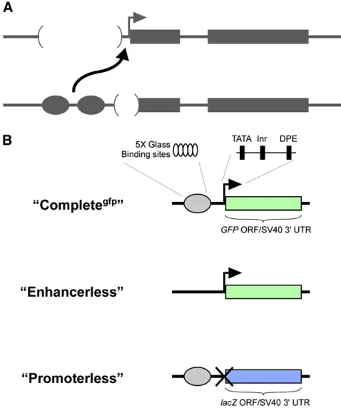
To assesscisexpression, we created a“Completegfp”reporter
(Figure 1). Additionally, we designed the Enhancerless con-struct to encode GFP and the Promoterless concon-struct to en-code b-galactosidase to differentiate potential expression of one transgene from the other. Importantly, neither the Enhan-cerless nor the Promoterless transgene is capable of reporter gene expression in isolation. We anticipated that pairing of the two nonfunctional transgenes would permit GMR to activate thehsp70 promoter intrans, producing GFP fl uo-rescence specifically in developing R cells.
We began by integrating our transgenes into an RMCE target site at polytene band 53F (Bateman and Wu 2008). As expected, the Completegfpreporter produced robust GFP
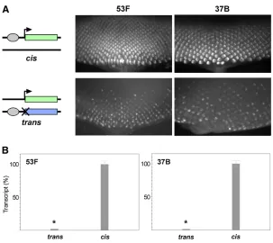
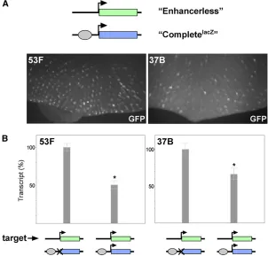
expression in mature retinal cells posterior to the morpho-genetic furrow of third-instar eye discs, whereas neither the Enhancerless nor the Promoterless transgene alone pro-duced detectable GFP (Figure 2A, data not shown). Next, we established crosses to produce progeny carrying paired Enhancerless and Promoterless constructs in trans to one another at 53F. Excitingly, we observed GFP fluorescence specifically in the photoreceptors of the developing retina, consistent with our prediction if GMR were to function in trans. To quantify relative levels of expression, we isolated RNA from whole eye-antennal discs expressing GFP under the control of GMR incisor intransand prepared cDNA for quantitative RT-PCR. We found that total levels of GFP tran-script in the eye-antennal disc were strongly diminished when GMR acted in trans, with 2.2% [95% confidence in-terval (C.I.), 2.0–2.3%] of transcript levels relative to discs where GMR acted incis(Figure 2B).
To ensure that our observations were representative of other genomic regions, we integrated the same constructs into RMCE targets located at polytene bands 37B and 38F and observed nearly identical patterns of expression in bothcisand trans(Figure 2A andSupporting Information,Figure S1). Ad-ditionally, quantitative RT-PCR showed that relative levels of GFP transcripts from constructs inserted at 37B were similar to those from transgenes at 53F, with 1.7% [95% C.I., 1.6– 1.8%] of total GFP transcript in eye discs where GMR acts in transrelative to those where activation is incis(Figure 2B). Notably, expression intrans was dependent on pairing of Enhancerless and Promoterless transgenes, as flies carry-ing an Enhancerless transgene at 53F and a Promoterless transgene at 37B produced no detectablefluorescence (data not shown). We conclude that the enhancer GMR, and there-fore the transcription factor Glass that is responsible for its expression, is capable of activating the hsp70 promoter in trans. Furthermore, as our transgenes carry only minimal en-hancer and promoter elements in addition to exogenous reporter sequences, we infer that, once homologous chromo-somes are paired, GMR and thehsp70promoter are capable of communicating in trans without the need for additional sequences (seeDiscussion).
GMR action in trans is variegated
In eye discs where GMR acts in trans, we noted that the pattern of GFP expression was uneven, or variegated, in cells
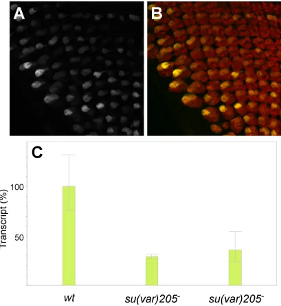
across the eye disc (Figure 2A). The variegated expression was unlikely to result from a position effect as has been observed for some transgenic insertions near heterochromatic regions, because we noted a similar pattern for GMR expression in transat all three genomic locations tested, whereas GMR ac-tion incisproduced no variegation. Furthermore, we found no evidence that the variegated expression produced when GMR acts intranswas altered by reduction of mRNA encoding the heterochromatin protein HP1, nor did we observe any evi-dence of variegated eye pigmentation for insertions of mini-white or a white transgene under the control of the hsp70 promoter in the same location (Figure S2,Figure S3, and data not shown). Thus, the variegated pattern appears to be spe-cific toGMRaction intrans.
To better understand the variegated phenotype, we used confocal microscopy to produce high-magnification three-Figure 1 Schematic view of constructs used to establish a transgenic system for transvection. (A) A schematic of typical alleles that show trans-vection at endogenous genes. In the top allele, the enhancers (circles) are deleted, while in the bottom allele, the promoter (bent arrow) is deleted. When paired, the remaining enhancers of the bottom allele act intrans
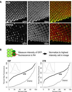
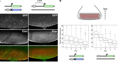
dimensional z-stacks of developing retinal cells expressing GFP under the control of GMR incisand intrans(Figure 3). The retinal photoreceptors undergo a stereotyped pattern of development as the morphogenetic furrow passes from pos-terior to anpos-terior across the eye disc, resulting in a repeating pattern of ommatidial clusters, each possessing eight photo-receptor subtypes, R1–R8. All subtypes are highlighted us-ing antibodies targetus-ing Elav, a neuronal-specific cell marker (Robinow and White 1988) (Figure 3). In discs where GMR acts incis, GFPfluorescence was evident in all of the R cells of each ommatidial cluster, with the highest levels of expres-sion in R3 and R4. When GMR acts intrans, expression in R3 and R4 once again predominated, but some of these cells expressed GFP strongly, some weakly, and some undetectably. To quantify the variegation, we assessed the relative GFP
fluorescence intensity specifically in R4 cells of the posterior-mostfive rows of ommatidial clusters in eye discs expressing GFP incisand intrans(seeMaterials and Methods) and plot-ted the data in percentile plots. For transgenes at position 53F, expression in cis showed a near-normal distribution, with 28.3% of R4 cells having less than half thefluorescent inten-sity of the brightest cells in the image and no cells having
,20% of the intensity of the brightest cells (Figure 4B). In contrast, expression intransshowed a strong shift in the dis-tribution of relative intensities, with 83.1% of R4 cells show-ing less than half the fluorescent intensity of the brightest cells; among these, 7.7% of cells showed no detectablefl uo-rescence (defined as ,1% of highest-intensity cells), which could reflect either weak expression that was below our limit of detection or a complete lack of enhancer activity. Analysis of transgenes inserted at 37B produced nearly identical results, with 83.4% of R4 cells that expressed GFP intransshowing
less than half the fluorescent intensity of the brightest cells and 15.9% of cells showing no detectablefluorescence. Thus, GMR action intransresults in weak expression in the majority of R4 cells, with sporadic“jackpot”events resulting in bright
fluorescence in a small proportion of the cell population.
Variegation is not reproduced by a distant enhancer in cis
We reasoned that the weak and variegated expression pro-duced by GMR action intransmight be related to changes in the distance between the enhancer and the promoter when the two communicate intransrelative to their interactions in cis. Indeed, in our Completegfptransgene, the enhancer and
promoter are separated by only 50 bp, which likely facilitates efficient enhancer–promoter communication. In contrast, we presumed that when GMR was carried on a separate chromo-some from the promoter, the two would be less likely to lie in close physical proximity; thus, one possible explanation for the variegated pattern of GFP activation is that the increased distance between enhancer and promoter leads to stochastic activation of the promoter as the probability of interaction is decreased.
As a test of whether a simple increase in distance between GMR and the hsp70 promoter can account for the variegated expression in trans, we created a variant of our Complete transgene wherein the GMR enhancer is separated from thehsp70promoter by 3.0 kb of DNA derived from the lambda phage genome (“Complete+3.0” transgene), thereby
increasing the distance between enhancer and promoter in cisrelative to our initial Completegfpconstruct. After
integrat-ing the Complete+3.0construct into position 53F, we assessed
total transcript levels by quantitative RT-PCR. Similar to the Figure 2 GMR activates thehsp70promoter intrans. (A) Schematics of genotypes and images of GFP expression in third-instar eye discs. Top row, GFPfluorescence resulting fromcis-activation by an insertion of the Completegfpon one homolog. Bottom row, GFPfluorescence fromtrans -expression of GFP inflies with the Enhancerless construct on one homolog and the Promoterless construct on the other. Transgenes were placed at an RMCE target at poly-tene position 53F (left) or 37B (right). The posterior of the eye disc is oriented to the bottom of each image. (B) Rel-ative quantification of GMR activation of GFP incisand in
case when GMR acts in trans, we observed a reduction in transcript levels to 15.2% (95% C.I., 12.8–18.1%) relative to the original Completegfpconstruct (data not shown),
consis-tent with the notion that increasing the distance between en-hancer and promoter decreases the efficiency of transcriptional activation. However, when we examined discs carrying Complete+3.0by microscopy, the pattern of GFP did not
ap-pear variegated; instead, we observed a“delayed”pattern of expression where GFPfluorescence was primarily observed in the posterior-most ommatidial clusters that were the earliest to commit to photoreceptor cell fates (Figure 4A).
To quantify this effect, we assessed the relativefluorescent intensities of R4 cells in the five posterior-most ommatidial rows as above; in this case, we isolated data from each row in separate box plots to better dissect changing levels of expression between early- and late-committing cells (Figure 4B). In discs where GMR acts intrans, we saw great variation in all rows; notably, some cells in the newly committed rows are among the brightest cells, whereas cells with undetectable levels offluorescence persist in the longest-expressing rows. In contrast, discs where GMR acts at a distance incisshow a strong trend for increased expression over time, with all nonexpressing
cells limited to the newly committed rows and all of the bright-est cells found in the early-committed cells. Importantly, the distributions of relative intensities in the posterior-most row are significantly different between the two genotypes (P ¼ 3.7·1029, Welch’st-test). In sum, our data indicate that in-creasing the distance betweenGMRand thehsp70promoter in cishas a negative impact on transcriptional activation, but does not lead to variegation.
Promoter targets in cis and in trans compete for GMR enhancer activity
In the experiments above, the GMR enhancer was provided with just one targethsp70promoter provided either incisor intrans. We next assessed how GMR would respond when given a choice between promoters in cis and in trans con-currently. First, we created a new Complete construct encod-ing b-galactosidase instead of GFP (CompletelacZ), which is
identical to the Promoterless construct except for the addition of a functionalhsp70promoter. After targeting CompletelacZto
RMCE sites at 53F and 37B, we verified that it was capable of robust expression ofb-galactosidase incis(Figure S4). We then placed the CompletelacZ construct in trans to the
Figure 3 GMR action in transis variegated. (A) Max-projected confocalz-stacks showing GMR-driven expression of GFP incis (top) or in trans(bottom). Constructs are identical to those in Figure 2 and are integrated at 53F. GFP is visualized by immunostaining, and ommatidial clus-ters are shown by Elav immunostaining. GFP expression is highest in cells R3 and R4 in both genotypes, withtrans -activation showing variegated expression. Posterior is ori-ented to the bottom of each image. (B) Quantification of relativefluorescent intensities. Top, rough schematic of cell positions in each ommatidial cluster; intensities of GFP fluorescence were measured for each R4 cell in the pos-terior-mostfive rows and normalized to the brightest cell of the image. Percentile plots show data for expression in
Enhancerless GFP construct and once again observed varie-gated GFP expression in the eye disc, indicating that GMR is capable of activating a promoter in transin the presence of a promoter incis(Figure 5A). Next, we prepared cDNA from discs carrying the Enhancerless construct intransto either the Promoterless or the CompletelacZconstruct and quantified
rel-ative levels of GFP transcript between the two genotypes via quantitative RT-PCR (Figure 5B). At both 53F and 37B, we observed a near twofold reduction in GFP transcript levels in flies carrying the Enhancerless transgene paired with CompletelacZrelative to those with the Promoterless construct
[50.1% (95% C.I., 45.8–56.4%) and 66.4% (95% C.I., 59.1– 74.7%) at 53F and 37B, respectively], consistent with the model that the promoter in cis competes for the enhancer’s activity and thereby interferes with GMR’s ability to act intrans. Our quantitative data suggest that GMR acts on both promoters incisand intransinflies carrying paired Enhan-cerless and CompletelacZconstructs. We reasoned that this
could happen in one of two ways:first, that the single copy of GMR in each photoreceptor would exclusively act on one promoter, with different cells choosing the promoter either in cis or in trans. Alternatively, the enhancer in each cell could have the capacity to activate both promoters, resulting in expression fromcis- andtrans-promoters in the same
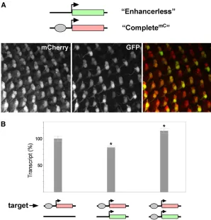
nu-cleus. To distinguish between these possibilities, we created a variant of our complete construct encoding the red fl uo-rescent reporter mCherry (CompletemC) so that when
CompletemC was paired with the Enhancerless construct,
we could easily observe expression incisvia redfluorescence and expression in transvia greenfluorescence. Inflies car-rying CompletemCalone at 53F, we observed prominent red
fluorescence in all R3 and R4 cells with lower levels offl uo-rescence in other cells of each ommatidial cluster, similar to the expression of GFP observed from Completegfp alone
(data not shown). We therefore focused our analysis on R3 and R4 where expression could be confidently scored. When CompletemCwas paired with the Enhancerless
con-struct at 53F, we never observed R3 or R4 cells exhibiting green fluorescence in the absence of red fluorescence (0/
switching of the enhancer between promoters occurs rapidly (seeDiscussion).
The data above support a model where promoters incis and in transwill compete for the activity of a single GMR enhancer when they are paired. As afinal test of this model, we assessed whether the presence of a promoter in trans could affect GMR activation of a promoter incis. To do so, we quantified levels of mCherry transcript generated from the CompletemCconstruct either alone or when paired with
the Enhancerless construct. Consistent with a competitive role for the promoter intrans, we observed a significant re-duction in mCherry transcript levels generated inciswhen the Enhancerless construct was paired with CompletemC
(83.3%; 95% C.I., 80.7–85.9%) relative to levels from the CompletemCconstruct alone (Figure 6B). To ensure that the
change in mCherry expression inciswas not due to possible topological changes when CompletemC is or is not paired to
another transgene, we also assessed mCherry transcript levels in flies carrying paired CompletemC and Completegfp
trans-genes, where the latter differs from the Enhancerless construct only by the addition of GMR. Notably, pairing with Completegfp
did not decrease transcript levels from CompletemC; in fact, we
observed a significant increase in mCherry transcripts by 14.5% (95% C.I., 10.0–19.4%) (Figure 6B), suggesting that the paired enhancers could augment expression from the CompletemC
promoter, perhaps via the concurrent action of one enhancer incisand the other intrans(seeDiscussion). In sum, our data support a model wherein, in each cell, an enhancer has the capacity to share its activity between promoters in cisand in trans, with an overall bias toward the promoter incis.
Discussion
Using a transgenic approach, we have demonstrated several key aspects of enhancer action in trans. First, we showed that the enhancer GMR, which had never before been tested for the ability to act intrans, can activate thehsp70promoter on a paired homolog, thereby adding a new enhancer– promoter pair to a growing list of those that can commu-nicate intrans. Second, because our transgenes carried only a simple enhancer, minimal promoter, and reporter sequen-ces derived from other organisms, it is unlikely that putative transvection-mediating sequences are required for an en-hancer to act in trans once homologs are paired. Third, because our approach provided single-cell resolution, we were able to show that GMR action in trans results in a variegated pattern of expression across the cells of the eye disc. Fourth, by simultaneously presenting GMR with multiple promoter targets, we showed that the enhancer can activate promoters incisand intranswithin the same cell. Finally, the use offluorescent reporters permitted us to precisely quantify relative levels of gene expression in the presence of varying regulatory components and to demon-strate that promoters in cis and in trans will each compete for the activity of an enhancer.
Although we expected GMR action in transto produce lower levels of transcript than those resulting from expression incis, we were surprised at the large divergence between the two modes of activation, with an50-fold difference detected at two different loci. Importantly, due to the variegated pat-tern of expression in discs where GMR acts intrans, the levels of GFP transcript assessed by quantitative RT-PCR represent Figure 5 A promoter incisdecreases GMR action intrans. (A) Top, schematic showing the Enhancerless construct paired with CompletelacZ. This combination of paired alleles produces variegated GFPfluorescence when inte-grated at 53F or 37B. (B) Relative quantification of GMR activation of GFP intranswith and without a promoter in
cisby quantitative RT-PCR. For both 53F and 37B, total levels of GFP transcript generated from the Enhancerless construct are decreased in discs with an intact promoter in
an average of mRNA from cells with little to no detectable expression and those with robust expression. It is possible that aspects of our transgenes could be optimized for higher ex-pression intrans; for example, we designed our transgenes to encode different proteins to differentiate expression incisand intrans, and the lack of homology over the coding regions may adversely affect the overall strength of trans-activation. How-ever, our results may be consistent with existing data from the
yellowlocus. Here, promoter mutations show that full pheno-typic complementation can be achieved with 11% of wild-type levels of yellowtranscript generated in cis, implying that en-hancer action intrans, which shows only partial complemen-tation, contributes an even lower amount of transcript (Morris et al.2004). In contrast, quantitative analyses of transvection at the genes encoding Gpdh and Men suggest that their enhancers can contribute up to 100% of normalcis-activity via enhancer action intrans, but that strength oftrans-activity varies greatly with allele structure (Gibsonet al.1999; Lum and Merritt 2011). Thus, it is likely that local chromosomal environment and enhancer identity are important factors in determining the strength of an enhancer’strans-activity.
GMR action in trans is variegated
When GMR acts in trans, the majority of R4 cells express GFP at low or undetectable levels, with occasional jackpot cells producing robustfluorescence. The variegated pattern is specific totransaction; we saw no evidence of variegation for any transgenic construct where a reporter was controlled
by an enhancer incis, including a construct where GMR was separated from thehsp70promoter by 3 kb. The latter con-struct, Complete+3.0, resulted in diminished levels of target
gene expression relative to Completegfp, suggesting reduced
transcriptional activation with increasing distance between the enhancer and the promoter as previously described (Dobi and Winston 2007). Although Complete+3.0did not show evidence
of variegation, in other cases, long-distancecis-interactions be-tween enhancers and promoters may produce variegated ex-pression, as previously reported for a transgenic insertion into the engrailed locus (Kwonet al.2009). It should be noted that some topological aspects of expression in cis may be dis-tinct from those required for enhancer action intrans, com-plicating straightforward comparisons of expression from the Complete+3.0transgene to that from paired
Enhancer-less and PromoterEnhancer-less constructs; for example, the intervening sequence between an enhancer and a promoter in cis must bend to permit enhancer–promoter contact (reviewed by Bulger and Groudine 2011), but no such intervening sequence exists between enhancers and promoters carried on different chromosomes. Thus, aspects of enhancer action intrans, in-cluding local topology and patterns of gene expression, are distinct from long-distance interactions incis.
b-globin genes, which are encoded on a different chromo-some (Noordermeeret al.2011). In this system, single-cell analysis showed that only a small number of cells have ele-vated levels of endogenousb-globin mRNA, suggesting sto-chastic activation of the nativeb-globin promoter. Similarly, expression of IFN-bin response to viral infection in humans is stochastic, with only a subset of the cells of a population showing detectable transcript (Zawatzkyet al.1985). A re-cent analysis has led to a model wherein IFN-bexpression depends on interchromosomal associations between multiple unlinked loci, which occur only in a subset of cells (Apostolou and Thanos 2008). In the case of GMR action intrans, cell– cell variability in the proximity of GMR and the hsp70 pro-moter may account for the variegated pattern of expression.
Because the majority of prior studies of transvection have focused on developmental phenotypes affecting adult struc-tures, it is not yet clear whether cell–cell variability in expres-sion will be common to other Drosophilagenes that undergo transvection. One prior study used immunostaining to compare levels ofUbxexpression directed by theContrabithorax1(Cbx1)
regulatory insertion in cis and in trans (Castelli-Gair et al. 1990). While this study demonstrated that expression intrans was limited to a reduced population of wing disc cells relative to expression incis, it is not clear whether this change reflects variegation or some other patterning change. It is likely that variables including local pairing dynamics, efficiencies of pro-moter activation and transcriptional elongation, and mRNA or protein stability will strongly influence whether variegation will be a consequence of enhancer action intrans.
Cis- and trans-promoters compete for enhancer activity
Our data show that GMR is biased toward a promoter incis relative to a promoter in trans, as evidenced by its relative strength of action incis vs. transas well as the strong reduction intrans-activation when acis-promoter is present. Given that the sequences of the promoters in our experiments were iden-tical, this bias likely reflects the closer proximity of the pro-moter incis, consistent with other studies relating distance to promoter competition (Dillonet al.1997; Kmitaet al.2002). We also found that a promoter intransnegatively affects ex-pression from a promoter incis, supporting a competitive role for thetrans-promoter to the detriment of thecis-promoter. To our knowledge, this represents the first demonstration of a negative impact on expression incisby a promoter intrans. We speculate that other transgenic insertions of strong promoters may attract the activity of compatible enhancers located nearby on a paired homolog, analogous to their rec-ognized ability to trap enhancer activity in cis (O’Kane and Gehring 1987).
Prior genetic analyses have shown that, for many genes, enhancer action intransis reduced in the presence of a pro-moter in cis (Martinez-Laborda et al. 1992; Casares et al. 1997; Gohl et al. 2008). Our data are consistent with the model that this reduction results from an enhancer sharing its activity between the two promoters. An apparent ex-ception occurs at the well-studiedyellowgene, where the
presence of a functional promoter incisabolishes all evidence of enhancer action intrans, a phenomenon termed“cis-preference” (Geyeret al.1990; Morriset al.1998, 1999). While this may reflect a novel mechanism, it is possible that cis-preference results from a highly skewed competition between promoters incisand intranssuch thattrans-interactions, while permitted, are so reduced that the few transcripts resulting from enhancer action intranshave no impact on phenotype. According to this reasoning, the ratio of enhancer activity between promoter targets incisand intrans, and thus the overall availability of an enhancer to act in trans, may vary among different enhancers and/or loci. Excitingly, these concepts can be eas-ily tested using a transgenic system similar to that used here. Our study demonstrates that a single enhancer can activate promoters incisand intransin the same cell. Due to the time delay required for detection of fluorescence and the perdur-ance of GFP and mCherry following translation, we were un-able to determine whether GMR periodically switches from one target to the other over the time that we assayed or, alternatively, whether transcription is actively promoted simultaneously from both promoters in those cells expressing reporters in cis and in trans to the enhancer. We assayed relative fluorescent intensities of newly committed R4 cells whenfluorescence wasfirst detectable and found no evidence that the intensity of one channel was negatively correlated with that of the other channel (data not shown), suggesting that, if switching takes place, it must do so on a relatively short timescale. An analysis using reagents and strategies better suited to short-term dynamics, including observa-tions of nascent transcripts (Kosman et al. 2004) or the use of destabilized fluorescent reporters (Li et al. 1998), may help to differentiate between these possibilities.
Finally, we note that our quantitative data show a signif-icant elevation of expression from our CompletemCreporter
when paired with the Completegfptransgene that carries its
own enhancer and promoter, suggesting a possible synergis-tic effect when“Complete”genes are paired. This synergism could result from the activities of both enhancers upon the promoter of the CompletemCconstruct, as would be predicted
if both enhancers acted on promoters in cis and in trans simultaneously. It is also possible that the nearby Completegfp
Transvection as a model for interchromosomal interactions
The study of transvection inDrosophilaenjoys a rich history of genetic and cytological studies from some of the pioneers of genetics. Our recognition of the prevalence of interchro-mosomal interactions has continued to grow in other model systems, expanding the possibilities of potential effects on gene regulation resulting from interactions in trans. While the mechanisms that identify and pair homologs in Drosoph-ilaare not yet fully understood, they are likely to differ from many of the forces that bring together nonhomologous loci in other systems such as vertebrates. However, irrespective of the pairing mechanism, once chromosomes are brought in close proximity, the potential fortrans-interactions between regulatory sequences is likely similar in different organisms, particularly given the strong evolutionary conservation of mechanisms for gene regulation incis. Transgenic analyses of transvection in Drosophilahave the potential to address the capacity of diverse enhancers and promoters to interact intransand allow precise quantification of subtle effects on gene expression. Furthermore, the varying dynamics of pair-ing at different genomic locations in theDrosophilagenome (Fung et al. 1998) afford the ability to contrast effects on transvection when chromosomes are stably or poorly paired. Thus, detailed analyses of transvection have the potential to inform our understanding of interchromosomal interactions in diverse systems.
Acknowledgments
We thank Jeff Settleman for the gift of pGMR, Chris Smith at the Mount Desert Island Biological Laboratory DNA Se-quencing Core, and the BloomingtonDrosophilaStock Cen-ter. We specially thank Ting Wu, Pam Geyer, Judy Kassis, Lori Wallrath, and Gio Bosco for stimulating conversations and ideas. This work was supported by grants from the Na-tional Center for Research Resources (5P20RR016463-12) and the National Institute of General Medical Sciences (8 P20 GM103423-12) from the National Institutes of Health.
Note added in proof: In this issue of GENETICS, Mellert and Truman (pp. 1129–1141) describe an independent study of enhancer action intransbetween transgenes inserted using site-specific transformation methods. In agreement with our data, they demonstrate that promoters incisand intransto an enhancer will compete for the enhancer’s activity, with a bias toward the promoter in cis. Furthermore, they report that enhancer action intransis limited to a smaller group of cells relative to activation incis, with evidence of stochastic activa-tion intransfor several enhancers.
Literature Cited
Akbari, O. S., E. Bae, H. Johnsen, A. Villaluz, D. Wonget al., 2008 A
novel promoter-tethering element regulates enhancer-driven gene
expression at the bithorax complex in the Drosophila embryo.
Development 135: 123–131.
Apostolou, E., and D. Thanos, 2008 Virus infection induces
NF-kappaB-dependent interchromosomal associations mediating
monoallelic IFN-beta gene expression. Cell 134: 85–96.
Bacher, C. P., M. Guggiari, B. Brors, S. Augui, P. Clerc et al.,
2006 Transient colocalization of X-inactivation centres
accom-panies the initiation of X inactivation. Nat. Cell Biol. 8: 293–299.
Bantignies, F., and G. Cavalli, 2011 Polycomb group proteins:
re-pression in 3D. Trends Genet. 27: 454–464.
Bartkuhn, M., and R. Renkawitz, 2008 Long range chromatin
in-teractions involved in gene regulation. Biochim. Biophys. Acta
1783: 2161–2166.
Bateman, J. R., and C. T. Wu, 2008 A simple polymerase chain
reaction-based method for the construction of
recombinase-mediated cassette exchange donor vectors. Genetics 180: 1763–
1766.
Bateman, J. R., A. M. Lee, and C. T. Wu, 2006 Site-specific
trans-formation of Drosophila via phiC31 integrase-mediated cassette
exchange. Genetics 173: 769–777.
Bischof, J., R. K. Maeda, M. Hediger, F. Karch, and K. Basler,
2007 An optimized transgenesis system for Drosophila using
germ-line-specific phiC31 integrases. Proc. Natl. Acad. Sci. USA
104: 3312–3317.
Bulger, M., and M. Groudine, 2011 Functional and mechanistic
diversity of distal transcription enhancers. Cell 144: 327–339.
Calhoun, V. C., and M. Levine, 2003 Long-range
enhancer-promoter interactions in the Scr-Antp interval of the Drosoph-ila Antennapedia complex. Proc. Natl. Acad. Sci. USA 100:
9878–9883.
Calhoun, V. C., A. Stathopoulos, and M. Levine, 2002
Promoter-proximal tethering elements regulate enhancer-promoter
speci-ficity in the Drosophila Antennapedia complex. Proc. Natl. Acad.
Sci. USA 99: 9243–9247.
Casares, F., W. Bender, J. Merriam, and E. Sanchez-Herrero,
1997 Interactions of Drosophila Ultrabithorax regulatory regions
with native and foreign promoters. Genetics 145: 123–137.
Castelli-Gair, J. E., J. L. Micol, and A. Garcia-Bellido, 1990
Transvec-tion in the Drosophila Ultrabithorax gene: a Cbx1 mutant allele induces ectopic expression of a normal allele in trans. Genetics 126:
177–184.
Chandler, V. L., and M. Stam, 2004 Chromatin conversations:
mechanisms and implications of paramutation. Nat. Rev. Genet.
5: 532–544.
Chen, J. L., K. L. Huisinga, M. M. Viering, S. A. Ou, C. T. Wuet al.,
2002 Enhancer action in trans is permitted throughout the
Drosophila genome. Proc. Natl. Acad. Sci. USA 99: 3723–3728.
Chung, J. H., M. Whiteley, and G. Felsenfeld, 1993 A 59element
of the chicken beta-globin domain serves as an insulator in hu-man erythroid cells and protects against position effect in
Dro-sophila. Cell 74: 505–514.
Dean, A., 2011 In the loop: long range chromatin interactions and
gene regulation. Brief. Funct. Genomics 10: 3–10.
Dillon, N., T. Trimborn, J. Strouboulis, P. Fraser, and F. Grosveld,
1997 The effect of distance on long-range chromatin
interac-tions. Mol. Cell 1: 131–139.
Dobi, K. C., and F. Winston, 2007 Analysis of transcriptional
ac-tivation at a distance in Saccharomyces cerevisiae. Mol. Cell.
Biol. 27: 5575–5586.
Duan, Z., M. Andronescu, K. Schutz, S. McIlwain, Y. J. Kimet al.,
2010 A three-dimensional model of the yeast genome. Nature
465: 363–367.
Duncan, I. W., 2002 Transvection effects in Drosophila. Annu.
Rev. Genet. 36: 521–556.
Fujioka, M., X. Wu, and J. B. Jaynes, 2009 A chromatin insulator
mediates transgene homing and very long-range
Fung, J. C., W. F. Marshall, A. F. Dernburg, D. A. Agard, and J. W.
Sedat, 1998 Homologous chromosome pairing in Drosophila
melanogaster proceeds through multiple independent
initia-tions. J. Cell Biol. 141: 5–20.
Galagan, J. E., and E. U. Selker, 2004 RIP: the evolutionary cost of
genome defense. Trends Genet. 20: 417–423.
Geyer, P. K., M. M. Green, and V. G. Corces, 1990 Tissue-specific
transcriptional enhancers may act in trans on the gene located in the homologous chromosome: the molecular basis of
trans-vection in Drosophila. EMBO J. 9: 2247–2256.
Gibson, J. B., D. S. Reed, S. Bartoszewski, and A. V. Wilks,
1999 Structural changes in the promoter region mediate
transvection at the sn-glycerol-3-phosphate dehydrogenase
gene of Drosophila melanogaster. Biochem. Genet. 37: 301–315.
Gohl, D., M. Muller, V. Pirrotta, M. Affolter, and P. Schedl,
2008 Enhancer blocking and transvection at the Drosophila
apterous locus. Genetics 178: 127–143.
Gondor, A., and R. Ohlsson, 2009 Chromosome crosstalk in three
dimensions. Nature 461: 212–217.
Groth, A. C., M. Fish, R. Nusse, and M. P. Calos, 2004 Construction
of transgenic Drosophila by using the site-specific integrase from
phage phiC31. Genetics 166: 1775–1782.
Hopmann, R., D. Duncan, and I. Duncan, 1995 Transvection in the
iab-5,6,7 region of the bithorax complex of Drosophila: homology
independent interactions in trans. Genetics 139: 815–833.
Horton, R. M., Z. L. Cai, S. N. Ho, and L. R. Pease, 1990 Gene
splicing by overlap extension: tailor-made genes using the
poly-merase chain reaction. Biotechniques 8: 528–535.
Kennison, J. A., and J. W. Southworth, 2002 Transvection in
Dro-sophila. Adv. Genet. 46: 399–420.
Kmita, M., N. Fraudeau, Y. Herault, and D. Duboule, 2002 Serial
deletions and duplications suggest a mechanism for the
collin-earity of Hoxd genes in limbs. Nature 420: 145–150.
Koeman, J. M., R. C. Russell, M. H. Tan, D. Petillo, M. Westphal
et al., 2008 Somatic pairing of chromosome 19 in renal onco-cytoma is associated with deregulated EGLN2-mediated oxygen-sensing response. PLoS Genet. 4: e1000176.
Kosman, D., C. M. Mizutani, D. Lemons, W. G. Cox, W. McGinnis
et al., 2004 Multiplex detection of RNA expression in Drosoph-ila embryos. Science 305: 846.
Kwon, D., D. Mucci, K. K. Langlais, J. L. Americo, S. K. DeVidoet al.,
2009 Enhancer-promoter communication at the Drosophila
engrailed locus. Development 136: 3067–3075.
Leiserson, W. M., and N. M. Bonini, and S. Benzer, 1994 Transvection
at the eyes absent gene of Drosophila. Genetics 138: 1171–1179.
Lewis, E. B., 1954 The theory and application of a new method of
detecting chromosomal rearrangements in Drosophila mela-nogaster. Am. Nat. 88: 225.
Li, X., X. Zhao, Y. Fang, X. Jiang, T. Duonget al., 1998 Generation
of destabilized green fluorescent protein as a transcription
re-porter. J. Biol. Chem. 273: 34970–34975.
Lieberman-Aiden, E., N. L. van Berkum, L. Williams, M. Imakaev, T.
Ragoczy et al., 2009 Comprehensive mapping of long-range
interactions reveals folding principles of the human genome.
Science 326: 289–293.
Lin, Q., 2003 The promoter targeting sequence facilitates and
restricts a distant enhancer to a single promoter in the
Drosoph-ila embryo. Development 130: 519–526.
Lum, T. E., and T. J. Merritt, 2011 Nonclassical regulation of
transcription: interchromosomal interactions at the malic
en-zyme locus ofDrosophila melanogaster. Genetics 189: 837–849.
Martinez-Laborda, A., A. Gonzalez-Reyes, and G. Morata, 1992 Trans
regulation in the Ultrabithorax gene of Drosophila: alterations in the
promoter enhance transvection. EMBO J. 11: 3645–3652.
McKee, B. D., 2004 Homologous pairing and chromosome
dy-namics in meiosis and mitosis. Biochim. Biophys. Acta 1677:
165–180.
Moon, N.-S., L. Di Stefano, E. J. Morris, R. Patel, K. Whiteet al.,
2008 E2F and p53 induce apoptosis independently during
Drosophila development but intersect in the context of DNA damage. PLoS Genet. 4: e1000153.
Morris, J. R., J. L. Chen, P. K. Geyer, and C. T. Wu, 1998 Two modes
of transvection: enhancer action in trans and bypass of a chromatin
insulator in cis. Proc. Natl. Acad. Sci. USA 95: 10740–10745.
Morris, J. R., P. K. Geyer, and C. T. Wu, 1999 Core promoter
elements can regulate transcription on a separate chromosome
in trans. Genes Dev. 13: 253–258.
Morris, J. R., D. A. Petrov, A. M. Lee, and C. T. Wu, 2004 Enhancer
choice in cis and in trans in Drosophila melanogaster: role of the
promoter. Genetics 167: 1739–1747.
Moses, K., and G. M. Rubin, 1991 Glass encodes a site-specific
DNA-binding protein that is regulated in response to positional signals
in the developing Drosophila eye. Genes Dev. 5: 583–593.
Naumova, N., and J. Dekker, 2010 Integrating one-dimensional and
three-dimensional maps of genomes. J. Cell Sci. 123: 1979–1988.
Noordermeer, D., E. de Wit, P. Klous, H. van de Werken, M. Simonis
et al., 2011 Variegated gene expression caused by cell-specific
long-range DNA interactions. Nat. Cell Biol. 13: 944–951.
O’Kane, C. J., and W. J. Gehring, 1987 Detection in situ of
geno-mic regulatory elements in Drosophila. Proc. Natl. Acad. Sci.
USA 84: 9123–9127.
Robinow, S., and K. White, 1988 The locus elav of Drosophila
melanogaster is expressed in neurons at all developmental
stages. Dev. Biol. 126: 294–303.
Ronshaugen, M., and M. Levine, 2004 Visualization of
trans-homolog enhancer-promoter interactions at the Abd-B Hox
locus in the Drosophila embryo. Dev. Cell 7: 925–932.
Sandhu, K. S., C. Shi, M. Sjolinder, Z. Zhao, A. Gondor et al.,
2009 Nonallelic transvection of multiple imprinted loci is
or-ganized by the H19 imprinting control region during germline
development. Genes Dev. 23: 2598–2603.
Sexton, T., E. Yaffe, E. Kenigsberg, F. Bantignies, B. Leblancet al.,
2012 Three-dimensional folding and functional organization
principles of the Drosophila genome. Cell 148: 458–472.
Siegal, M. L., and D. L. Hartl, 1996 Transgene coplacement and
high efficiency site-specific recombination with the Cre/loxP
system in Drosophila. Genetics 144: 715–726.
Singer, S. D., J.-M. Hily, and Z. Liu, 2009 A 1-kb bacteriophage
lambda fragment functions as an insulator to effectively block
enhancer–promoter interactions in Arabidopsis thaliana. Plant
Mol. Biol. Rep. 28: 69–76.
Spilianakis, C. G., M. D. Lalioti, T. Town, G. R. Lee, and R. A.
Flavell, 2005 Interchromosomal associations between
alterna-tively expressed loci. Nature 435: 637–645.
Stevens, N. M., 1908 A study of the germ cells of certain Diptera
with reference to the heterochromosomes and the phenomena
of synapsis. J. Exp. Zool. 5: 359–374.
Williams, A., C. G. Spilianakis, and R. A. Flavell, 2010 Interchromosomal association and gene regulation in trans.
Trends Genet. 26: 188–197.
Xu, N., C. L. Tsai, and J. T. Lee, 2006 Transient homologous
chro-mosome pairing marks the onset of X inactivation. Science 311:
1149–1152.
Zawatzky, R., E. De Maeyer, and J. De Maeyer-Guignard,
1985 Identification of individual interferon-producing cells by
in situ hybridization. Proc. Natl. Acad. Sci. USA 82: 1136–1140.
Zhang, Y., R. P. McCord, Y. J. Ho, B. R. Lajoie, D. G. Hildebrand
et al., 2012 Spatial organization of the mouse genome and its
role in recurrent chromosomal translocations. Cell 148: 908–921.
Zhou, J., and M. Levine, 1999 A novel cis-regulatory element, the
PTS, mediates an anti-insulator activity in the Drosophila
em-bryo. Cell 99: 567–575.
GENETICS
Supporting Information http://www.genetics.org/content/suppl/2012/05/29/genetics.112.140954.DC1
Comparing Enhancer Action in
Cis
and in
Trans
Jack R. Bateman, Justine E. Johnson, and Melissa N. Locke
Figure S1 GMR acts in trans at polytene position 38F. GFP fluorescence is shown for discs where GMR acts in cis (A) or in trans (B-‐C). The constructs carried are (A), Completegfp alone; (B), Enhancerless construct paired with an insertion of a precursor to the Promoterless construct with a compromised promoter (see below); (C), Enhancerless paired with CompletelacZ, which carries an intact promoter. In (B), the precursor is identical to the Promoterless
construct except that the hsp70 promoter flanked by loxP sites has not been excised (See Materials and Methods). Quantitative RT-‐PCR shows that the precursor construct expresses lacZ in cis to a level of ~5% of CompletelacZ (data
not shown), indicating that the loxP sites compromise the function of the promoter. We therefore consider the expression in (B) to be analogous to that of Enhancerless paired with Promoterless.
Figure S2 Variegation of GMR action in trans is not sensitive to reduction in HP1. (A, B) Max-‐projected confocal z-‐ stacks showing discs carrying Enhancerless and Promoterless constructs at 53F in a background where Su(var)205 (encoding HP1) expression is reduced in the eye disc via the expression of a hairpin under the control of ey-‐GAL4. (A) anti-‐GFP staining only; (B) merged image showing GFP in green and anti-‐Elav staining in red. Flies carrying the hairpin P[TRiP.HMS00278] targeting Su(var)205 were obtained from the Bloomington Drosophila Stock Center. (C)
Figure S3 Lack of variegation of mini-‐white expression from RMCE targets at 53F, 37B, or 38F. Each image represents a fly carrying a single copy of the P[attP.w+.attP] target cassette, which is marked with mini-‐white. No variegation is evident.
Figure S4 Expression of lacZ in cis. X-‐gal stained discs carrying constructs CompletelacZ (top) or Promoterless (bottom)
alone at 53F. In the top disc, the staining reaction was stopped after 20 minutes, whereas for the bottom disc, staining was carried out overnight. Quantitative RT-‐PCR indicates that expression from the Promoterless construct is <1% that of CompletelacZ (data not shown).

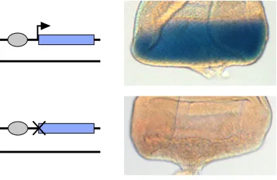
![Figure
S3
Lack
of
variegation
of
a
fly
carrying
a
single
copy
of
the
P[attP.w+.attP]
target
cassette,
which
is
marked
with
mini-‐white
expression
from
RMCE
targets
a](https://thumb-us.123doks.com/thumbv2/123dok_us/1561530.1191798/17.612.88.310.100.541/figure-variegation-carrying-single-target-cassette-expression-targets.webp)