1556-6811/11/$12.00 doi:10.1128/CVI.00500-10
Copyright © 2011, American Society for Microbiology. All Rights Reserved.
Efficacy of the Canine Influenza Virus H3N8 Vaccine To Decrease
Severity of Clinical Disease after Cochallenge with Canine
Influenza Virus and
Streptococcus equi
subsp.
zooepidemicus
䌤
Laurie J. Larson,
1Jamie Henningson,
1Patricia Sharp,
1Bliss Thiel,
1Muralidhar S. Deshpande,
2Tamara Davis,
2Huchappa Jayappa,
2Terri Wasmoen,
2Nallakannu Lakshmanan,
2* and Ronald D. Schultz
1Department of Pathobiological Sciences, School of Veterinary Medicine, University of Wisconsin—Madison, 2015 Linden Drive West, Madison, Wisconsin 53706,1and Intervet/Schering-Plough Animal Health,
21401 West Center Road, Elkhorn, Nebraska 680222
Received 18 November 2010/Returned for modification 26 January 2011/Accepted 11 February 2011
Since first emerging in the North American canine population in 2004, canine influenza virus (CIV) subtype H3N8 has shown horizontal transmission among dogs, with a high level of adaptation to this species. The severity of disease is variable, and coinfection by other respiratory pathogens is an important factor in the degree of morbidity and mortality. The first influenza vaccine for dogs, an inactivated vaccine containing CIV subtype H3N8, was conditionally approved by the U.S. Department of Agriculture (USDA) for licensure in May 2009 and fully licensed in June 2010. This study evaluates the efficacy of this vaccine to reduce the severity of illness in dogs cochallenged with virulent CIV andStreptococcus equisubsp.zooepidemicus.
Canine influenza virus (CIV) was first observed in racing greyhounds in Florida in 2004. These dogs exhibited signs of respiratory disease, ranging from fever and cough to peracute death due to hemorrhagic pneumonia. The virus isolate, A/
Canine/Florida/43/2004, shared⬎96% sequence identity with
contemporary equine influenza A virus (H3N8) (5). Sustained dog-to-dog transmission of the virus has led to the widespread distribution of CIV in the United States (13). Dogs housed in groups or exposed to transient populations of other dogs, such as dogs at the racetrack, shelter, or dog daycare environments, are at the greatest risk of exposure to CIV. These dogs are also at great risk of exposure to other respiratory pathogens, such as Streptococcus equi subsp. zooepidemicus, which has been isolated from shelter dogs with acute fatal hemorrhagic pneu-monia (12, 14). A vaccine against CIV (H3N8) has been de-veloped and was licensed in 2009 (Intervet/Schering-Plough Animal Health, Elkhorn, NE) (7). In the present study,
coin-fection with both CIV andS. equi subsp. zooepidemicus did
lead to higher morbidity in canine infectious respiratory dis-ease (CIRD) complex caused by these pathogens. On the basis of comparative clinical signs and pathology, vaccination with canine influenza H3N8 provided protection from disease in the face of dual challenges.
MATERIALS AND METHODS
Animals.Approval from the institutional animal care and use committee was obtained before the study began. Thirty-two male and female dogs aged 7 to 10 weeks and seronegative for CIV were used in this study. Animals were grouped
by litter and randomly assigned to 1 of 4 treatment groups. Group 1 contained 6 unvaccinated dogs to be challenged with CIV alone. Group 2 contained 6
un-vaccinated dogs to be challenged withS. equisubsp.zooepidemicusalone. Group
3 contained 10 unvaccinated dogs to be challenged with both CIV andS. equi
subsp.zooepidemicus. Group 4 contained 10 dogs that were vaccinated against
CIV and then challenged with both CIV andS. equisubsp.zooepidemicus. All
dogs were housed in separate rooms by group at biosafety level 2 in the isolation unit of the University of Wisconsin School of Veterinary Medicine Charmany Instructional Facility, which is an AAALAC-accredited facility. Standard animal
husbandry was practiced, and food and water were availablead libitum.
Vaccination.One group of 10 dogs (group 4) was vaccinated subcutaneously with 1 ml of inactivated H3N8 CIV vaccine containing an aluminum-based adjuvant (Intervet/Schering-Plough Animal Health) on study days 0 and 21. There was no placebo group in this study; however, the remaining 22 dogs were kept unvaccinated throughout the study.
Samples.Blood samples for serology were collected from all dogs on study days 0, 34, and 49. Nasal swabs for bacterial isolation were collected in tryptose phosphate broth from dogs in groups 2, 3, and 4 on study days 34, 38, 41, 44, 47, and 49. Nasal swabs for viral isolation were collected in Dulbecco’s modified Eagle medium containing gentamicin and amphotericin B from dogs in groups 1, 3, and 4 on study day 34 and then daily on study days 36 to 45. Swabs for bacterial isolation were always collected prior to swabs for viral isolation on the days when they coincided. On study day 49, all dogs were humanely euthanized, lungs were scored for consolidation (pneumonia), and samples of lung tissue were collected into formalin for histopathologic examination. Sterile saline was infused into large bronchi and collected for bacterial isolation at this time as well. Individuals handling samples and scoring lung lesions were not aware of the study groups.
Viral challenge. Canine influenza virus isolate A/Canine/Florida/14/2006 (H3N8) previously shown to be virulent in susceptible dogs (6, 11) was prepared
to contain 7.5 log1050% tissue culture infective dose (TCID50) per dose. All dogs
in groups 1, 3, and 4 were challenged with virus given by aerosol on study day 35.
Bacterial challenge.Two isolates ofS. equisubsp.zooepidemicuswere ob-tained from dogs that died of severe CIRD complex in humane animal shelters in Nevada and Wisconsin. Primary culture was made on blood agar plates and
typed. The resulting colonies were frozen at⫺80°C for storage. Before
inocula-tion into dogs, these isolates were restreaked onto blood agar to ensure purity
and viability. The resultingS. equisubsp.zooepidemicuscolonies were mixed in
equal volumes, cultured for 24 h, and prepared to contain 1⫻109CFU per ml.
Using an intranasal cannula, 0.5 ml of bacterial culture was instilled into each nostril of all dogs in groups 2, 3, and 4 on study day 38, which was 3 days after the CIV challenge.
* Corresponding author. Mailing address: Intervet/Schering-Plough Animal Health, 29160 Intervet Lane, P.O. Box 318, Millsboro, DE 19966. Phone: (302) 934-4222. Fax: (302) 934-4204. E-mail: nallakannu [email protected].
䌤Published ahead of print on 23 February 2011.
559
on August 17, 2020 by guest
http://cvi.asm.org/
Clinical observations.Beginning on study day 33 and continuing until study day 49, all dogs were observed daily for clinical signs of respiratory disease and abnormal body temperature by a veterinarian who was not aware of treatment group assignments. Each clinical sign was assigned a score based on severity. Ocular and nasal discharge was scored as follows: 0 for no discharge, 0.5 for serous discharge, 1.0 for mild mucopurulent discharge, and 2.0 for severe mu-copurulent discharge. Coughs was scored as follows: 0 for no cough; 0.5 for mild cough; 1.0 for moderate, persistent cough; and 2.0 for severe cough accompanied by choking or retching sounds. Sneezing, dyspnea, and depression were scored as follows: 0 for absent and 2 for present. Body temperature was scored as follows:
0 for a temperature of⬍39.5°C and 2 for a temperature ofⱖ39.5°C.
Sample processing.Serum samples collected on study days 0, 34, and 49 were tested for antibodies against CIV by hemagglutination inhibition (HI) assay as described previously (11). Live CIV titers in nasal swabs and challenge material were determined by virus isolation and titration on Madin-Darby canine kidney (MDCK) cells as described previously (11). Bacteria were isolated from nasal swabs and lung washes via plating on blood agar, and identification to the species level was done by Gram staining and metabolic assays (16).
Pathology.On study day 49, all remaining dogs were euthanized, and lungs were evaluated for gross lesions. Each lung lobe was scored for percent consol-idation, and weighted lung lesion scores and total score for each dog were calculated as described previously (6, 7). Fresh lung tissues and lung lavage samples were collected for bacterial isolation, and lung tissues in buffered for-malin were collected for histopathology.
Statistical analysis.Median lung scores, summed clinical scores, area under the curve for virus shedding, the number of days of virus shedding, and the number of days of bacterial shedding were compared between treatment groups by Wilcoxon rank sum tests. Statistical analysis was performed using SAS version
9.1.3 (SAS Institute, Cary, NC).Pvalues of⬍0.05 were declared statistically
significant.
RESULTS
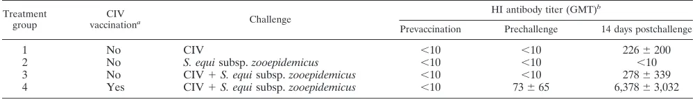
Serology. All vaccinated dogs in group 4 seroconverted to CIV following vaccination (Table 1). The hemagglutination inhibition (HI) antibody titers ranged from 10 to 160, with a geometric mean titer (GMT) of 73 just before challenge. The majority (80%) of vaccinates developed an HI titer of 40 or greater (data not shown). All nonvaccinated dogs in groups 1, 2, and 3 were CIV seronegative at the time of CIV challenge
(HI titer of⬍10). Following challenge, antibody titers in
vac-cinated dogs increased significantly (GMT⬎6,378) compared
with unvaccinated cohorts, demonstrating the efficacy of the vaccine in priming the immune system against virulent CIV. The minimum HI antibody titer after challenge was 2,560 in vaccinated dogs (data not shown). Nonvaccinated dogs chal-lenged with CIV in groups 1 and 3 seroconverted following CIV challenge, with a GMT of 226 and 278, respectively. All dogs in group 2 remained seronegative for CIV at the time of necropsy, confirming that they were not exposed to CIV and biosecurity procedures were efficient.
Clinical signs.All dogs were monitored and scored for body temperature and clinical signs of respiratory disease. In group 1 (CIV alone), 2 of the 6 dogs developed clinical fever
(ⱖ39.5°C) for only 1 day. This was in contrast to previous
experimental challenge studies (6, 7), where none of the dogs challenged with CIV developed fever. One of the 2 dogs also
developed severe depression. In group 2 (S. equisubsp.
zooepi-demicusalone), 1 of the 6 dogs developed fever for only 1 day.
In group 3 challenged with both CIV andS. equisubsp.
zooepi-demicus, 90% (9 of 10) of the dogs developed fever and a majority (70%) exhibited fever for at least 2 days. Fifty percent (5 of 10) of the vaccinated dogs (group 4) also developed fever following dual challenge; however, the fever lasted for only 1 day.
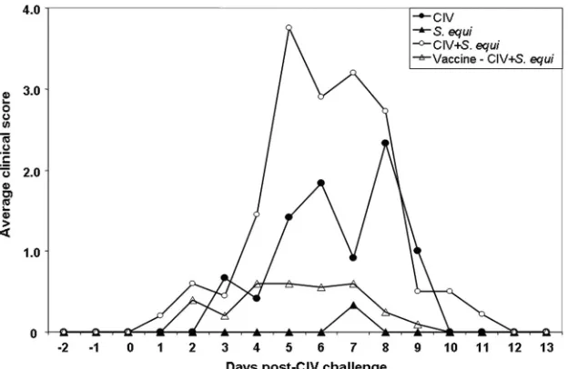
All dogs challenged with CIV and CIV plus S. equisubsp.
zooepidemicus(groups 1, 3, and 4) exhibited a range of clinical signs starting from 2 days postchallenge (Fig. 1). Coughing and dyspnea were the predominant clinical signs. All dogs in group 1 (6 dogs) and group 3 (10 dogs) exhibited various degrees of coughing, which lasted an average of 2.7 days and 4.5 days, respectively. On the other hand, only 60% (6 of 10) of the vaccinated dogs in group 4 exhibited a mild cough, which lasted an average of 1.3 days. One dog in group 1 was euthanized at 9 days postchallenge due to respiratory distress and pneumo-nia. One dog in group 3 died at 8 days postchallenge due to
severe pneumonia. None of the 6 dogs challenged withS. equi
subsp.zooepidemicusalone exhibited any clinical signs. Dogs
challenged with CIV plusS. equisubsp.zooepidemicusshowed
significantly higher clinical scores than dogs challenged with
CIV alone (median score of 17.8 versus 7.0, respectively;P⫽
0.012). The CIV vaccine significantly reduced the clinical
scores induced by CIV plusS. equisubsp.zooepidemicus
(me-dian score, 2.5; P ⬍ 0.0001). The results suggest that CIV
causes severe respiratory disease in dogs that could lead to
mortality. S. equi subsp. zooepidemicus acts as a secondary
pathogen and enhances CIV-induced disease. In this study,S.
equisubsp.zooepidemicus did not induce any clinical disease
on its own.
Virus shedding.Nasal virus shedding was monitored in all CIV-challenged dogs (groups 1, 3, and 4) on the day before challenge and then every day from day 1 through day 10 post-challenge. The average virus titer for each group, expressed as
log10TCID50/ml, was plotted against days postchallenge (Fig.
2). Fifty percent of the dogs in groups 1 and 4 and 100% of dogs in group 3 started shedding CIV in nasal secretions from day 1 postchallenge. By day 2 postchallenge, all dogs in groups 1, 3, and 4 were positive for nasal virus shedding. Viral shed-ding in groups 1 and 3 reached its peak between day 4 and day 5 after challenge with CIV, followed by a precipitous drop on day 6 (Fig. 2). Peak viral shedding in group 4 was on day 2 post-CIV challenge, followed by a precipitous drop by day 3.
TABLE 1. Seroconversion in four groups of dogs
Treatment group
CIV
vaccinationa Challenge
HI antibody titer (GMT)b
Prevaccination Prechallenge 14 days postchallenge
1 No CIV ⬍10 ⬍10 226⫾200
2 No S. equisubsp.zooepidemicus ⬍10 ⬍10 ⬍10
3 No CIV⫹S. equisubsp.zooepidemicus ⬍10 ⬍10 278⫾339 4 Yes CIV⫹S. equisubsp.zooepidemicus ⬍10 73⫾65 6,378⫾3,032
aCIV, canine influenza virus.
bThe hemagglutination inhibition (HI) antibody titer (geometric mean titer关GMT兴) is shown. The mean⫾standard deviation are shown for some values.
560 LARSON ET AL. CLIN. VACCINEIMMUNOL.
on August 17, 2020 by guest
http://cvi.asm.org/
Dogs in group 4 continued to shed virus at low levels until day 7 postchallenge. Both nonvaccinates and vaccinates stopped shedding CIV in their nasal secretions by day 8 postchallenge. The mean estimates for the area under the curve were
signif-icantly higher for groups 1 and 3 than for group 4 (P⫽0.0075
and P⬍ 0.0001, respectively). The mean number of days of
viral shedding in groups 1 and 3 was also significantly higher
than for group 4 (5.7 days and 5.8 days versus 4 days; P ⫽
0.0356 andP⫽0.0033, respectively). The results demonstrate
that the CIV vaccine significantly reduced nasal virus shedding in vaccinated dogs as reported previously (7).
Bacterial shedding.All dogs in all groups were negative for
S. equisubsp.zooepidemicusshedding at the time of challenge
with CIV orS. equisubsp.zooepidemicus(Fig. 3). Interestingly,
none of the 6 dogs in group 2 challenged withS. equisubsp.
zooepidemicusalone was positive for nasal shedding ofS. equi
subsp.zooepidemicusat any time point after challenge.
How-ever,S. equisubsp.zooepidemicuswas isolated from the lung
lavage sample from 1 dog (17%) in group 2. In contrast, 100%
of the dogs in group 3, challenged with both CIV plusS. equi
subsp.zooepidemicus, were positive forS. equisubsp.
zooepi-demicusin their nasal secretions at 3 and 6 days afterS. equi
subsp.zooepidemicuschallenge; the majority (67%) continued
to shed bacteria for at least 9 days after challenge. The majority
(67%) of dogs in group 3 were also positive forS. equisubsp.
zooepidemicusin their lung washes (11 days afterS. equisubsp.
zooepidemicus challenge), suggesting that the bacteria colo-nized and multiplied in the lower respiratory tract. The major-ity (60%) of CIV-vaccinated dogs challenged with CIV plus
S. equisubsp.zooepidemicus also shedS. equi subsp. zooepi-demicus in their nasal secretions 3 days afterS. equi subsp.
zooepidemicuschallenge. From postchallenge day 6 onwards,
the number of dogs sheddingS. equisubsp.zooepidemicuswas
substantially less in group 4 compared to group 3. Only 20% of
the vaccinated dogs were positive forS. equi subsp.
zooepi-demicusin their lung lavage samples (Fig. 3).
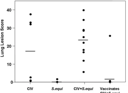
Lung pathology.Lung consolidation/pneumonia is the major pathological lesion in all influenza infections. All dogs (100%)
in the CIV and the CIV plus S. equi subsp. zooepidemicus
challenge groups (groups 1 and 3) exhibited various degrees of lung consolidation, whereas only 3 dogs (30%) in the CIV-vaccinated group (group 4) developed lung consolidation, 2 of which were very mild (Fig. 4). Only 1 dog (17%) in the group
challenged withS. equisubsp.zooepidemicusalone (group 2)
exhibited lung lesions, and they were mild. In group 1, lung lesions were predominantly observed in cranial lobes, whereas the lesions appeared to be present in all lung lobes in group 3. Lung lesions were characterized by hemorrhages and reddish consolidation and hepatization. Lung lesion scores in groups 1,
FIG. 1. Clinical disease. All dogs were challenged with CIV on study day 35 and monitored daily from day⫺2 through 13 days postchallenge for clinical signs. One dog in group 1 was euthanized at 9 days postchallenge, and one dog in group 3 died at 8 days postchallenge. The average clinical scores for each treatment group were plotted against time (days after CIV challenge).
FIG. 2. Virus shedding. All dogs in groups 1, 3, and 4 were chal-lenged with CIV on study day 35 (day 0 after CIV challenge). Nasal swabs were collected on the day before challenge (day⫺1) to confirm that the dogs were free of CIV shedding. Nasal virus shedding was monitored in challenged dogs daily for 10 days (days 1 to 10 postchal-lenge). One dog in group 1 was euthanized at 9 days postchallenge, and one dog in group 3 died at 8 days postchallenge. The average virus titers for each treatment group, expressed as log10TCID50/ml, were
calculated and plotted against time (days after CIV challenge).
on August 17, 2020 by guest
http://cvi.asm.org/
3, and 4 ranged from 0.5 to 32.97 (median score, 17.27), 5.53 to 39.74 (median lung score, 23.29), and 0 to 25.54 (median score, 0), respectively. Lung scores in group 4 were significantly lower
than in group 1 (P ⫽ 0.0019) and group 3 (P ⫽ 0.0002).
Although the lung scores in group 3 were higher than group 1,
they were not significantly different (P⫽0.5622). Data
dem-onstrate that the CIV vaccine aids in the prevention of lung
lesions induced by CIV alone and CIV plus S. equi subsp.
zooepidemicus.
Histopathologic changes were most severe and extensive in the dual-challenged, unvaccinated group. Examples of histo-pathologic lesions are shown in Fig. 5. Bronchioles showed signs of necrosis and in many instances were filled with neu-trophils (Fig. 5D). Alveolar septa were thickened, and alveolar lumens were filled with inflammatory cells. Signs of hemorrhage
were noted. In contrast, lung sections of dual-challenged, vacci-nated dogs showed mild pathology, with mild edema and slight thickening of alveolar septa in some instances (Fig. 5E). The group of dogs challenged with CIV only showed moderate pathology, including some evidence of hemorrhage and thick-ened alveolar septa (Fig. 5B); however, changes were generally less severe than those found in the unvaccinated dogs
chal-lenged with both CIV andS. equisubsp.zooepidemicus. Single
challenge withS. equisubsp.zooepidemicusproduced only mild
pathological changes (Fig. 5C), if any.
DISCUSSION
Canine infectious respiratory disease (CIRD) complex is an important disease in dogs that is caused by a number of viral and bacterial pathogens (1, 3, 4, 8, 9). In recent years, CIV and
S. equi subsp. zooepidemicus have been isolated frequently from cases of canine respiratory disease, particularly in kennels and in shelter dogs (2, 3, 5, 12, 14). This study was performed to investigate the pathogenesis of dual infection with CIV and
S. equisubsp.zooepidemicusin dogs. Additionally, an inacti-vated-CIV H3N8 vaccine, the only U.S. Department of Agri-culture (USDA)-licensed vaccine in the United States (manu-factured and marketed by Intervet/Schering-Plough Animal Health), was evaluated for efficacy in reducing the severity of
the disease caused by experimental challenge with CIV plusS.
equisubsp. zooepidemicus.
S. equisubsp. zooepidemicus is a normal inhabitant of the upper respiratory tracts of many mammals (15, 17) and has been isolated from dogs with wound infections, septicemia (15), and acute necrotizing hemorrhagic pneumonia (10).
Ad-ditionally,S. equisubsp.zooepidemicus has been isolated
fre-quently from the lower respiratory tracts of clinically healthy dogs and dogs with CIRD (3). In the current study, the dogs
experimentally infected with S. equi subsp. zooepidemicus
alone did not exhibit any clinical signs of respiratory disease.S.
FIG. 3. Bacterial isolation from nasal swabs and lung washes. All dogs in groups 2, 3, and 4 were challenged with CIV on study day 35 (day 0) and withS. equisubsp.zooepidemicuson study day 38 (day 3 after CIV challenge). Nasal swabs for bacterial isolation were collected on the day before CIV challenge (day⫺1) and 3, 6, 9, 12, and 14 days postchallenge. At the time of necropsy, lung wash samples were also collected. The results are expressed as the percentage of dogs positive forS. equisubsp.zooepidemicus.
FIG. 4. Lung lesions. The gross lung lesion scores are shown. One dog in group 1 was euthanized at 9 days postchallenge, and one dog in group 3 died at 8 days postchallenge. All remaining dogs were eutha-nized at the end of the study (study day 49). Lung lesions were scored for consolidation, and the total lesion score for each dog was calcu-lated.
562 LARSON ET AL. CLIN. VACCINEIMMUNOL.
on August 17, 2020 by guest
http://cvi.asm.org/
equisubsp.zooepidemicus could be isolated from the lung of
only 1 dog (17%) infected withS. equisubsp.zooepidemicus
alone, suggesting thatS. equi subsp.zooepidemicus does not
cause respiratory disease in healthy dogs.
However, severity of disease caused by CIV increased in the
presence ofS. equisubsp.zooepidemicus. Unlike the dogs
in-fected with CIV alone, the majority of dogs inin-fected with CIV plusS. equisubsp.zooepidemicusexhibited clinical fever that
lasted longer and had more-severe clinical signs, particularly a
cough, that lasted longer. S. equi subsp. zooepidemicus was
isolated with increased frequency in both nasal secretions and
lung wash samples of dogs infected with CIV andS. equisubsp.
zooepidemicus. Furthermore, lung lesion scores were more
se-vere in dogs infected with CIV and S. equi subsp.
zooepi-demicus. These data strongly suggest thatS. equisubsp. zooepi-demicusis an opportunistic pathogen that contributes to the
FIG. 5. Examples of histopathologic lung lesions. Lung sections from a healthy dog (A), from a dog challenged with CIV only (B), from a dog challenged withS. equisubsp.zooepidemicus(C), from a dog challenged with CIV andS. equisubsp.zooepidemicus(D), and from a CIV-vaccinated dog challenged with CIV andS. equisubsp.zooepidemicus(E) are shown.
on August 17, 2020 by guest
http://cvi.asm.org/
severity of CIRD. Clinical disease and lung lesions were more severe in dual infection compared with infection with either
CIV alone orS. equisubsp.zooepidemicusalone.
In naturalS. equisubsp.zooepidemicusoutbreaks in shelter
and kenneled dogs, lung lesions were characterized by acute hemorrhagic pneumonia (12, 14). The lung lesions observed in the current study in dual-challenged dogs were much milder than the lesions in dogs during natural outbreaks. This could be attributed to multiple factors contributing to disease and pathology in the shelter environment compared with the cur-rent study, where the dogs were housed in a clean, controlled environment.
The canine influenza virus H3N8 vaccine used in this study has previously been shown to reduce the severity of clinical disease, virus shedding, and lung lesions following experimen-tal CIV challenge (7). In the current study, the CIV (H3N8) vaccine significantly reduced the severity of clinical disease, viral shedding, bacterial shedding, and lung pathology in dogs
infected with CIV and S. equi subsp.zooepidemicus. Hence,
CIV vaccine could be used to aid in protecting dogs against CIV as well as against opportunistic bacterial pathogens, such asS. equisubsp.zooepidemicus. These findings are important because most, if not all, dogs will be infected with a variety of pathogens commonly found in the canine respiratory tract
(e.g.,S. equisubsp.zooepidemicus), and concomitant infection
with CIV could further complicate respiratory disease and may result in mortality. Therefore, vaccination of dogs at risk for infection, such as those housed in shelters or kennels or those that are frequently boarded or go to dog day care and dog parks, is the best way to protect against CIV as well as against other secondary pathogens.
To achieve protection, it is important that dogs are vacci-nated at least 3 weeks before exposure to CIV. This killed vaccine should be given in 2 doses no less than 2 weeks apart. Immunity can be expected approximately 7 days after the sec-ond dose. To ensure protection, dogs at risk for CIV, such as those that routinely spend time in multidog facilities (i.e., dog day care, boarding facilities, dog shows, other training facili-ties), should be given 2 doses of CIV (H3N8) vaccine 2 weeks apart and then held for 7 days before being placed in contact with other dogs.
ACKNOWLEDGMENTS
We thank Claudia Hirsch and employees of the Charmany Instruc-tional Facility for technical assistance and passionate caring of the animals and Diane Sweeney for assistance with the data analyses.
Funding for this study was provided by Intervet/Schering-Plough Animal Health.
REFERENCES
1.Buonavoglia, C., and V. Martella.2007. Canine respiratory viruses. Vet. Res.
38:355–373.
2.Byun, J. W., S. S. Yoon, G. H. Woo, B. Y. Jung, and Y. S. Joo.2009. An
outbreak of fatal hemorrhagic pneumonia caused by Streptococcus equi
subsp.zooepidemicusin shelter dogs. J. Vet. Sci.10:269–271.
3.Chalker, V. J., H. W. Brooks, and J. Brownlie.2003. The association of
Streptococcus equisubsp. zooepidemicus with canine infectious respiratory
disease. Vet. Microbiol.95:149–156.
4.Chalker, V. J., et al.2004. Mycoplasmas associated with canine infectious
respiratory disease. Microbiology150:3491–3497.
5.Crawford, P. C., et al.2005. Transmission of equine influenza virus to dogs.
Science310:482–485.
6.Deshpande, M., O. Abdelmagid, A. Tubbs, H. Jayappa, and T. Wasmoen.
2009. Experimental reproduction of canine influenza virus H3N8 infection in
young puppies. Vet. Ther.10:29–39.
7.Deshpande, M. S., et al.2009. Evaluation of the efficacy of a canine influenza virus (H3N8) vaccine in dogs following experimental challenge. Vet. Ther.
10:103–112.
8.Erles, K., and J. Brownlie.2005. Investigation into the causes of canine infectious respiratory disease: antibody responses to canine respiratory coro-navirus and canine herpesvirus in two kennelled dog populations. Arch.
Virol.150:1493–1504.
9.Ford, R. B.2006. Canine infectious tracheobronchitis, p. 54–61.InC. E. Green (ed.). Infectious diseases of the dog and cat, 3rd ed. Saunders-Elsevier, St. Louis, MO.
10.Garnett, N. L., et al.1982. Hemorrhagic streptococcal pneumonia in newly
procured research dogs. J. Am. Vet. Med. Assoc.181:1371–1374.
11.Jirjis, F. F., et al.2010. Transmission of canine influenza virus (H3N8)
among susceptible dogs. Vet. Microbiol.144:303–309.
12.Kim, M. K., et al.2007. Outbreak and control of haemorrhagic pneumonia
due toStreptococcus equisubspecieszooepidemicusin dogs. Vet. Rec.161:
528–530.
13.Payungporn, S., et al.2008. Influenza A virus (H3N8) in dogs with
respira-tory disease, Florida. Emerg. Infect. Dis.14:902–908.
14.Pesavento, P. A., K. F. Hurley, M. J. Bannasch, S. Artiushin, and J. F. Timoney.2008. A clonal outbreak of acute fatal hemorrhagic pneumonia in
intensively housed (shelter) dogs caused byStreptococcus equisubsp.
zooepi-demicus.Vet. Pathol.45:51–53.
15.Quinn, P. J., M. E. Carter, B. Markey, and G. R. Carter.1999. The
strep-tococci and related cocci, p. 127–136.InP. J. Quinn, M. E. Carter, B.
Markey, and G. R. Carter (ed.), Clinical veterinary microbiology. Mosby, Edinburgh, Scotland.
16.Schultz, R. D., H. W. Dunne, and C. E. Heist.1969. Effects of sublethal concentrations of penicillin G on groups C, E, K, and L streptococci. Am. J.
Vet. Res.30:1451–1459.
17.Timoney, J. F., J. H. Gillespie, F. W. Scott, and J. E. Barlough (ed.).1988.
The genusStreptococcus, p. 181–187.InHagan and Bruner’s microbiology
and infectious disease of domestic animals. Comstock Publishing Associates, London, United Kingdom.
564 LARSON ET AL. CLIN. VACCINEIMMUNOL.