FINE
STRUCTURE
MAPPING, COMPLEMENTATION,AND
PHYSIOLOGY OF ESCHERZCHZA COLZ hfl MUTANTS
JAMES W. GAUTSCH AND DANIEL L. WULFF
Department of Molecular Biology and Biochemistry, University of California at Irvine, Irvine, California 92664
Manuscript received October 17, 1973
Revised copy received March 12,1974
ABSTRACT
Six of seven hfl mutations of Escherichia coli K12, characterized by high frequencies of lysogenization by phage lambda and XcIII mutants, are shown to be tightly linked to, but not within, the purA locus. All six hfl mutations are recessive to wild type in hfi+/hfl merodiploids and all lie in a single com- plementation group, located just counterclockwise from the pur.4 locus. All six mutations confer a slightly increased resistance to penicillin and rifamycin and a slightly increased sensitivity to sodium dodecyl sulfate. Some cases of intragenic complementation and intragenic recombination were observed. It is
argued that the hfl+ gene determines the synthesis of a protein which an- tagonizes lysogenization by phage lambda. It is further argued that the func- tion of the AcIIZ gene product is to negate the antagonistic effect of this hfi+
protein.
N
1971 BELFORT and WULFF described a mutant of Escherichia coli K12 whichI
was lysogenized with very high frequencies by phage lambda and hcZZZ mu- tants. Subsequent studies showed that this mutant, called hfl-2 for high frequency of lysogeny, was also slightly resistant to penicillin and rifamycin, this latter effect being due to a change in permeability, and slightly more sensitive to so- dium dodecyl sulfate (BELFORT and WULFF 1973a). Further studies showed that the host hfl mutation was cZZZ-specific, which is to say that it acted as if it supplied a cZZZ-like function in abundance to infecting lambda phage particles (BELFORT and WULFF 1973b). But the mechanism by which it acted was unclear. One pos-sibility was that the hfl-2 mutation represented a change in function of some protein within the host so that it could substitute for the XcZZZ gene product, much in the way in which certain leuD mutations may be suppressed by gene substitu- tion (KEMPER and MARGOLIN 1969). Another possibility was that the hfl-2 mu- tation represented a loss of function, that is, a mutation from an active protein, which might be called the Hfl+ protein, to an inactive protein. The
Hfl+
protein would in some way antagonize the establishment of lysogeny by lambda, al- though the mode of action of the Hfl+ protein could be quite indirect. Accordingto this second model, it would be further argued that the normal
, W I Z
gene product promotes lysogenization by negating the antagonistic effect of this Hflfprotein. The first hypothesis, that of a change of function in the hfl-2 mutant,
436 J. W. GAUTSCH A N D D. L. WULFF
predicts that hfl mutants should be dominant over the hfl+ allele in merodiploids. The second hypothesis, that of loss of function in the hfl-l mutant, predicts that hfl mutcnts should be recessive to the hfl+ allele in merodiploids.
Another interesting facet of the earlier studies on the hfl-1 mutation was the extremely tight linkage of hfl-1 to the purA locus
(BELFORT
and WULFF 1973a). With the purA mutation in strain ES4, 100% co-transduction was observed (no recombinants in several hundred transductants) and with the purA mutation in strain KG20 there was 97% co-transduction. This raised the possibility that the hfl-I mutation lay within the purA locus itself.T o answer these questions we isolated several hfz mutants and did both fine structure mapping and complementation studies with them. The results indicate that the hfl mutation represents a loss of function and that the hfl gene lies to the left (counterclockwise) of all purA mutants tested.
MATERIALS A N D METHODS
Strains: Bacterial strains used in this study are derivatives of Escherichia coli K12; hfl+
strains are described in Table 1, haploid hfl strains in Table 2 and merodiploid hfl strains in Table 3. The methods used to obtain these strains are described more fully below.
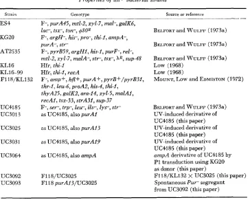
TABLE 1
Properties of hfl+ bacterial strains
Strai-1 _____
ES4
KG20
AT2535
KL16
KLI 6-99
F118/KL132
UC4185 UC3013
UC3025
UC3031
UC3064
UC3092
UC3093
Genotype Source or reference
F-, purA45, mtl-2, xyl-7, mal-, galK6, l a c , tsx-, ton-, @OR
F-, argU-, h i s , pro-, ihi-2, ampA-, purA-, s t r
F-, pyrB59, argH2, his-I, purF-, rel-, mtl-2, xyl-7, malA-, s t r , t s r , AR, sup-48 Hfr, thi-I
Hfr, thi-I, recA
F-, amp+, hfl+, purA+, pyrB+/pyrB3f, thr-I, leu-6, proAZ, his-4, thi-I,
thyA25, galKZ, ara-14, xyl-5, malAI, recAI, tsx-33, strA31, sup-37 F-, s e r , trp-, l e r , ilv-, l y s , str-
as UC4185, also purAI
as UC4185, also purAI3
as UC4185, also purAI9
as UC4185, also ampA
F118/UC3025 F118 purAI3/UC3025
BELFORT and WULFF (1973a)
BELFORT and WULFF (1973a)
BELFORT and WULFF (1973a)
Low (1968)
Low (1968)
MOUNT, Low and EDMISTON (1972)
BELFORT and WULFF (1973a) UV-induced derivative of
UC4185 (this paper) UV-induced derivative of
UC4185 (this paper) UV-induced derivative of
UC4185 (this paper)
ampA derivative of UC4185 by
PI transduction using KG20 as donor (this paper)
GENETICS OF hfl MUTANTS
TABLE 2
hfl deriuatiues of strain UC4185
43 7
Column I* hfl strains Strain hfl allele
UC2014 1
UC3413 53
UC2168 58
UC2043 59
uc204.F 65
UC2169 66
UC2167 29
Column 2:
k f l puril strains Strain purA allele
_ _ _ ~
UC3035 23
uc304.0 28
UC3041 29
UC3043 31
uc301.5 33
Column 3%
kfl purA ampA strains
__
Strain
UC3V58 UC3414 UC3MO Uc3061 UC3062 UG30863
~~
* The strains in Column 1 are all hfl strains derived from UC4185 by NG mutagenesis, with the exception of strain UC3413. Strain UC3413 is a Trp+ Hfl- R i f recombinant of the cross Hfr KL16 x UC3403. Strain UC34Q3 was derived from UC4185 by NG mutagenesis. (All strains have the same growth factor requirements as UC4185 except UC2014, which is also wr and p a n , and UC3413, which is trp+.)
t
The strains in Column 2 are all UV-induced purA mutants of the corresponding strains i n Column 1.$ The strains i n Column 3 are ampA purA derivatives of the corresponding strains in Column 2. They were derived by PI transduction, using UC3064 as donor and selecting for Amp-. Strain UC3414 was derived from UC3413 by P1 transduction, using strain UC3057 as donor and selecting for Amp-. It is hfl-53 purAl9 ampA.
Media: TB broth, LB broth, EMBO agar, and TC buffer have been described previously (BELFORT and WULFF 1973a). For isolation of purA mutants we used a medium of 56/2 buffer
(Low 1973) plus glucose (0.4%) and required supplements. For other phases of this study re- quiring buffer or minimal medium we used the medium E of VOGEL and BONNER (1956), with
or without glucose (0.4%) and required supplements. Supplements were at the following con- centrations: amino acids, 20 ,pg/ml; bases, 10 pg/ml; vitamins, 0.1 pg/ml. Agar agar #3 (Con- solidated Laboratories, Chicago), autoclaved separately, was added to a final concentration of
1.0% for solid minimal media. Adenine (IO pg/ml) was added to TB broth and LB broth for growth of Pur- strains. In growing P1 transducing stocks on purA mutants, adenine was added to LB plates and LB top agar to a concentration of 40 pg/ml. Penicillin G (potassium salt) was added to a concentration of 75 pg/ml to TB agar or minimal agar for testing the ampA marker. Isolation of additional Hfl- mutants: Seven Hfl- mutants, all deirved by N-methyl-“-nitro-
TABLE 3
Construction of strains with hfl markers on the F118 episome
Method of obtaining hfl
marker on the episome‘
Strain Genotype
Method P1 donor Recipient
1 or 2 strain strain UC3097 F118 ampA hfl-l/UC3035 1 UC3058 F118/UC3035 UC3095 F118 amp+ h/i’-53/UC3025 2 UC3413 F118 purA13/UC3095 UC30S6 F118 amp+ h/i’-58/UC3025 2 UC2168 F118 purA13/UC3025 UC3098 F118 ampA hfl-59/UC3035 1 UC3061 F118/UC3035 uc3099 F118 ampA h/i’-SS/UC30U, 1 UC3062 F118/UC3040 UC3101 F118 ampA hfl-&/UC3045 1 UC3063 F118/UC3M
438
N-nitrosoguanidine (NG) mutagenesis of Hfl+ strain UC1.185, were included i n the study. Strain UC2014, which contains hfl-I, is the original Hfl- mutant (BELFORT and WULFF 1971) and five of the six additional Hfl- mutants were isolated by D. WIEBE in this laboratory by the same method. The remaining Hfl- mutant, strain UC31.33, was found among NG-induced Rif- mutants of strain UC4185. It grew very slowly and was crossed with Hfr strain KL16 to yield a fast-growing Trpf Hfl- Rif- recombinant, designated strain UC3413. The Hfl- and Rif- characters of this strain transduce independently of each other and therefore represent two independent mutational events.
Measurement of percent lysogeny: Stationary phase host strains were centrifuged and re- suspended at 2
x
10s cells/ml in 0.02 M MgCl,. Equal volumes of cell suspension and phage in TB broth were mixed and, after 30 minutes absorption at 30". anti-X serum was added to a final Kt of 5. The infected cells were diluted and plated on TB plates at 37" by the soft-agar method, using strain C600 as indicator bacteria, to enumerate the number of infective centers. T o deter- mine the number of lysogens the diluted infected cells were also plated on EMBO plates which had been seeded with ca. IO9 Xc126 particles per plate. After 48 hours at 30", the white lysogenic colonies were easily distinguished from the darker non-lysogenic colonies ( GOTTESMAN and YARMOLINSKY 1968). All experiments were done with phage carrying the temperature sensitive cI857 repressor, which is active at 30" but not 37".PI transductions: These were done as described previously (BELFORT and WULFF 1973a), ex- cept that recipient strains were first grown in a medium containing medium E, glucose (0.4%),
required supplements and 1% LB broth, and transductants were selected on solid medium con- taining medium E buffer instead of 56 buffer. If transductants which were non-lysogenic for phage P I were required, the multiplicity of infecting P I transducing phage particles per re- cipient cell was reduced from 10 to between 0.1 and 1. (Non-lysogenic transductants are more frequent at low multiplicities of infection (LURIA, ADAMS and TING (1960.) For transduction in which Amp- was the selected marker, TB broth and TB agar plus penicillin was sometimes sub- stituted f o r minimal medium and minimal agar plus penicillin.
Testing selected colonies for the Hfl phenolype: This was done as described previously using Xclll phage (BELFORT and WULFF 1973a) except that about 200 phage particles in .02 M MgC1, and bacterial colonies resuspended in TB broth were mixed for 30 minutes at room temperature for adsorption, followed by addition of 1 ml TB top agar and pouring onto small (15 X 60 mm) plates containing TB agar.
Isolation and mapping of purA mutants: Strains were grown in supplemented minimal me- dium to approximately 108 cells/ml, irradiated with ultraviolet light to about 1% survival, di- luted IO-fold into supplemented minimal medium plus adenine and aerated for 8 hours to allow growth and heterozygote segregation. Mutants with an absolute requirement for adenine are purA or pur& whereas adenine-requiring mutants in other loci are satisfied by guanine as well (STOUTHOMER, DE HAAN and NIJKAMP 1965). The cultures were washed by centrifugation and suspended i n supplemented minimal medium plus guanine (2.5 pg/ml) at IO7 cells/ml and aerated for two hours. Penicillin was added (75 pg/ml) and the cultures aerated for 18 hours. Survivors were plated on minimal agar containing a limiting amount of adenine (1 pg/ml). Following incubation for 2-3 days, small colonies were transferred to minimal agar containing adenine (10pg/ml) and minimal agar containing guanine (10 pg/ml) with sterile toothpicks. About 10% of the small colonies which survived the penicillin selection had an absolute require- ment for adenine when the starting strain was UC4185. However, because of varying degrees of penicillin resistance among the hfl strains, adenine-requiring mutants of hfl strains repre- sented 1 % to less than 0.1 % of the small colony survivors of penicillin selection.
Isolation of recA strains: To obtain reCA derivatives of various bacterial strains, the Hfr recA strain KL16-99 was mated with appropriate recipient strains in LB broth as described previously
(BELFORT and WULFP 1973a), followed by selection for Lys+ S t r or T r p + S t r . Recombinants were judged to be of the RecA- phenotype if they (1) showed increased UV sensitivity and/or (2) allowed XcI26 but not hbiolO to form plaques when used as indicator bacteria (SIGNER, MANLY and BRUNSTETTER 1969).
GENETICS OF hfl MUTANTS 439
Freshly grown F-prime and F- colonies were transferred by sterile sticks to 0.15 m l medium E
buffer to a concentration of about lO*-lO9 cells/ml. About 0.02 m l of each suspension was spread (by 0.1 ml pipettes a t right angles and crossing each other) on a minimal agar designed to allow only the desired mating result to grow. The new merodiploids appeared as isolated colonies below the area of the cross, and as confluent growth a t the junction. The resulting merodiploids were further purified by streaking on the same medium.
It was discovered that F’ ampA/curzpA+ merodiploids have an A m p - phenotype, which al- lowed us to select for the ampA marker in F-prime matings. However, when selection was for
AmpA- in this type of mating, it was found, in most cases, that if the F-prime donor carried ampA purA on the episome, no transfer would occur. The reason for this is unknown, but it may be related to a similar problem in PI-mediated transduction (ERICKSSON-GRENNBERG 19681, where ampA purA donor strains did not generate ampA purA transductants. If the F-prime donor was ampA purA+, and selection was for ampA, transfer and growth of the resulting merodiploid occurred at normal frequencies.
Charcoal-filtered casamino acids, which contain subminimal amounts of aromatics, were added to selection plates (0.4%) in instances where tryptophan and adenine were used i n selec- tion. This decreased the time necessary for merodiploid growth from 2-3 days to 1 day.
Construction of F-prime strains with episomal hfl markers: Two different methods were used: In method 1, strains of the form F118/hfl-y purA trp recA+ were made by crossing F118/132 with the corresponding F- hfl-y purA trp recA+ strain and selecting for Pur+. These mero- diploids were then used as recipients in P1 transduction, using an ampA hfl-x purA donor P1
phage stock and selecting for Amp-. Transductants which were Amp- Hfl- Pur+ were tested for the ability to transfer a n F118 amPA hfl-x purA+ episome to a n F- recipient which was ampilt. hfl-y purA+ trp+ recA, where selection was for Amp- Trp+. Strains with this capabil- ity (Table 3) were then used as donor strains to construct merodiploids of the form F118
hfl-x/hfl-z recA f o r complementation tests.
In method 2 we obtained a rare PI18 purAlS/purAl3 segregant from a n F118/purAl3
merodiploid by the method of Low (1973). The frequency of P u r segregants was about 1 in 250, but only 1 of 200 Pur- segregants turned out to be a suitable F-prime donor. This PI18 purAl3/purAl3 merodiploid was then used as a recipient in P1 transduction, using ampA+ hfl-x purA+ donors and selecting for P u r + . Transductants were tested €or the ability to transfer a n F118 a m p A f hfl-x purA+ episome to a n F- recipient which was ampA+ hfl-y purA trp+ recA, where selection was for Pur+ Trp+. Strains with this capability (Table 3) were then used as donor strains to construct merodiploids of the form Fl18 hfl-x/hfl-z recA for Complementation tests.
RESULTS
Properties of hfl strains and linkage to purA: All six additional
Hfl-
mutants isolated in this study show increased frequencies of lysogenization upon infection by lambda and lambdacZZZ
mutants. However, none shows as large an increase as with the original hfl-l strain, UC2014. At an average multiplicity of infection of 0.4 phage per bacterium, XcZ857cZZZc0, lysogenized strain UC4185 ( h f l f ) at a frequency of 0.8% and strain UC2014 (hfl-1) at 61%. Four of the six additionalHfl-
strains (with alleles hfl-53, hfl-58, hfl-59 and hfl-66) were lysogenized at frequencies ranging from 15% to 39%. The remaining twoHfl-
strains were lysogenized at considerably lower frequencies, with strain UC2167 (hfl-29) at 3.8% and strain UC2044 (hp-65) at 2.2%. Lambda wild type, AcZZZco, and hc17440 J. W. GAUTSCH A N D D. L. WULFF
TABLE 4
Co-transduction of hfl markers with purA markers in struins ES4 and KG20
Donor s'rain Percent
Pur" which are
Hfl- with recipient strains
uc2014 UC2167 UC3413 UC2168 UC2043 UC2044 UC2169
hfl allele
1
29
53 58 59
65
66
ES4
100 0 96 88 96 85
94
KG20
91
81. 83 98 97 77
ES4 and 97% co-transduction with the purA marker of strain KG20 (BELFORT and WULFF 1973a). We therefore tested the six additional
Hfl-
strains for linkage to these two purA markers. All but one of the Hfl- mutants were found to be tightly linked to the purA locus, with co-transduction frequencies ranging from 77% to 98% (Table 4 ) . The remaining Hfl- mutant, strain UC2167 (hfl-29) was unlinked to purA. It was lysogenized at a frequency of only 3.8% by xcZ857 clllco, and has not been characterized further.The original hfl-1 mutation, when transferred into strain ES4 by phage P1- mediated transduction, conferred a slight increase in rifamycin resistance and penicillin resistance and a slight increase in sensitivity to sodium dodecyl sulfate
( BELFORT and WULFF 1973a). Each of the other five purA-linked Hfl- mutations
was tested in this regard after transfer to strain ES4. All were found to behave like the hfl-l mutation.
Functional independence of the hfl and purA genes: With such high co-trans- cluction frequencies between the hfl locus and the purA locus, we envisioned the possibility that the hfl mutants lay within the purA gene. Yet hfl could not repre- sent a mutation to loss of the purA function, for none of the Hfl- strains required adenine for growth. In addition, of some 50 independently isolated UV-induced purA mutants, none showed an Hfl- phenotype The possibility remained, how- ever, that the Hfl- mutation was to be associated with a change of the purA func- tion which did not impair the ability of the purA gene product to function in the adenine biosynthetic pathway. If this were the case, then mutation of an
Hfl-
PurA+ strain to PurA- should concurrently result in reversion of the Hfl- pheno- type to Hfl+. Such an expectation, however, was not fulfilled: Using penicillin selection, a total of 15 UV-induced adenine-requiring mutants were isolated in five of the six purA-linked hfl mutant strains. All of these mutants retained their Hfl- phenotypes. Thirteen of these adenine-requiring mutants were tightly linkedto the hf2 locus by P1-transduction and could be classified as purA mutants
( Table 5). This classification was verified for the two purA derivatives of strain
GENETICS OF hfl MUTANTS
TABLE 5
Isolation of purA hfl muianis
44
1
Starting strain
hfl allele
Number of
adenine-requiring
mutants isolated Hfl- phenotype Number with
Number tightly linked to hfl-locus
by P1 transduction
uc2014 1
UC3413 53
UC2168 58
UC204.3 59 u c 2 w 65
UC2160 66
Total 15
3 0 4
1
2 5 15
__
2
0 4
1
2
4 13 ~
cessful in isolating adenine-requiring mutants of strain UC3413 (hfl-53), but found that a n ampA hfl-53 purA derivative of this strain obtained by Pl-trans- duction retained the
Hfl-
phenotype.Mapping hfl mutants: Mapping was by P1 -transduction with donors which were ampA hfl purAx and recipients which were amp+ hfl+ purAy, where purAx and purAy are two different purA mutations which are separable by recombina- tion. Selection was for Pur+ and distribution of the unselected Hfl and Amp phenotypes was noted. The expected results are shown in Figure 1. If purAx lies to the right of purAy, most of the Pur+ transductants will be
Hfl- if
the order is ampA-hfZ-purA and Hfl+ if the order is ampA-purA-hfZ. IfpurAx lies to the left of purAy, just the opposi:e results are expected. (The purA mutants were first ordered with respect to ampA with reciprocal 3 factor crosses by the method of MARGOLIN ( 1963). With two hfZ mutants, hfl-58 and hfl-65, the cross was of the form shown in Case 2 of Figure 1. I n both cases a majority of recombinants were Hfl+ (Table 6), indicating a map order of ampA-hfl-purA. With three hfl mu- tants, hfl-1, h@-59 and hfl-66, the cross was of the form shown i n Case 1 of Figure 1. I n all three cases a majority of recombinants wereHfl-
(Table 6) indicating the same map order: ampA-hfl-purA. The distribution of the outside ampA marker among Pur+ recombinants in these crosses is also consistent with this map order (Table 6). Mutant hfl-53 was not mapped with respect to ampA and purA.W e have never observed a recombinational event between the purA mutation in strain ES4 and the hfl-1 locus, even after screening several hundred trans- ductants. W e therefore wished to know if the purA locus in ES4 lies at the left- hand extremity of the purA gene. Unfortunately we were unable to map the ES4 purA gene using ampA as a n outside marker, as we did with the other purA mu- tants, because we were unable to grow a stock of transducing phage on a n ampA ES4 derivative. However, we could tell if the ES4 purA marker lay to the right
442 J. W. GAUTSCH A N D D. L. WULFF
purAx
R D
: ampA+ PurAY
CASE I ampA
-
hf I-
purAa r A
,
I
p r A x hflampA+ P U ~ A Y R
CASE I ompA-purA-hfl
a 7 A h:f' pyrA;!
,
R
ampA+ P r A Y
CASE 2 ampA-hfl-purA
amp A purAx hfl
D
R
I ;
\
,"PA+ Pur AY CASE 2 ampA
-
purA- hf IFIGURE 1.-Mapping hfl mutants using purA donors and recipients. In Case 1 the recipient purA allele is closer to ampA than the donor purA allele. In Case 2 the donor purA allele is closer to ampA. Mapping by this method requires prior ordering of the purA alleles with respect
to ampA.
This indicates that the purA marker of ES4 lies to the right of both purA28 and purA3I. The failure to observe crossovers between the purA of ES4 and hfl-1 therefore would seem to be due to some kind of crossover suppression phenome- non, rather than an extremely close proximity of these two loci.
Properties of hfl+/hfl merodiploids: Having established that the purA-linked hf2 mutants lay outside of and to the left of the purA locus, we next wished to establish whether hfl was dominant o r recessive to hfl+ in merodiploids. The F118 F-prime factor described by Low (1973) spans a 5-minute region of the E coli chromosome from pyrB to malB, including the purA, hfl and ampA loci, and is ideal for this purpose.
GENETICS O F hfl MUTANTS
TABLE 6
Ordering of hfl markers with respect to ampA and purA
443
Unselected markers Donor or Case 1 or 2 Selected
Strain Genotype” recipient (Figure 1 ) marker Hfl Amp Number
UC3058
Uc3031
UC3061 UC3013
u c 3 w 3 UC3031
UC306Q UC3031
UC3062 UC3031
ampA hfl-I purA23 amp+ hfl+ purA19
ampA hfl-59 purA29 amp+ hfl+ purAl
ampA hfl-66 purA33 amp+ hfl+ purA19
ampA hfl-58 purA28 amp+ hfl+ purA19
ampA hfl-65 purA31 amp+ hfl+ purAl9
D R D R D R D R D R
1 Pur+ -
+
+
-
1 Pur+ -
+
+
1 Pur+ -
+
+
+
2 Pur+
+
I
2 Pur+
+
-
-
29 9 10 0 32 6 8 02 $4
9 12 3 23 4 12 0 29 4 13 2
* The order of t h e purA alleles was first determined by the method of MARGOLIN (1963). It is ampA-(purA28, purA3i) -purAi9-purAi- (purA33, purA23) -purA29.
TABLE 7
Ordering of purA marker in ES4 with respect to purA28 and purA31
Unselected markers Donor or Selected
Strain Genotype recipient marker Hfl Amp Number
+
+
w
Uc3060 ampA hfl-58 purA28 D Pur+
+ -
e
ES4 amp+ hfl+ purA45 R
-
+ 61
-
-+
+
11UC3062 ampA hfl-65 purA32 D Pur+
+ -
0ES4 amp+ hfl+ purA45 R
-
f 50
-444 J. W. GAUTSCH .4ND D. L. WULFF
constructed as described in MATERIALS AND METHODS (Table 3, Method
2).
All the F' hfl+/hfl and theF8
hfl/hfl+ merodiploids tested had a nHfl+
phenotype. These included F118 hfZ+/hfZ recA inerozygotes with hfl-2, hfZ-58, hfl-65 and hfL66,
F118 hflflhfl recA+ merozygotes with hfl-53 and hfl-59, and F118 hfl/hfl+ recA+ merozygotes with hfl-53 and hfl-58.The genetic structures of these merezygotes were verified in several ways: The structures of the F118 hfl+ pur+/hfl purA recA merozygotes were substantiated by their ability to transfer the Pyrf marker to the pyrB strain AT2535 and by their Pur+ phenotype. Alternate structures are unlikely since the recA mutation reduces recombination between episome and chromosome to a negligible level (LOW 1968). Similar observations were made with the F118 hflf pur+/hfl purA recA+ merozygotes. Although we cannot be quite so sure that a rare recombina- tional event between chromosome and episome did not occur i n these recA+ strains to form 211 F118 hfZ+/hfZ+ recombinant, we do know that when we looked for such recombinants in constructing an F118 purA/purA recA+ deriva- tive (Table 1 and MATERIALS AND METHODS), they were quite rare. The structures of the PI18 hfl purA+/hfl+ purA recA+ merozygotes were substantiated by showing that bacteria purified from the plate on which the
Hflf
phenotype was determined were still Pur+ and were still able to transfer this Pur+ characteristicTABLE 8
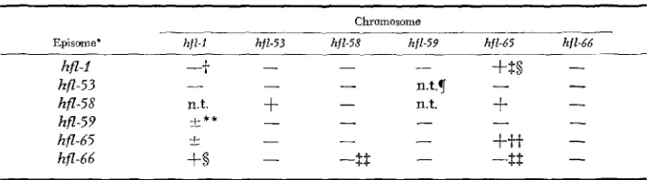
Construction and complementation properties of F118 h fl-x/h fl-y recA merodiploids
Chromosome
-- Episome' hfl-1 hfl-53 hfl-58 hfl-59 hfl-65 hfl-66
fSB
-
hfl-1
-+
hfl-53 - -
-
n.t.v-
-
hfZ-58 n.t.
+
hfZ-59 +-** -
+tt
-
hfl-65 t
-$$
hfl-66
+8
- - -
-
-
n.t.
+
-
-
-
-
- - -
-
-
-8
-
* Merodiploids with hfl-1, hfl-59, hfZ-65 and hfl-66 on the episome were constructed by mating the appropriate merodiploids in Table 3 with F- amp+ hfl-y t r p + recA derivatives of strains in Table 2 Column 1 and selecting f o r Amp-Trp+. Merodiploids with hfl-53 and hfl-58 on the episome were Constructed by mating the appropriate merodiploids in Table 3 with F- hfz-y purA trp+ reCA derivatives of strains in Table 2 Column 2 (except the F- hfl-53 recipient, which was strain UC3414) and selecting for Pur+ Trp+.
f - = Turbid XcIII plaques (Hfl- phenotype).
$
+
= Clear XcIII plaques (Hfl+ phenotype).This Hfl+ F' hfl-x/hfr-y recA merodiploid was mated with an F- hfl-66 recA strain to obtain a F' hfi7-x/hfZ-6&recA merodiploid. The new merodiploid had an Hfl- phenotype, as would be expected from examination of the Table.
q n.t.
= Not tested.* * f = Slightly turbid XcIII plaques.
j-t This Hfl+ F' hfz-65/hfZ-65 recA merodiploid was mated with an F- hfl-66 recA strain to obtam a I?' hf7-65/hfz-66 recA merodiploid. The new merodiploid, which hac the expected Hfl- phenotype, was then mated with an F- hfl-65 recA merodiploid to obtain a F' hfl-65/hfl-65 recA
merodiploid once more. Like the original merodiploid, the new merodiploid had an Hfl+
phenotype.
GENETICS OF hfl MUTANTS 445 to a n F- purA strain. The presence of the hfl marker on the episomes of these merozygotes was demonstrated by their successful use as doEors in producing the merozygotes in lines 2 and 3 of Table 8.
We can, therefore, conclude with some confidence that all six hfl mutants are recessive to hfl+
.
This conclusion was strengthened by the subsequent finding that all six mutants lay in a single complementation group.For complementation studies, strains carrying different hfl mutations on the F118 episome were constructed as described in MATERIALS A N D METHODS (Table 3). These strains were then mated with recA derivatives of F- hfl strains to form rpcA merodiploids of the form F118 hfl-xlhfl-y (Table 8), Most combinations resulted in an Hfl- phenotype (Table 8), indicating that all six hfl mutants lie in a single complementation group. There were a few examples of apparent in- tragenic complementation, notably F118 hfl-58/hfl-53, F118 hfl-66/hfl-l, and, to a marginal extent, F118 hfl-59/hfl-l (Table 8). I n all three cases, the reciprocal pairs did not complement, which is possibly due to a gene dosage effect stemming from multiple copies of episome per chromosome. The mutation hfl-65 has only a marginal hfl phenotype in quantitative lysogenization tests and it should not be surprising that merodiploids containing the hfl-65 mutation frequently show apparent complementation. The fact that hfl-65 apparently “complements” itself could be understood in terms of a marginally stable protein synthesized in greater quantities in an F118 hfl-65/hfl-65 merodiploid than in the correspond- ing F- hfl-65 haploid strain.
Zntragenic recombination between hfl mutants: Since all six purA-linked hfl mutants lie in a single complementation group, we did not attempt to order the different hfl mutants. W e did show that intragenic recombination occurs, which demonstrates that the hfl mutants occur at different sites on the chromosome. For example. when UC3061 (ampA hfl-59 purA29) was the donor and UC3035 (ampA+ hfZ-1 purA23) the recipient, 88 Pur+ recombinants were
Hfl-
like the parents and 22 Pur+ recombinants were Hfl+ recombinants as well. The 88Hfl-
Pur+ recombinants included 71 Amp+ and 1 7 Amp- colonies. All 22Hfl+
Pur+ double recombinants were Ampf, which indicates an order of ampA-hfZ-59- hfl-l-purA. The reciprocal cross was not done because we did not have suitably marked purA derivatives and crosses with a pur+ donor gave considerably lower frequencies of recombination between hfl mutants. This latter observation, aswell as the lower frequencies of recombination shown i n Table 4 than in Table 6,
is an example of the well knowii phenomenon of high negative interference between closely linked inarkers (CHASE and DOERMANN 1958).
DISCUSSION
446 J. W. GAUTSCH A N D D. L. WULFF
than hfl+ strains. (3) Lambda wild type, XcZZZ and k l 7 show a reduced plaque- forming ability on these mutants.
(4)
These mutants all show a slightly in- creased resistance to penicillin and rifamycin. (5) These mutants are all slightly more sensitive to sodium dodecyl sulfate. These similar phenotype properties, the recessive nature of hfl mutations to hfl+ in merodiploids, and the complementa- tion behavior of hfl mutants suggest that the hfl mutation represents a loss of function and that the hflf gene is a classical gene, probably coding for the syn- thesis of a protein. W e propose to call the purA-linked hfl locus the hflA locus.An estimate of the distance between the hflA and purA loci may be obtained from the formula of Wu (1966), relating co-transduction frequencies to map distance, which is being used in construction of the E. coli K12 genetic map
(TAYLOR
and TROTTER 1972). From Table 4 i t may be calculated that the average co-transduction frequency between hflA and purA mutants is 92%, from which a 0.054 minute separation between the hflA and purA loci may be calculated. Assuming 5000 genes in the E . coli chromosome and a total rcap length of 90 minutes, this would mean that the hflA and purA loci were only 3 genes apart, which is to say that there were only 2 intervening genes, Although calculations based on co-transduction frequencies are necessarily imprecise, they do empha- size the close proximity o€ the hflA and purA loci. It is possible that the two genes are adjacent to one another. Thirteen purA mutants of UC4185 isolated in this study did not revert to wild type and may be deletions. However, all of these mutants had a n Hfl+ phenotype and all recombined with both purA28 and purA29, which are at the left and right ends of our purA fine structure map (Table 6 ) . Thus, while it. is conceivable that non-lethal dektims may extend from the purA locus into the hfZA locus, we did not find any.Our finding of isolated instances of intragenic complementation in F118 hfl-x/ hfl-y merodiploids fortifies our conclusion that hflA locus is a classical gene. In- deed, all instances of intragenic complementation deal with classical gene prod- ucts, namely enzymes (SCHLESINGER and LEVINTHAL 1965; FINCHAM 1966). Intragenic complementation is known to involve interactions of subunits in multi- meric proteins. However, care must be taken in interpreting the data on the hf2A locus because in no instance was intragenic complementation reciprocal. This could be caused by unequal amounts of synthesis of the hfZA product from episomal and chromosomal genes.
If the hfl+ gene indeed determines the synthesis of an Hflf protein, then how
are the high frequencies of lysogenization of hflA strains by A + phage and XcZZZ
mutants to be interpreted? The obvious interpretation is that the Hfl+ protein normally antagonizes lysogenization, and that the product of the XcZZZ gene negates this antagonistic effect, although its mode of action could be indirect. The idea of a phage protein acting to inhibit a host protein is not new. ROBERTS (1969) has suggested that the N protein of inhibits the host termination factor, rho, al- lowing transcription to continue past normal termination signals.
GENETICS OF hj?. MUTANTS 447
scription from this promoter must occur in the absence of the lambda cZZZ gene product in an hflA host.
How
could theHfl+
protein antagonize transcription? One possibility is that it controls the concentration of some small molecule which interacts at the promoter with RNA polymerase to control transcription. This small molecule could be a nucleotide, but could not be adenosine 3’:5’-cyclic monophosphate(BELFORT
and WULFF 1973). A second possibility is that theHfl+
protein is normally bound to RNA polymerase but is not essential for its activity. ( A number of such proteins may exist(TRAVERS
andBUCKLAND
1973) .)Such a polymerase molecule would be hypothesized to be unable to transcribe from the pre promoter unless
XCIII
protein were present to negate somehow the inhibitory effect of theHfl+
protein.It will be of great interest to identify the function of the
Hfl+
protein. While the existence of the hflA mutants implies that theHfl+
protein is not an essential host protein, it is possible that all hflA mutants have at least partialHfl+
activity and that someHfl+
activity is required for the host cell to function. Indeed, all six of the hflA mutants retain theirHfl-
phenotypes when transduced into thesuf strain ES4, which suggests that none of them are amber mutants. The fact that the percent lysogenization of the six hflA mutants by a XcZZZ phage strain varies over a wide range indicates that most o r all hflA mutants have at least partial
Hfl+
function. While this might mean that a totally inactiveHfl+
protein is lethal to the host, it also could merely be a consequence of most NG-induced mutants having partial activity, combined with a very sensitive test for detecting hfZA mutants.This work was supported by NSF grant No. GB 32194 to D. W.
LITERATURE CITED
BELFORT, M. and D. L. WULFF, 1971 A mutant of Escherichia coli that is lysogenized with high frequency. pp. 739-742. In: The Bacteriophage Lambda. Edited by A. D. HERSHEY.
Cold Spring Harbor, New York.
-
, 1973a A genetic and biochemical investigation of the Escherichia coli mutant hfl-1 which is lysogenized a t high frequency by bacteriophage lambda. J. Bacteriol. 115: 299-306. -, 1973b An analysis of the processes of infec- tion and induction of Escherichia coli mutant hfl-1 by bacteriophage lambda. Virology 5 5 :CHASE, M. and A. H. DOERMANN, 1958 High negative interference over short segments of the genetic structure of bacteriophage T4. Genetics 43: 332-353.
ECHOLS, H. and L. GREEN, 1971 Establishment and maintenance of repression by bacteriophage lambda: The role of the cI, cII, and cIII proteins. Proc. Natl. Acad. Sci. U.S. 68: 2190-2194.
ERIKSSON-GRENNBERG, K. G., 1968 Resistance of Escherichia coli to penicillins. 11. An improved mapping of the ampA gene. Genet. Res. 12: 147-156.
FINCHAM, I. R. S., 1966
GOTTEsMaN, M. and M. YARMOLINSKY, 1968
KEMPER, J. and P. MARGOLIN, 1969
LIEBERMAN, I., 1956 183-192.
Genetic Complementation. W. A. Benjamin Press, New York.
Integration negative mutants of lambda. 5. Molec.
Suppression by gene substitution for the leuD gene of
Enzymatic synthesis of adenosine-5‘-phosphate from inosine-5’-phosphate. Biol. 31 : 487-505.
Salmonella typhimurium. Genetics 6 3 : 263-279.
448 J. W. GAUTSCH A N D D. L. WULFF
Low, B., 1968 Formation of merodiploids in matings with a class of Rec- recipient strains of
Escherichia coli K12. Proc. Natl. Acad. Sci. U.S. 60: 160-167.
-,
1973 Rapid map- ping of conditional and auxotrophic mutations in Escherichia coli K12. J. Bacteriol. 113:798-812.
LURIA, S . E., J. N. ADAMS and R. C. TING, 1960 Transduction of lactose utilizing ability among strains of E. coli and S. dysenteriae and the properties of the transducing phage particles. Virology 12 : 348-390.
Genetic fine structure of the leucine operon in Salmonella. Genetics 43:
441-457.
Dominant mutations ( l e x ) in Escherichia coli K-12 which effect radiation sensitivity and frequency of ultraviolet light-induced mu-
tations. J. Bacteriol. 112: 886-893.
REICHARDT, L. and A. D. KAISER, 1971 Control of lambda repressor synthesis. Proc. Natl. Acad.
ROBERTS, J. W., 1969
SCHLLSINGER, M. J. and C. LEVINTHAL, 1965
zyme interactions. Ann. Rev. Microbiol. 19: 267-284. SIGNER, E. R., K. F. MANLY and M. BRUNSTETTER, 1969
of bacteriophage lambda. Virology 39: 137-141.
STOUTHAMER, A. H., P. G. DE HAAN and H. J. J. NIJKAMP, 1965
Escherichia coli K12. Genet. Res. 6: 442-453. TAYLOR, A. L. and C. D. TROTTER, 1972
Rev. 36: 504-524.
TRAVERS, A. and R. BUCKLAND, 1973 Biol. 243: 257-260.
VOGEL, H. J. and D. M. BONNER, 1956
W u , T. T., 1966 MARGOLIN, P., 1963
MOUNT, D. W., K. B. Low and S. J. EDMISTON, 1972
Sci. U.S. 68: 2185-2189.
Termination factor for RNA synthesis. Nature 224: 1168-1174. Complementation a t the molecular level of en-
Deletion mapping of the cIII-N region
Mapping of purine markers in
Linkage map of Escherichia coli strain K12. Bacteriol.
Heterogeneity of E. coli RNA polymerase. Nature New
Acetyl-ornithase of Escherichia coli: Partial purification
A model for three-point analysis of random general transduction. Genetics 54:
Corresponding editors: D. KAISER and H. ECHOLS and some properties. J. Biol. Chem. 218: 97-106.