der Fakultät für Chemie und Pharmazie
der Ludwig-Maximilians-Universität München
Functional characterization of rabies virus in
mouse embryonic stem cell-derived neurons
Maximilian Ferdinand Eizinger
aus
II Erklärung
Diese Dissertation wurde im Sinne von § 7 der Promotionsordnung vom 28. November 2011 von
Herrn Prof. Dr. Karl-Klaus Conzelmann betreut und von Herrn Prof. Dr. Klaus Förstemann von der
Fakultät für Chemie und Pharmazie vertreten.
Eidesstattliche Versicherung
Diese Dissertation wurde eigenständig und ohne unerlaubte Hilfe erarbeitet.
München, den 28.05.2018
Max Eizinger
(Unterschrift des Autors)
Dissertation eingereicht am: 26.02.2018
1. Gutachter: Prof. Dr. Klaus Förstemann
2. Gutachter: Prof. Dr. Karl-Klaus Conzelmann
III
“Science is the process that brings us from confusion to understanding…”
Brian Greene
IV
Danksagung
Ich möchte mich sehr herzlich bei allen bedanken, die mich während dieser sehr interessanten,
anspruchsvollen und schöne Zeit begleitet und unterstützt haben.
An aller erste Stelle möchte ich mich bei Prof. Dr. Karl-Klaus Conzelmann bedanken, der mir das Vertrauen gab in seiner Arbeitsgruppe meine Doktorarbeit anzufertigen. Ich möchte mich dafür
bedanken, dass ich an diesem höchst interessanten und herausvordernden Themen arbeiten
durfte. Indem er mir die Freiheiten gab meinen eingenen Ideen nachzugehen und seine Türe für
Fragen und Diskussion immer offen stand, kreierte Prof. Dr. Karl-Klaus Conzelmann hervorragende
fachliche sowie menschliche Rahmenbedingungen. Des Weiteren möchte ich mich bei Prof. Dr.
Klaus Förstemann bedanken, der sich freundlicherweise bereit erklärt hat diese Arbeit als Fachvertreter der Biochemie zu betreuen. Zudem möchte ich den Mitgliedern der
Prüfungskomission Prof. Dr. Christian Wahl-Schott, PD Dr. Dietmar Martin, PD Dr. Barbara Adler und Prof. Dr. Karl-Peter Hopfner für Ihre Mühen bedanken.
Ein ganz besonderer Dank gilt meinen lieben Arbeitskollegen, die diese aussergewöhnlich gute
Arbeitsatmosphere ermöglicht haben. Allen voran möchte ich mich bei Alex bedanken der mir bereits während der Bachelorarbeit die ersten Schritten in der Laborwelt beigebracht hat und sich
während der Doktorarbeit für Fragen, Diskussionen und hilfreiche Tipps immer zeitgenommen
hat. Merci beaucoup Chloé für das hervorragende Teamwork bei der Stammzellenkultur. Mein Dank gilt selbstverständlich auch Blex, Verena und Johanna für ihre Unterstützung im Laboralltag, wenn nötig die passenden aufmunternden Worte und die vielen lustigen Kaffeepausen. Vielen
Dank auch an Doro die mich über all die Jahre immer wieder mit sehr feinen Kuchen versorgt hat. Auch den Alumnis möchte ich ganz herzlich danken. Marco für die vielen spannenden Diskussionen; Daniel für die vielen Stunden beim „soggen“; Konstantin, Kerstin, Tobi, Nadin und Martina für ihre große Hilfsbereitschaft am Anfang meiner Doktorarbeit.
Mit der größte Dank gilt meinen Eltern für ihre großartige Unterstützung und ihren Zuspruch. Selbsvertändlich möchte ich mich auch bei Susi, Luki, Isabell und Jürgen für ihre Unterstützung bedanken.
V
Contents
Contents ... V
1 Summary ... 1
2 Introduction ... 3
2.1 Rabies virus ... 3
2.1.1 Rabies disease ... 3
2.1.2 Taxonomy of RABV ... 4
2.1.3 Route of RABV infection ... 5
2.1.4 Virion structure and protein function ... 6
2.1.5 RABV transcription and replication ... 9
2.1.6 The envelope proteins ... 11
2.1.6.1 The matrix protein ... 11
2.1.6.2 The glycoprotein... 12
2.1.7 Retrograde axonal transport, transsynaptic spread and monosynaptic tracing of RABV ... 14
2.2 Membrane scission and fusion machineries ... 16
2.2.1 The ESCRT machinery ... 16
2.2.2 SNARE proteins ... 19
2.2.3 Neurotoxins ... 21
2.3 mESCs and mESC-derived neurons at a glance ... 23
2.3.1 mESCs at a glance ... 23
2.3.2 Introduction of mESC-derived neurons ... 24
3 Material and Methods ... 26
3.1 Materials ... 26
3.1.1 Laboratory equipment ... 26
3.1.2 Dishes, well plates and miscellaneous ... 27
3.1.3 Cell lines and Bacteria strains ... 28
3.1.4 Chemicals ... 29
3.1.5 Buffers and solutions... 30
3.1.6 Medium and cell culture additives ... 33
3.1.7 Kits ... 35
3.1.8 Enzymes ... 35
VI
3.1.10 Secondary antibodies ... 36
3.1.11 Oligonucleotides... 37
3.1.12 Plasmids ... 37
3.1.13 Recombinant viruses ... 41
3.2 Methods ... 42
3.2.1 Working with DNA ... 42
3.2.1.1 Polymerase Chain Reaction (PCR) ... 42
3.2.1.2 Agarose gel electrophoresis ... 43
3.2.1.3 Restriction enzyme digestion ... 43
3.2.1.4 Gel purification of DNA fragments ... 44
3.2.1.5 Ligation of DNA fragments ... 44
3.2.1.6 Transformation of plasmid DNA into competent bacteria ... 45
3.2.1.7 Mini preparation of plasmid DNA from bacteria ... 45
3.2.1.8 Midi preparation of plasmid DNA from bacteria ... 45
3.2.1.9 Sequencing of DNA ... 46
3.2.2 Cell culture ... 46
3.2.2.1 Cultivation of mammalian adherent cell lines ... 46
3.2.2.2 Cultivation of mESC in a feeder free culture ... 46
3.2.3 Freezing and thawing of mESCs ... 47
3.2.4 Transfection... 47
3.2.5 Immunofluorescence imaging ... 48
3.2.6 Denaturing polyacrylamide gel electrophoresis (SDS-PAGE) ... 49
3.2.7 Western Blotting ... 50
3.2.8 Immunodetection ... 50
3.2.9 Generation of recombinant rabies virus by reverse genetics (virus rescue) ... 50
3.2.9.1 Rescue of recombinant full-length SAD ... 51
3.2.9.2 Rescue of recombinant SAD ΔM eGFP ... 52
3.2.9.3 Rescue of recombinant SAD ΔG eGFP (SAD G) ... 52
3.2.10 Virus stock production ... 53
3.2.11 Titration of virus ... 53
3.2.12 Fluorescence labeling of RABV positive cells ... 54
3.2.13 Multistep growth curves ... 54
3.2.14 Generation of SAD ΔG (EnvA) ... 54
3.2.15 Virion purification by ultracentrifugation ... 55
3.2.16 Trans-complementation of SAD ΔG ... 55
VII
3.2.17.1 RNA Isolation ... 56
3.2.17.2 Reverse transcription ... 56
4 Results ... 57
4.1 Generation of an in vitro model to investigate RABV in neurons in a mouse free system ... 57
4.1.1 Differentiation of mESCs into glutamatergic pyramidal neurons ... 57
4.1.1.1 Feeder free mESC culture system ... 59
4.1.1.2 “Priming” of naive pluripotent mESCs ... 59
4.1.1.3 Formation of embryoid bodies... 60
4.1.1.4 Transformation of EBs to neurospheres ... 60
4.1.1.5 Differentiation of neurospheres to glutamatergic pyramidal neurons ... 63
4.1.1.6 Characterization of mESC derived neurons ... 64
4.1.2 Generation of an in vitro model to study the monosynaptic spread of RABV ... 65
4.2 Transsynaptic spread of RABV ... 68
4.2.1 Role of RABV M in transsynaptic spread ... 68
4.2.2 Role of the ESCRT machinery in the neuronal transfer of RABV ... 70
4.2.3 Role of the SNARE machinery in the transsynaptic spread of RABV ... 76
4.2.3.1 Effect of dominant-negative Syntaxin-1, -3 and -4 on the transsynaptic spread of RABV ... 76
4.2.3.2 Effect of neurotoxins on transsynaptic spread of SAD... 79
4.2.4 EM studies to visualize the transsynaptic spread of RABV ... 85
4.3 Manipulation of the RABV glycoprotein ... 86
4.3.1 Manipulation of the SAD G ectodomain ... 86
4.3.2 Manipulation of the cytoplasmic tail ... 88
4.3.3 Characterization of SAD G-TM-3xKBS-RTeGFP in mESC-derived neurons ... 91
4.4 Generation of a less cytotoxic RABV for long-term studies of RABV infected neurons in vitro and in vivo ... 94
4.4.1 Generation of persistently infected HEK293T cells ... 94
4.4.2 Characterization of non-cytotoxic SAD eGFP ... 95
4.4.3 Deep sequencing of non-cytotoxic SAD eGFP ... 98
4.4.4 Characterization of potential non-cytotoxic viruses ... 100
4.4.5 Reduced cytotoxicity of SAD mut52/54 eGFP and SAD mut52/54/12845 in mESC-derived neurons ... 102
5 Discussion ... 105
5.1 Establishment of a feeder-free mESC culture and a differentiation protocol of mESCs into mESC-derived glutamatergic pyramidal neurons ... 107
VIII
5.2.1 M-dependent spread of RABV in neurons ... 108
5.2.2 The ESCRT machinery contributes to the neuronal spread of RABV ... 108
5.2.3 Destruction of SNARE complexes attenuates transsynaptic spread of RABV ... 111
5.2.3.1 Dominant-negative syntaxin-1, -3, and -4 hinder the neuronal transfer of SAD ... ... 111
5.2.3.2 TeNT and BoNT/A hinder the neuronal transfer of RABV ... 112
5.2.4 Visualization of the spread of RABV in neurons by EM ... 114
5.2.5 Prospective approaches for studying the neuronal transfer of RABV ... 115
5.2.6 Working hypothesis for the neuronal transfer of RABV ... 116
5.3 Insertion of a trafficking signal downstream of the transmembrane domain is a potential site for rerouting SAD G ... 117
5.4 In vitro evolution experiment of SAD eGFP ... 119
5.4.1 SAD eGFP in vitro evolution resulted in a non-cytotoxic virus ... 119
5.4.2 Future outlook ... 121
6 Appendix ... 122
6.1 List of oligonucleotide sequences ... 122
6.2 Cloning strategies of plasmids produced in this thesis ... 124
6.3 List of abbreviations ... 128
7 List of figures and tables ... 133
7.1 List of figures ... 133
7.2 List of tables ... 134
1
1
Summary
While rabies virus (RABV) is the causative agent of the rabies disease that kills approximately
59,000 people per year, RABV is also a superb tool to reveal neural circuits due to its exclusive
spread to presynaptically connected neurons. Since the mechanism behind the anterograde
transport of RABV to dendrites and the exclusive retrograde transsynaptic spread of RABV in the
CNS are still poorly understood, this thesis aimed to provide an insight into the molecular
mechanism of the neuronal virus transmission, which would have a tremendous impact in terms
of improving RABV as a neuronal tracer, understanding the RABV pathology and developing
therapeutic strategies to fight RABV infection.
The first and overarching aim of this thesis was the introduction of a high quality, feeder-free mESC
culture system, followed by the establishment of a highly reliable differentiation protocol of
mESCs into a nearly homogenous neuronal cell population of glutamatergic pyramidal neurons,
forming chemical synapses. Further, by generating a neuronal mix population, which contains a
small portion of mESC-derived neurons that enable the primary infection of this neuron with an
EnvA pseudotyped G-gene deficient RABV and the in situ trans-complementation of the G-gene
deficient RABV, an in vitro model to investigate the mono- transsynaptic spread of RABV ΔG was
established.
Using the mESC-derived neurons and reverse genetics to generate a recombinant RABV encoding
for a mutated late domain within the matrix protein (M) it was shown that the ESCRT system is
involved in the neuronal transfer of RABV. Further, this thesis provides some evidence that
34PPPEYVPL41 represents rather an extended motif of the PPEY late domain, than overlapping late
domains consisting of PPEY and YVPL. In a second approach, which included recombinant RABVs
encoding for dominant-negative syntaxins and the transduction of mESC-derived neurons with
lentiviral vectors encoding for neurotoxin light chains a participation of SNARE proteins in the
transsynaptic spread of RABV was illustrated.
Since the retrograde axonal transport of RABV is G-dependent, an insertion site for external
trafficking signals within G should be identified in order to change G trafficking and thereby the
subcellular localization of G. While modifications of the ectodomain destroyed the functionality
of G, the insertion of the kinesin light-chain binding sequence (KBS) downstream of the
transmembrane domain resulted in infectious virus particles. Further, the recombinant SAD
G-TM-3xKBS-2 RT exhibited a homogeneous distribution, which differed clearly from granular, probably
PSD-associated distribution of G wildtype. Thus, G-TM-3xKBS-RT might be the first step to generate an
redirected anterograde transsynaptic tracer.
In order to evolve a less cytotoxic SAD virus for long-term studies of infected neurons, a persistent
infection model for SAD in HEK293T cells, which usually succumb to viral infection, was generated.
The following analysis of the virus pool produced by the surviving HEK293T cells by NGS revealed
only six notable mutations that occur in different frequencies, reflecting the existence of a mixed
virus population. Interestingly, two point mutations within the non-coding leader region were
sufficient to turn SAD eGFP into a non-cytotoxic virus, not only in HEK293T cells but also in
mESC-derived neurons.
In summary, this thesis provides an insight into the molecular mechanism behind the transsynaptic
spread of RABV, creates first hints for generating an anterograde transsynaptic tracing model, and
3
2
Introduction
2.1
Rabies virus
2.1.1 Rabies disease
Numerous writings which date back until the antique describe dogs and humans suffering from
rabies disease, most likely due to its disturbing symptoms, its long period of suffering and its
fatality after the onset of symptoms (Wilkinson, 1977). Already Hippocrates described supposedly
human rabies as he wrote, “persons in a frenzy drink very little, are disturbed and frightened,
tremble at the least noise, or are seized by convulsion” (Jackson, 2013). Aristotle wrote that the
disease drives the dog mad, is fatal to the afflicted dog itself and any animal, which was bitten by
the dog. Wrongly, Aristotle wrote that humans are not affected by the disease (Jackson, 2013;
Wilkinson, 1977). Celsus already introduced the term hydrophobia as a symptom of rabies in
humans and he identified that the causative agent of rabies is in the saliva of the biting animal
(Jackson, 2013).
Nowadays it is well-known, that rabies is a viral zoonotic disease caused by RABV, which still kills
approximately 59000 people per year according to the center of disease control and prevention
(CDC.) With the exception of the Antarctica, rabies is distributed all over the world. Whereas all
mammals are susceptible to RABV infection, Carnivora and Chiroptera represent the main
reservoirs of the virus (reviewed in (Rupprecht et al., 2002)).
In most cases, humans are infected with RABV through an animal bite or scratch, which leads to
the exposure of muscle cells with the RABV containing saliva (Davis et al., 2015). After the
exposure to RABV, rabies exhibits a mean incubation period of one up to two months. However,
extreme incubation periods of as short as seven days (after a dog bite into the brachial plexus) up
to more than six years (after ABLV infection) are documented (Hemachudha et al., 2002). The
incubation period depends probably on the virus load, virus type and the site of infection.
Nevertheless, incubation times of up to six years are still puzzling.
The pre-exposure vaccination (an active immunization) or the post-exposure prophylaxis (PEP),
which consists of wound cleansing, passive vaccination with human rabies immune globulin and
active vaccination with cell culture rabies vaccine, protects efficiently against RABV (Manning et
al., 2008). However, if RABV exposed humans are not vaccinated and the PEP is not carried out
4 symptoms, rabies proceeds to almost 100 % fatal (Koyuncu et al., 2013). Until now, only a single
patient, which had neither a pre-exposure vaccination nor a PEP, survived a RABV infection after
the onset of symptoms (Willoughby et al., 2005).
According to the progression of the acute neurological phase, rabies is grouped into classic rabies
and non-classic rabies. The classic human rabies is subdivided into furious (encephalitic) rabies
and paralytic (dumb) rabies (Hemachudha et al., 2002). Two-thirds of the classic human rabies
cases are furious rabies cases, which lead to death within seven days after the onset of symptoms.
The symptoms of the furious rabies start with hyperactivity, which is intensified by thirst, fear,
light, noise and other stimuli (Hemachudha et al., 2002). The disease progresses with the onset of
fluctuating consciousness, phobic or inspiratory spasm (hydrophobia, aerophobia), and autonomic
dysfunction (hypersalivation, sweating) (Hemachudha et al., 2002). The victims die because of
cardiac arrest, circulatory insufficiency, or respiratory failure (Davis et al., 2015). The paralytic
rabies kills the patients within 14 days on average (Hemachudha et al., 2002). It causes an
ascending paralysis, followed by coma and death (Davis et al., 2015).
The non-classic rabies occurs in patients, which were infected by bats or in patients, which were
infected by a dog-bite in Thailand. The non-classic rabies has no characteristic features, however
observed clinical features include neuropathic pain and radicular pain (reviewed in (Hemachudha
et al., 2002)).
2.1.2 Taxonomy of RABV
The RABV genome is a single stranded, non-segmented, negative-sense ribonucleic acid (RNA)
genome. RABV belongs to the order Mononegavirales, which is also designated as non-segmented
negative strand RNA viruses (NNSV), and is a prototype of the Rhabdoviridae family. However,
recently the family Rhabdoviridae was expanded by genera Dichorhavirus and Varicosavirus,
which are bi-segmented plant viruses (Afonso et al., 2016; Shi et al., 2018).
The order Mononegavirales is subdivided into the families Bornaviridae, Filoviridae,
Mymonaviridae, Nyamiviridae, Paramyxoviridae, Pneumoviridae, and Rhabdoviridae (International Committee on Taxonomy of Viruses (ICTV) 28.11.2017). Since RABV virions have a
bullet-/rod-shaped structure (Figure 1C) it is grouped into the family Rhabdoviridae, deduced from
the greek word rhabdos, meaning rod (Wunner and Conzelmann, 2013).
The family Rhabdoviridae is subdivided into 18 genera named Almendravirus, Curiovirus,
Cytorhabdovirus, Dichorhavirus, Ephemerovirus, Hapavirus, Ledantevirus, Lyssavirus,
Novirhabdovirus, Nucleorhabdovirus, Perhabdovirus, Sigmavirus, Sprivivirus, Sripuvirus,
5 the genus Lyssavirus. This name has its origin in the greek mythology since Lyssa (meaning “rage”)
was the goddess of madness (reviewed in (Wunner and Conzelmann, 2013)).
Dependent on the passaging history, RABV strains are subdivided into street strains (wildtype) and
fixed strains. Street strains are isolated directly out of infected animals or humans, whereas fixed
strains were generated by passaging the viruses repeatedly in cell culture or in animals (Davis et
al., 2015).
In this thesis, all RABV constructs are based on the fixed Street Alabama Dufferin (SAD) L16 strain,
which was the first negative-sensed RNA virus generated by reverse genetics. Indeed, SAD L16
virus emerges from the cDNA encoding for SAD B19 (Schnell et al., 1994). SAD virus was isolated
from a rabid dog, passaged on mouse brain cells, and adapted on BHK-21 cells. The passaging of
SAD, which was initially passaged on mouse brain cells, on a cloned BSR cell line resulted in SAD
B19, which is used as a live vaccine virus (Vos et al., 1999). Another fixed RABV strain, which was
used in this work is CVS-N2c. CVS-N2c was generated by passaging the mouse adapted fixed strain
CVS-24 (challenged virus standard) on a mouse neuroblastoma cell line. CVS-N2c exhibits a higher
neurotropism as well as a reduced cytotoxicity compared to SAD B19 (Morimoto et al., 1998;
Reardon et al., 2016). Another strain used in this thesis, is the street strain THA, which was isolated
from a human bitten by a rabid dog, and passaged only for a few times on a mouse neuroblastoma
cell line (Thongcharoen et al., 1990).
2.1.3 Route of RABV infection
In most of the human rabies cases, people are exposed to RABV by a bite or scratch of a rabid
animal (Davis et al., 2015). Thereby, RABV containing saliva gets in contact with the peripheral
muscle tissue. RABV can bind to the muscular nicotinic acetylcholin receptor (nAChR), which is
located at the postsynaptic membrane of a neuron muscular junction (NMJ), and enter the muscle
cell via a receptor-mediated endocytosis (Kalamida et al., 2007; Lafon, 2005). The presence of the
nAChR at the postsynaptic membrane of the NMJ indicates that not motor neurons, but muscle
cells are the primarily infected cells. Presumably, the infection of muscle cells enables the
amplification of RABV. The additional replication cycle might increase the probability for a
successful infection of the motor neuron (Lafon, 2005). After virus transcription and replication,
RABV is released into the synaptic cleft of a NMJ, binds to the p75 nerve growth factor receptor
(p75NTR) or the neuronal cell adhesion molecule (NCAM) on the surface of the presynaptic motor
neuron and internalizes via receptor-mediated endocytosis into the first order neuron (Ugolini,
2011). In the motor neuron, the entire enveloped virus travels within an endosomal vesicle along
microtubules by a directed retrograde axonal transport to the cell body (Klingen et al., 2008).
6 than the transport of a standard p75NTR-vesicle. Further, a p75NTR-knockdown in dorsal root
ganglions suggested that the p75NTR-dependent RABV transport is faster and more directed than
the p75NTR-independent axonal transport of RABV (Gluska et al., 2014).
The acidification of the endosomal lumen causes the glycoprotein-dependent fusion of the viral
membrane with the endosomal membrane and results in the release of the ribonucleoprotein
(RNP) complex into the cytoplasm of the host cell (Gaudin, 2000; Roche and Gaudin, 2004). After
the release of the RNP into the cytoplasm, viral transcription and replication takes place within
spherical cytoplasmic inclusion bodies named Negri bodies (Lahaye et al., 2009; Nikolic et al.,
2017). Finally, new RABV particles assemble and spread to presynaptically connected neurons.
The retrograde transport directs RABV directly to the CNS. After the invasion of the CNS, at late
stage of disease, RABV spreads centrifugally from the CNS to extraneuronal organs like the heart
or salivary glands (Jackson et al., 1999; Ugolini, 2011).
2.1.4 Virion structure and protein function
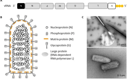
RABV virions have an average length of 180 nm, an average diameter of 75 nm and form a
bullet-shaped structure (Figure 1C). The genome is a single-stranded, non-segmented, negative-sense
RNA genome. It is 12 kb long and has an unmodified 3´-hydroxyl end and a 5´-triphosphate end
(Wunner and Conzelmann, 2013). The genome encodes for five monocistronic genes in the
following order: nucleoprotein (N), phosphoprotein (P), matrix protein (M), glycoprotein (G) and
the RNA-dependent RNA polymerase, also named large protein (L). The 3´-end of the viral genome
is flanked by a non-coding leader (Le) RNA region and the 5´-end is flanked by a non-coding trailer
7
Figure 1: Organization of a Rabies virus particle.
A, Schematic representation of the 12 kb long, negative-sense RNA genome with its triphosphorylated 5´-end (adapted from (Ghanem and Conzelmann, 2016)). B, Schematic representation of the bullet-shaped RABV virion (adapted from dissertation of Marco Wachowius, 2016). C, SAD L16 virions were purified via sucrose cushion centrifugation and were subjected to uranyl acetate negative staining, followed by transmission electron microscopy. The pictures were taken together with Daniel Aberle in collaboration with Otto Beringhausen (LMU, Gene Center).
The RABV genome organization
The RABV genome order 3´-Le-N-P-M-G-L-Tr-5´ is conserved in the family Rhabdoviridae and in
the order of Mononegavirales (reviewed in (Pfaller et al., 2015)). However, many rhabdoviruses
and other virus families of the Mononegavirales can encode for accessory genes (Davis et al., 2015;
Pfaller et al., 2015). Within the RABV genome, the five monocistronic genes are separated by
conserved gene border signals, which encode for a transcription stop signal, a polyadenylation
signal for the upstream gene, an intergenic sequence (IGS) and a transcription restart signal for
the downstream gene (reviewed in (Wunner and Conzelmann, 2013)).
The nucleoprotein (RABV N)
The nucleoprotein is 450 amino acids long and has a molecular weight of about 57 kDa (Wunner
and Conzelmann, 2013). RABV N enwraps the viral genome into a permanent, helical nucleocapsid
and thereby shields the viral RNA from nucleases and the recognition by the immune system, e.g.
the Toll-like receptors and RIG-I like receptors during the virus entry (Albertini et al., 2006).
8 replication by the viral RNA-dependent-RNA polymerase, which is a complex of RABV L and its
cofactor RABV P. Moreover, the amount of RABV N plays an important role in the transition from
viral transcription to replication (reviewed in (Albertini et al., 2008)).
The phosphoprotein (RABV P)
The phosphoprotein is 297 amino acids in size, exhibits in SDS-PAGE a molecular weight of 37 kDa
and fulfills multiple functions. First, RABV P is the noncatalytic cofactor of the RNA-dependent RNA
Polymerase RABV L. Second, RABV P binds freshly synthesized RABV N (N0), prevents RABV N
polymerization and unspecific binding to cellular RNA. Thus, RABV P enables the interaction of N0
with the freshly transcribed viral RNA (reviewed in (Wunner and Conzelmann, 2013)). Third, RABV
P suppresses innate immune signaling in infected cells by interfering with the induction of
interferon and by the inhibition of interferon signaling. The current model is that RABV P blocks
on the one hand the phosphorylation of the transcription factor IRF-3 by TBK1 and IKKε, thereby
inhibiting the transcription of type I interferon genes (Brzozka et al., 2005; Marschalek et al.,
2009). On the other hand, RABV P blocks the interferon signaling by retaining the activated STATs
in the cytoplasm. Thus, P inhibits the transcription of the antiviral interferon-stimulated genes
(ISGs) (Brzozka et al., 2006).
The large protein (RABV L)
RABV L is 2127 amino acids long and exhibits a molecular weight of 244 kDa. Together with its
noncatalytic cofactor P, RABV L forms the active RNA-dependent RNA polymerase, which is
responsible for the viral transcription, the cotranscriptional modifications (5´-capping,
methylation and 3´-polyadenylation of the mRNA) and the viral replication (reviewed in (Wunner
and Conzelmann, 2013)).
The matrix protein (RABV M)
The matrix protein is 202 amino acids in size and has a molecular weight of 25 kDa. RABV M is a
peripheral membrane protein, which is located under the lipid bilayer envelope and bridges G with
the RNP complex. It is a multifunctional viral protein. Its tasks include the regulation of viral RNA
synthesis, virus assembly, formation of the typical bullet-shape structure, virus budding and
interaction with cellular proteins like Nedd4 (neuronal precursor cell-expressed developmentally
gene 4) (Finke et al., 2003; Harty et al., 1999; Mebatsion et al., 1999). See below for a more
9 The glycoprotein (RABV G)
The mature RABV G is 505 amino acids long, highly glycosylated and has a molecular weight of 65
kDa in SDS-PAGE. RABV G is a type I transmembrane protein, which enables receptor binding and
pH-dependent fusion of viral membrane with endosomal membrane. See below for a more
detailed description of the functions of RABV G.
2.1.5 RABV transcription and replication
Negri bodies are spherical cytoplasmic inclusion bodies that resemble liquid organelles (Nikolic et
al., 2017). They contain, next to cellular proteins, particularly the viral proteins N, P, L, all five viral
messenger RNAs (mRNAs), genomic RNA and antigenomic RNA. The presence of these viral
components indicate that Negri bodies represent the site of viral transcription and replication
(Lahaye et al., 2009).
As mentioned above, the RNP complex represents the template for viral transcription and
replication. The 3´-end leader region represents the genomic promoter for the transcription of
monocistronic mRNAs and the replication of the antigenome/complementary RNA (cRNA).
However, in the antigenome the 3´-end trailer region acts as the antigenomic promoter that
directs exclusively the replication of the full-length vRNA (reviewed in (Wunner and Conzelmann,
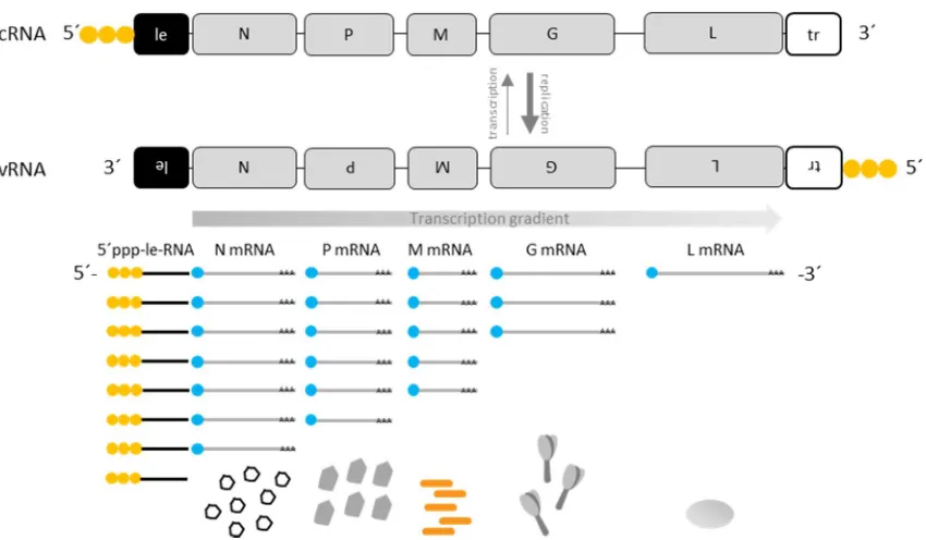
2013)). The primary transcription starts by binding of the RNA-dependent RNA polymerase
complex (L-P complex) to the genomic promoter, followed by the transcription of the 58
nucleotides (nts) long, non-coding, 5´-triphosphorylated, non-polyadenylated leader RNA.
Subsequently, the monocistronic mRNAs of N, P, M, G and L are transcribed into 5´-end capped
and 3´-end polyadenylated mRNAs (reviewed in (Wunner and Conzelmann, 2013)). The
transcription of the genes downstream of N (P, M, G and L) depend on the reinitiation of the
transcriptase after the IGS at the transcription restart signal. The decreasing probability for the
transcriptional reinitiation results in a gradient of viral mRNA transcripts, according to the gene
order in the genome. The steepness in mRNA levels in RABV is more striking than in other
Rhabdoviridae (e.g. vesicular stomatitis virus), since the length of IGS increase in RABV from a dinucleotide in between N-P, to pentanucleotides between P-M and M-G, up to 19-28 nucleotides
between G-L. This causes a steeper mRNA gradient compared to VSV, which has only two
10
Figure 2: Rabies virus replication cycle.
The schematic representation of the RABV transcription and replication cycle was adapted from the dissertations of Alexander Ghanem (2012) and Marco Wachowius (2016). First, the non-segmented negative-sense genomic vRNA is transcribed. The transcription starts at the 3´-end of the vRNA and generates a non-coding, 5´-end triphosphorylated, non-polyadenylated leader RNA, followed by the transcription of five monocistronic 5´-end capped and 3´-end polyadenylated mRNAs. In between the viral genes are non-coding regions, which encode for a transcriptional stop signal, a polyadenylation signal, an IGS and a transcription restart signal. The decreasing chance of reinitiation of the L-P complex at the transcription restart signal, in combination with the increasing length of IGSs results in a transcriptional gradient. RABV N and RABV M concentrations regulate the transition from transcription to replication, which results in the antigenome (cRNA). The stronger antigenomic promoter in the trailer leads to a more efficient replication of the cRNA, leading to a vRNA/cRNA ratio of 49:1 that is indicated by the bold replication arrow.
The transition from transcription to replication, in which the cis-acting elements in the gene
borders must be skipped, is still topic of ongoing research and the gained knowledge bases
especially on the vesicular stomatitis virus (VSV). The nucleoprotein concentration seems to play
an important role in the regulation of the transition from transcription to replication. The binding
of nucleoproteins to the nascent leader RNA results in a skipping of the transcriptional stop and
start signals and an N-encapsidation of the nascent cRNA (Blumberg et al., 1981). Furthermore,
the existence of a tripartite replicase complex is discussed, consisting of N, P and L, which initiates
the replication at the genome 3´-end (Banerjee, 2008; Curran and Kolakofsky, 2008; Whelan,
2008). Next to the nucleoprotein concentration, Finke and colleagues demonstrated that the
increasing concentrations of matrix protein causes a downregulation of the transcription and
11 In the cRNA, the antigenomic promoter is located within the trailer region. Interestingly, the
antigenomic promoter exhibits a much stronger signal for replication than the genomic promoter
of the vRNA. That leads to a ratio of genome to antigenome of 49:1 (Finke and Conzelmann, 1997).
In turn, the vRNA is used for further transcription, replication, and virus assembly. In Figure 2, the
RABV replication cycle is schematically illustrated.
2.1.6 The envelope proteins
The matrix protein and the glycoprotein form together with the host cell derived double
membrane the envelope of RABV. Both proteins are multifunctional. In the following, the
functions of M and G, especially concerning budding and transsynaptic spread, are explained in
more detail.
2.1.6.1 The matrix protein
RABV M is 202 amino acids long and has a molecular weight of about 25 kDa. The matrix protein
is located on the cytoplasmic site of the viral envelope (Mebatsion et al., 1999). For VSV M, the
binding of the matrix protein to the lipid bilayer bases on electrostatic as well as hydrophobic
interactions (Ye et al., 1994). In the case of RABV M, an additional palmitoylation anchor might
contribute to the binding of M to the membrane (Gaudin et al., 1991).
Cryo-electron microscopy studies of VSV virions have shown that the matrix protein forms a helical
mesh, in which a matrix protein is located within a groove of the helical nucleocapsid. Inside of
that groove, the matrix protein interacts with nucleoproteins of the upper and lower helical turn
and thereby stabilizes the RNP. Additional homotypic interactions between the matrix protein and
its lateral neighbor as well as with the matrix protein of the upper helical turn have stabilizing
effects for the condensed RNP complex (Ge et al., 2010). Furthermore, RABV M interacts with the
cytoplasmic tail of the glycoprotein and thus links the glycoprotein with the RNP complex
(Mebatsion et al., 1999).
As already mentioned above, RABV M regulates the transition of viral transcription to replication.
Finke and colleagues showed that the transcription rate is enhanced in M gene-deletion mutants
and that the transcription rate is regulated in an M dose-dependent manner (Finke et al., 2003).
Furthermore, they identified aa R58 as a critical residue concerning the downregulation of viral
transcription and activation of viral replication (Finke and Conzelmann, 2003). Remarkably, the
ability of supporting virus assembly and budding is not impaired by a R58G mutation, indicating
that the regulation of viral RNA synthesis and the support of virus assembly and budding are two
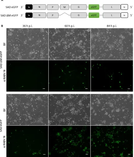
12 Mebatsion and colleagues demonstrated that the deletion of the M gene (SAD ΔM) results in a
500.000-fold reduction of virus titers in the supernatant of infected BSR cells. In addition, the few
released SAD ΔM virions do not exhibit the normal bullet-shape structure, but a long rod-shape
structure, which could be compensated by the trans-complementation with M, pointing out the
important role of the matrix protein in the condensation of the RNP complex (Mebatsion et al.,
1999). Interestingly, the deletion of the glycoprotein (SAD ΔG) caused only a 30-fold reduction in
virus titers and the virions exhibited the typical bullet-shape structure (Mebatsion et al., 1996).
These findings emphasize the central role of RABV M in virus assembly and budding.
With PPxY and YxxL, two late domain (L-domain) motifs could be identified in the matrix protein,
which overlap as followed: 35PPEYVPL41 (Okumura and Harty, 2011). The PPxY motif interacts with
the HECT E3 ubiquitin ligase Nedd4 and the YxxL motif usually mediates an interaction with the
ESCRT-III associated adaptor protein ALIX (apoptosis-linked gene 2-interacting protein X) (Chen
and Lamb, 2008). However, Wirblich et al. 2008 suggested that exclusively the PPxY motif plays a
crucial role in the RABV release (Wirblich et al., 2008).
2.1.6.2 The glycoprotein
The RABV glycoprotein is translated as a 524 aa long precursor protein, which comprises a 19 aa
long signal peptide (SP) that is cleaved off in the ER lumen. Further, the ectodomain is
N-glycosylated depending on the RABV strain on one up to three sites, followed by oligomerization
to a homotrimer in the Golgi apparatus. The final mature glycoprotein is a type I transmembrane
protein, consisting of an N-terminal, highly glycosylated ectodomain (aa 1 -439), a hydrophobic 22
aa long transmembrane domain that anchors the trimeric spikes in the plasma membrane and
after budding in the virion envelope, and a 44 aa long cytoplasmic domain (Conzelmann et al.,
1990; Tordo et al., 1986; Whitt et al., 1991; Wunner and Conzelmann, 2013).
The correct folding and thereby the functionality and stability of the RABV G is dependent on the
correct glycosylation. Especially the N-glycosylation of Asparagine 319, which is conserved among
all Lyssaviruses is essential for correct folding and the transport to the plasma membrane (Wojczyk
et al., 2005; Wunner and Conzelmann, 2013).
During the entry process, three different RABV G conformations have been identified. First, RABV
G binds to one of its cell receptors in its native pre-fusion conformation (pH 7). After
receptor-mediated endocytosis, the acidification in the early endosome leads to the transition of RABV G
into the activated conformation, in which a hydrophobic part in the glycoprotein is able to interact
with the host cell membrane. During the endosomal maturation, the endosome lumen gets more
13 resulting in the G-mediated fusion of the virus membrane with the endosomal membrane.
Remarkably, by incubation of the post-fusion state glycoprotein at a pH above 7, G can revert into
the pre-fusion conformation of the glycoprotein. That is a unique feature of the rhabdoviral
glycoproteins (reviewed (Albertini et al., 2012)).
Ectodomain of RABV G
As mentioned above, the N-glycosylated ectodomain is responsible for receptor binding, low
pH-dependent fusion of the viral membrane with the endosomal membrane, the retrograde axonal
transport and the transsynaptic spread of RABV (Etessami et al., 2000; Mazarakis et al., 2001;
Wunner and Conzelmann, 2013)
Until now, the muscular nicotinic acetylcholine receptor (nAChR) (Lentz et al., 1983), the neural
cell adhesion molecule (NCAM) (Thoulouze et al., 1998) and the p75 neurotrophin receptor
(p75NTR) (Tuffereau et al., 1998) have been described as RABV receptors (Lafon 2005). However,
the role of each single receptor in the RABV life cycle is still highly controversially discussed. The
muscle-type nAChRs are located almost exclusively at the postsynaptic membrane of
neuromuscular junctions (NMJ) (Kalamida et al., 2007; Lentz et al., 1983). However, in the brain
the neuronal nAChRs are located in presynaptic, perisynaptic and postsynaptic areas (Kalamida et
al., 2007). NCAM is concentrated in synaptic regions and at the NMJs (Lafon, 2005), where it is
located at the presynaptic membrane as well as at postsynaptic membranes within junctional folds
of NMJ (reviewed in (Lafon, 2005)). It plays a central role in the invasion of the nervous system
through RABV. p75NTR is expressed in neuronal tissues, is located at the presynaptic membrane
and is suggested to be involved in the trafficking of RABV (reviewed in (Lafon, 2005). Furthermore,
highly sialylated gangliosides, phospholipids, glycolipids and glycoproteins might play a role in the
attachment and entry of RABV in fibroblasts (Lafon, 2005; Superti et al., 1984; Superti et al., 1986;
Wunner et al., 1984).
The ectodomain comprises two major so called “antigenic sites”, which are the main targets for
virus neutralizing antibodies. Antigenic site II is a discontinuous epitope that is composed out of
two clusters. The first cluster is located between aa 34-42 and the second cluster between aa
198-200 (Prehaud et al., 1988). Antigenic site III is a continuous epitope, which is located between aa
330 and aa 338 (Seif et al., 1985). Interestingly, substitution experiments have shown that R333
of the antigenic site III is responsible for the pathogenicity of RABV (Coulon et al., 1998; Seif et al.,
1985). Another noteworthy prominent site in the RABV G is the nAChR binding site. The nAChR
binding site was identified with help of an alignment with snake venom curaremimetic
neurotoxins - a potent nAChR antagonist – and an antagonist assay of the binding of
14 competitively the binding of α-bungarotoxin to nAChR and therefore might represent the nAChR
binding site of RABV G (Lentz, 1990; Lentz et al., 1983; Lentz et al., 1987; Lentz et al., 1984)
Transmembrane domain and C-tail of RABV G
The 22 aa long hydrophobic, single alpha-helical transmembrane (TM) domain contains a
palmitoylation site at Cys 461 that most likely stabilizes the trimeric anchor. It is also discussed
that the palmitic acid might support virus budding by enabling the interaction of the C-tail with
the matrix protein (reviewed in (Wunner and Conzelmann, 2013)).
The C-terminal, cytoplasmic domain (C-tail) interacts with the M mesh and supports the virion
assembly (Mebatsion et al., 1999). Nevertheless, Mebatsion et al. demonstrated that a
recombinant virus lacking the glycoprotein C-tail was still able to spread (Mebatsion et al., 1996).
However, the C-tail contains a specific signal that is crucial for efficient G-incorporation and
therefore used for pseudotyping of RABV with chimeric glycoproteins e.g. HIV-1 Env with RABV
C-tail (Mebatsion and Conzelmann, 1996).
2.1.7 Retrograde axonal transport, transsynaptic spread and monosynaptic tracing of RABV
The first hint for a microtubule dependent axonal transport of RABV was the observation that
colchicine blocks the axonal transport of RABV (Ceccaldi et al., 1989). Interestingly, a direct
interaction of RABV P and LC8 dynein light chain– a component of the dynein motor protein
complex, which moves along microtubules to the minus end – was detected (Raux et al., 2000).
However, closer examinations revealed that the deletion of the LC8 binding site in the
phosphoprotein had no effect on retrograde axonal transport of the virus, rather on attenuation
of primary transcription and replication in neurons (Mebatsion, 2001; Tan et al., 2007).
Pseudotyping of lentiviral vectors with RABV G resulted in a retrograde axonal transport of the
lentiviral vectors, demonstrating that RABV G is responsible and sufficient for the retrograde
axonal transport (Mazarakis et al., 2001). Further analyses showed that the complete RABV
particle is transported within an endosomal vesicle along microtubules in the retrograde direction,
although this does not exclude the transport of single viral components along the axon (Klingen
et al., 2008).
Next to the active axonal transport, the glycoprotein is essential for the transsynaptic spread of
RABV, which occurs exclusively at chemical synapses and not at electrical synapses via gap
junctions (Ugolini, 2011). However, the exact molecular mechanism of the transsynaptic spread
15 The specific unidirectional retrograde transsynaptic spread of RABV via exclusively chemical
synapses make RABV to an ideal tool for neuronal tracing.
The major problem of the usage of non-modified RABV as a transsynaptic tracer is its capability of
unlimited transsynaptic spread (Ghanem and Conzelmann, 2016).
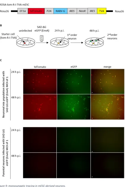
In 2007 Wickersham and colleagues established a system for mono-transsynaptic (also termed
monosynaptic) tracing, which has become the gold standard for mapping direct synaptic
connections. The system is based on an EnvA pseudotyped G gene-deficient RABV virus and a
starter cell that expresses the EnvA receptor TVA (tumor virus A), the RABV glycoprotein and the
red fluorescent protein (DsRed2) (Wickersham et al., 2007b). EnvA is the glycoprotein of the avian
sarcoma leucosis virus (ASLV) that infects avian cells via its receptor TVA. Pseudotyping of SAD ΔG
eGFP (EnvA) guarantees the specific targeting of the TVA-expressing mammalian neuronal starter
cells, as TVA is naturally not expressed in mammals (Wickersham et al., 2007b). After the primary
infection, the starter neuron expresses next to DsRed2 also the virus encoded eGFP, which enables
an identification of the starter cell. The G gene-deficient virus is trans-complemented within the
starter cell and infects presynaptically connected neurons of the starter cell. The monosynaptically
connected neurons can be identified by the eGFP fluorescence and the absence of the DsRed2
fluorescence. Since the secondary infected neurons do not express the glycoprotein, which is
essential for the transsynaptic spread of SAD ΔG eGFP, no further round of transsynaptic spread
occurs, resulting in a retrograde monosynaptic tracing system (Wickersham et al., 2007b)
16
2.2
Membrane scission and fusion machineries
2.2.1 The ESCRT machinery
The endosomal sorting complex required for transport (ESCRT) complex regulates budding events
away from the cytoplasm. Its most prominent functions are in the biogenesis of multivesicular
bodies (MVBs), the cell abscission during cytokinesis and the budding of enveloped viruses like
HIV-1 (Hurley, 2015). The ESCRT machinery, which is responsible for the MVB biogenesis is
composed of the peripheral membrane protein complexes ESCRT-0,-I, -II and –III, ALIX and the
AAA+ ATPase vacuolar protein-sorting associated protein 4 (VPS4) (reviewed in (Hurley and
Hanson, 2010)).
ESCRT-0 is responsible for the clustering of ubiquitinated cargos, ESCRT-I and ESCRT-II are
responsible for budding (meaning vesicle is still attached to the membrane) and ESCRT-III is
essential for membrane scission (reviewed in (Hurley and Hanson, 2010)).
During MVB biogenesis the heterotetramers ESCRT-0, ESCRT-I and ESCRT-II bind
monoubiquitinated membrane proteins at the endosomal membrane. ESCRT-I and ESCRT-II form
a bud neck, followed by recruitment of ESCRT-III. ESCRT-III in turn recruits deubiquitinases and
catalyzes the scission of the vesicle (reviewed in (Hurley and Hanson, 2010; Schmidt and Teis,
2012)). Additionally, ESCRT-III recruits VPS4, which disassembles the ESCRT-III complex by ATP
hydrolysis, releases ESCRT subunits into the cytoplasm, and thereby recovers the ESCRT machinery
17
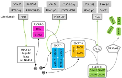
Figure 3: The ESCRT machinery
The illustration above was adapted form (Bieniasz, 2006; Chen and Lamb, 2008) and the dissertation of Anika Kern (2011). Schematic representation of the ESCRT components, which are involved in the MVB biogenesis and which are hijacked by the late domain of multiple enveloped viruses. Grey arrows indicate interactions of ESCRT components, green arrows indicate the interaction between the respective late domain and its ESCRT component, and black arrows indicate the interaction of ubiquitinated cargos and the ESCRT machinery. See text above for a detailed description of the illustration.
Interestingly, only for the MVB formation all five ESCRT complexes are necessary, while for the
separation of two daughter cells or the egress of HIV ESCRT-I, ESCRT-III, VPS4 and ALIX are
sufficient (Hurley and Hanson, 2010).
As already mentioned, many enveloped viruses use the ESCRT machinery for viral budding. It was
shown that four peptide motifs (P(S/T)AP, YP(x)nL, PPxY and FPIV) can mediate the interaction with
specific components of the ESCRT machinery and thereby support the ESCRT-dependent virus
budding (Figure 3). As these motifs play a role in the late stage of the virus life cycle, they are
named late (L) domains (reviewed in (Chen and Lamb, 2008; Freed, 2002; Votteler and Sundquist,
2013)). Interestingly, some viruses like EBOV encode for overlapping late domains (PTAPPEY)
(Chen and Lamb, 2008). It is also suggested that RABV with PPEYVPL encodes for an overlapping
late domain, which is composed of PPxY that binds Nedd4 and YxxL that binds ALIX (Okumura and
Harty, 2011). Another interesting feature of late domains is that some of them are exchangeable.
Parent et al. demonstrated that the late domains of the retroviruses RSV and HIV are exchangeable
18 The late domain PTAP of HIV-1 is located in the p6 region of the precursor polyprotein Gag
(Gottlinger et al., 1991; Huang et al., 1995). It was shown that PTAP binds Tsg101 (a subunit of
ESCRT-I), thereby recruits ESCRT-I, ESCRT-III and VPS4 to the site of viral budding at the plasma
membrane, followed by the release of the HIV virions (Pornillos et al., 2003) (reviewed in (Chen
and Lamb, 2008; Votteler and Sundquist, 2013). The tetrapeptide FPIV was identified in the matrix
protein of the parainfluenza virus-5. It was demonstrated that FPIV can replace PTAP in HIV-1
(Schmitt et al., 2005). However, the cellular interaction partner of FPIV motif is unknown (Chen
and Lamb, 2008). The YP(x)nL motif was identified by Puffer and colleagues in the Gag p9 region
of the equine infectious anemia virus (EIAV), which is the only lentivirus that does not contain the
P(S/T)AP motif (Puffer et al., 1997; Strack et al., 2003). YP(x)nL binds ALIX/AIP1, which in turn
interacts with Tsg101 (ESCRT-I subunit) and CHMP4 (ESCRT-III subunit) and thereby enables the
EIAV budding (Strack et al., 2003). The PPxY late domain was first described in the Gag p2 region
of the rous sarcoma virus (RSV) (Xiang et al., 1996). PPxY interacts with the membrane-localized
Nedd4, a HECT E3 ubiquitin ligase. Nedd4 ubiquitinates cargo proteins and thereby directs them
to the MVB sorting pathway (Sette et al., 2010). The binding of Nedd4 to the viral matrix protein
via the PPxY late domain, causes the mono-ubiquitination of the viral matrix proteins, as well as
the recruitment of the ESCRT machinery and the virus release (Han et al., 2014; Harty et al., 2001).
Like the majority of the Retroviridae, also matrix proteins of the Filoviridae and Rhabdoviridae
contain the PPxY late domain. Interestingly, the EBOV VP40 matrix protein contains overlapping
late domain sequences, consisting of PTAP and PPxY (PTAPPxY). In 2004, Irie and colleagues
demonstrated that the VSV (PPPY) late domain can be replaced by the overlapping EBOV late
domains (PTAPPEY), resulting in an 11-fold higher budding of VSV virus-like particles (VLPs),
indicating that L domains are exchangeable in terms of the efficiency to hijack the cellular ESCRT
machinery (Irie et al., 2004). The knockdown of Tsg101 and the overexpression of a
dominant-negative VPS4A, which does not bind and does not hydrolyze ATP, reduced the budding efficiency
of VSV VLPs with the EBOV late domain. However, neither the knockdown of Tsg101 nor the
overexpression of the dominant-negative VPS4A resulted in a reduction of VSV VLPs with its
natural PPxY late domain (Irie et al., 2004).
In contrast, Kielian and colleagues suggested that dominant-negative VPS4, in this approach
expressed by an inducible cell line, inhibits the viral budding of the VSV (30-fold). Additionally,
they showed that the proteasome inhibitor MG-132 that decreases the level of free ubiquitin in
the cell causes a 300-fold reduced budding of VSV, which indicates that VSV buds in an
ubiquitin-dependent way (Taylor et al., 2007). The effect of MG-132 is in agreement with previous
observations by Harty et. al., who also detected a 20 –fold reduction of VSV titers and a 16-fold
19 RABV contains an overlapping late domain near the N-terminus, which is composed of the PPxY
(interacts with Nedd4) motif and the YxxL (interacts with ALIX/AP1) motif. The entire overlapping
late domain is the heptapeptide 35PPEYVPL41. Wirblich and colleagues demonstrated that aa P35
is the most important residue for the late domain activity since mutation of this residue caused a
nearly 100-fold reduction in infectious viral titers, whereas the point mutations of P36, Y38, L41A,
and the mutations of all four aa in one construct caused at the most a 10-fold reduction.
Additionally, these data point out that the YxxL motif has no beneficial effect for RABV budding
(Wirblich et al., 2008). In summary, regardless of the conflicting findings of Harty and Kilian with
respect to the participation of VPS4 in RABV budding, Wirblich demonstrated an important role of
the PPEY motif in RABV budding.
2.2.2 SNARE proteins
Cellular soluble N-ethylmaleimide-sensitive factor attachment protein receptor (SNARE) proteins
are essential for the attachment and fusion of vesicles in general. In neurons, SNARE proteins are
essential for the presynaptic vesicle fusion and therewith e.g. the neurotransmitter release as well
as postsynaptic vesicle fusion, which is important for the postsynaptic plasticity (Harris et al., 2016;
Jurado et al., 2013).
Until now, the human SNARE protein superfamily consists of 38 members (Hong and Lev, 2014).
Interestingly, 31 out of the 38 SNARE proteins have a C-terminal transmembrane domain, while
the remaining SNARE proteins are located to the membrane by a palmitoylation (SNAP25, Snap23,
Stx9/19 and Stx11), prenylation (YKT6), or via interactions with other membrane bound SNARE
proteins (SNAP29, SNAP47) (Hong, 2005; Hong and Lev, 2014). However, all SNARE proteins
contain a 60-70 aa long, coiled-coil SNARE motif (Lou and Shin, 2016). A SNARE complex is
composed of four SNARE motifs, which assemble to a four-helix bundle (Lou and Shin, 2016). The
four-helix bundle is composed of 15 layers of interacting hydrophobic amino acids and a conserved
hydrophilic layer (0-layer) in its center, which is composed of three glutamines (Q) and one
arginine (R) residue (Lou and Shin, 2016; Sudhof and Rizo, 2011).
According to the presence of the hydrophilic residue of the SNARE motif in the center of the
four-helix bundle, SNARE motifs are grouped into Qa-SNAREs, Qb-SNAREs, Qc-SNAREs, and R-SNAREs.
The syntaxin family is specified as Qa-SNARE, the SNAP25 family is referred to as Qbc-SNARE, since
it has two SNARE motifs, and the VAMP family is referred to as R-SNARE (Hong, 2005; Lou and
Shin, 2016).
However, the SNARE proteins can be also functionally categorized into vesicular SNAREs
20 subfamily consists of VAMPs and its relatives. The t-SNAREs can be subdivided into the subfamily
of syntaxins and the subfamily of SNAP25 (Hong, 2005).
The syntaxin family (t-SNAREs/Qa-SNAREs) consists of 15 members. Syntaxins are type II
transmembrane proteins with the exception of syntaxin-19 and syntaxin-11, which have no
transmembrane domain (Hong and Lev, 2014; Teng et al., 2001). All syntaxins exhibit a specific
cellular localization, e.g. endosomes, TGN, plasma membrane. Syntaxin-1, -2, -3 and -4 are located
at the plasma membrane (Bennett et al., 1993; Teng et al., 2001). Interestingly, in the peripheral
nervous system (PNS) and CNS two isoforms of syntaxin-1 are coexpressed (syntaxin-1A and
syntaxin-1B) (Bennett et al., 1992; Bennett et al., 1993). While syntaxin-1A knockout mouse
exhibited a normal life span, the knockout of syntaxin-1B is lethal (Wu et al., 2015).
Whereas the presynaptic membrane of neurons is already well characterized with respect to the
SNARE composition, very little is known about the postsynaptic membrane. However, during the
last few years, postsynaptic functions for syntaxin-4 (Kennedy et al., 2010), syntaxin-3 (Jurado et
al., 2013) and syntaxin-1 (Hussain et al., 2016) were documented.
The synaptosome-associated protein of 25 kDa (SNAP25) subfamily (t-SNAREs) is classified as
Qbc-SNAREs since the family members have two SNARE motifs (Qb and Qc). The SNAP25 subfamily is
composed of Snap23, -25, -29, and -47. As already mentioned, Snap25 and Snap23 are associated
with the target membrane by multiple palmitoylations, whereas SNAP29 and SNAP47 have no
palmitoylation site and bind by interactions with other SNARE proteins to the target membrane
(Holt et al., 2006; Steegmaier et al., 1998).
The v-SNAREs (Vamp1, -2, -3, -4, -5, -7, -8) and the t-SNAREs YKT6 and Sec22b are structurally
classified into R-SNAREs. Vamp1, -2, -3, -4, -5, -7, and -8 exhibit an N-terminal R-SNARE motif, a
transmembrane domain and an intravesicular tail (Filippini et al., 2001). The Vamps can be
subdivided into brevins and longins. Whereas brevins exhibit a short, non-conserved N-terminal
region (Vamp1, -2, -3, -4, -5, -8), longins exhibit an approximately 110 aa long, highly conserved
longin-domain. The longin Vamps can be subdivided into the three subfamilies Vamp7, Ykt6 and
Sec22b (Daste et al., 2015; Filippini et al., 2001).
A SNARE complex assembly requires one of each structural SNARE motifs (R-, Qa-, Qb- and
Qc-SNARE) for a heterotypic membrane fusion (Sudhof and Rothman, 2009). The first and best
characterized SNARE complex is located at the presynaptic membrane, responsible for the
neurotransmitter release into the synaptic cleft and consists of the R-SNARE Vamp2, the
Qa-SNARE syntaxin-1 and the Qbc-Qa-SNARE Snap25. Briefly, the N-terminal end of the R-Qa-SNARE motif of
Vamp2 interacts with the N-terminal end of the Qa-SNARE motif of syntaxin-1 and the two
SNARE-motifs of the Qbc-SNARE Snap25, resulting in a trans-SNARE complex. The following zippering of
21 lower the energy barrier, and thereby enables the membrane fusion (van den Bogaart et al., 2013;
Zhang, 2017). The membrane fusion transforms the trans-SNARE complex to a cis-SNARE complex,
which in turn is disassembled by the ATPase NSF. R-SNAREs and Q-SNAREs are recycled and ready
for a new membrane fusion cycle (reviewed in (Lou and Shin, 2016; Sudhof and Rizo, 2011; Sudhof
and Rothman, 2009)). However, the fusion of synaptic vesicles with the membrane is a highly
complex process in that is not only regulated by SNARE proteins, but also by SM proteins
(Sec1/Munc18), Rab GTPases (Ras-related in brain GTPase), tethering factors, complexin and
synaptotagmin to mention only the most prominent (Baker and Hughson, 2016; Baker et al., 2015;
Stenmark and Olkkonen, 2001)
Figure 4: Illustration of the formation of a cis-SNARE complex.
Simplified illustration of the formation of a cis-SNARE complex at the presynaptic membrane adapted from (Dulubova et al., 2007). The cylinders represent the SNARE motifs. The synaptic vesicle interacts via the N-terminal end of the R-SNARE motif of Vamp2 with the N-terminus of the Qa-SNARE motif of Stx1 and the Qb,c-SNARE motifs of Snap25 resulting in a trans-SNARE complex (not depicted). The zippering of the four SNARE-motifs pulls the synaptic vesicle and the presynaptic membrane together, decreases the energy barrier and herewith drives the membrane fusion, resulting in a transformation of the trans-SNARE complex into a cis-SNARE complex. The components of the cis-SNARE complex are disassembled and recycled by the ATPase NSF and the vesicles are recycled and refilled with neurotransmitters(not depicted) (Sudhof and Rizo, 2011).
2.2.3 Neurotoxins
Clostridium botulinum toxins and tetanus toxin are bacterial neurotoxins that are described as
powerful tools for studying synaptic vesicle exocytosis, as they inhibit the fusion of synaptic
vesicles with the presynaptic membrane without changing the morphological structure of the
synapse (Sudhof and Rizo, 2011). Additionally, studies using neurotoxins delivered the first hint
22 postsynaptic plasticity at excitatory synapses (Kennedy et al., 2010; Lledo et al., 1998; Lu et al.,
2001).
The seven botulinum neurotoxins (BoNT/A-H) and the tetanus neurotoxin (TeNT) form the family
of Clostridial neurotoxins (CNTs). With LD50 values between 0.1 up to 1 ng/kg body weight, CNTs
represent the most toxic substances. CNTs are zinc metalloproteases, which hydrolyze specific
peptide bonds of SNARE proteins and thereby causing a blockade of neurotransmission (Binz et
al., 2010; Schiavo et al., 2000). CNTs are heterodimers consisting of a heavy chain (HC) and a light
chain (LC), which are connected by a disulfide bond. The HC is responsible for receptor binding
and translocation of the LC into the cytoplasm. The LC harbors the metalloprotease activity (Peng
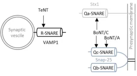
et al., 2013). TeNTLC hydrolyses the peptide bond between Gln76 and Phe77 of Vamp1, 2, and
-3, BoNT/A-LC cleaves Snap25 at position Gln197-Arg198 and BoNT/C-LC leads to the proteolysis
of Snap25 at position Arg198-Ala199 as well as of syntaxin-1A and syntaxin-1B between amino
acids Lys253-Ala254 and Lys252-Ala253, respectively (see Figure 5) (Schiavo et al., 1992; Wheeler
and Smith, 2013).
Figure 5: Cleavage site of CNTs at the neuronal SNARE complex.
23
2.3
mESCs and mESC-derived neurons at a glance
2.3.1 mESCs at a glance
During the mouse embryogenesis, three different stages of potency are passed: totipotency,
pluripotency and multipotency.
Totipotent stem cells can give rise to an entire functional organism. During mouse embryogenesis,
cells are totipotent beginning with the fertilization of the oocyte, resulting in a zygote, up to the
eight-cell stage of the morula. The morula evolves to a blastocyst, which consists of the outer
trophoblast cells and undifferentiated, pluripotent inner cells, named the inner cell mass (ICM).
The pluripotent mouse embryonic stem cells (mESCs) are isolated out of the ICM. Pluripotent stem
cells can differentiate into the three primary germ layers (ectoderm, mesoderm, and endoderm)
and into primordial germ cells. However, pluripotent stem cells cannot form an entire organism
by themselves. Pluripotent stem cells differentiate into multipotent stem cells, which differentiate
to more restricted specialized cells (reviewed in (Biehl and Russell, 2009)).
First, mESCs were cultured on mitotic inactivated mouse fibroblasts (feeder layer) in the presence
of calf serum. However, this conditions are very different from the physiological environment of
the cells. Further, nearly all pluripotent ES cell “lines”, which were derived by the cultivation on
feeder cells in the presence of calf serum, derived from the mouse strain 129 or its hybrids
(reviewed in (Martello and Smith, 2014)). Over the last decades, the requirements to keep
pluripotent, self-renewing mESC in culture were revealed. The first discovered differentiation
inhibitor was the leukemia inhibitor factor (LIF). LIF is produced by the feeder cells, where it is
mainly located at the cell surface. It activates the transcription factor Stat3 and thereby promotes
the mESC self-renewal property. However, in a feeder-free culture system with calf serum and LIF,
the ES cell culture contains some differentiated cells, which were absent in the presence of feeder
cells, indicating that feeder cells provided another important factor for the ES cell self-renewal. In
addition, the withdraw of the calf serum results within six days in the differentiation of mostly
neural precursors and neurons. A little bit conflicting seems the finding that LIF also activates
Janus-associated kinases (JAKs), which activates the extracellular signal-related kinase (Erk) that is
antagonistic to the ES self-renewal. Interestingly, the combination of LIF with the bone
morphogenetic protein (BMP) was able to inhibit the differentiation of the self-renewal ES cells in
absence of feeders and serum. Further investigations identified the fibroblast growth factor 4
(FGF4) as an autoinductive stimulation factor of the mitogen-activated protein kinase (ERK1/2).
Furthermore, the Wnt signaling pathway plays an important role in the ES cell self-renewal as it
24 demonstrated that ES cells could be cultivated in the absence of serum and LIF using
two-small-molecule inhibitors (2i) (Ying et al., 2008). PD0325901 inhibits MEK1 and MEK2 of the MEK/ERK
signaling pathway that would stimulate the differentiation of the ES cells. CHIR99021 inhibits the
glycogen synthase kinase-3 (GSK3) of the Wnt signaling pathway, thereby prevents the
phosphorylation of β-Catenin, which accumulates in the nucleus, binds the transcription repressor
Tcf3 and dissociates it from its binding site and leads to the expression of pluripotency factors.
The combination of LIF, PD0325901 and CHIR99021 (2i+LIF) results in a very robust cultivation of
self-renewal, naive pluripotent mESCs (reviewed in (Martello and Smith, 2014; Nichols and Smith,
2012; Wray et al., 2010)).
2.3.2 Introduction of mESC-derived neurons
The chance to study RABV in vitro in immortalized cell lines enabled big achievements regarding
the decipherment of the RABV biology. Fundamental insights into the RABV transcription,
replication, the functional characterization of the viral proteins and the suppression of the innate
immune system by RABV were gained. However, several specific aspects of the RABV biology, e.g
neuronal transfer, can only be addressed in neurons, which represent the natural target cells of
the neurotropic virus.
A difficulty in addressing neurobiological and neurovirological questions in vitro is the post-mitotic
nature of neurons. That is the reason why people use undifferentiated or differentiated
neuroblastoma cell lines (mouse N2A, human NS20Y), primary neuronal cells, or brain slices for
their investigations in vitro.
In 1995, three groups independently of each other reported that cultured mESCs can be
differentiated into neurons and glial cells (reviewed in (Wobus and Boheler, 2005)). Unfortunately,
mESCs tend to differentiate spontaneously even in the presence of LIF, which makes it quite
difficult to differentiation mESCs into a homogenous neuronal cell population (Bibel et al., 2007).
In most cases, the differentiation of mESCs into neuronal cultures results in a heterogeneous cell
population, which is composed of different neuronal subtypes and non-neuronal cells (Bibel et al.,
2004). However, over the last years big achievements were made regarding the development of
numerous in vitro differentiation protocols of mESC into distinct neuronal subtypes (reviewed in
(Garcia et al., 2012b)). It is now possible to differentiate mESCs into GABAergic neurons,
dopaminergic neurons, glutamatergic neurons, motor neurons, as well as non-neuronal cells. The
characterization of the mESC-derived neurons with molecular markers and electrophysiological
examinations demonstrated that the mESC-derived neurons resemble neurons of the intact brain
tissues regarding its action potentials and neurochemical profiles. Further, it was shown that
25 explants in vitro (Garcia et al., 2012a; Plachta et al., 2004) (reviewed in (Garcia et al., 2012b)).
However, the in vitro differentiation of mESCs into neurons is still a delicate process.
Due to the similarity of mESC-derived neurons with neurons in vivo, mESC-derived neurons
represent a powerful tool to investigate neurobiological questions in general as well as in this