5
XI
November 2017
International Journal for Research in Applied Science & Engineering Technology (IJRASET
)
ISSN: 2321-9653; IC Value: 45.98; SJ Impact Factor:6.887 Volume 5 Issue XI November 2017- Available at www.ijraset.com
427
©IJRASET (UGC Approved Journal): All Rights are ReservedBrain Tumor Detection Using Image Processing.
ShubhamTripathi1, Prof. Mrs. Priya Charles2,
1,
Department of electronics and telecommunication, 2
Dr. DY Patil Institute of Engineering, Management & Research, SavitribaiPhule Pune University Akurdi, Pune, India
Abstract: The basic idea of this project is to develop application software to detect the presence of brain tumour in MRI images. This project aims to develop accurate determination of tumor in the brain tumour. Here we are using image processing techniques to detect exact position of tumour. Actually we are performing morphological operations on MRI segmented and enhanced image.
Keywords: MRI, Morphological, Feature Extraction, Diagnosis
I. INTRODUCTION
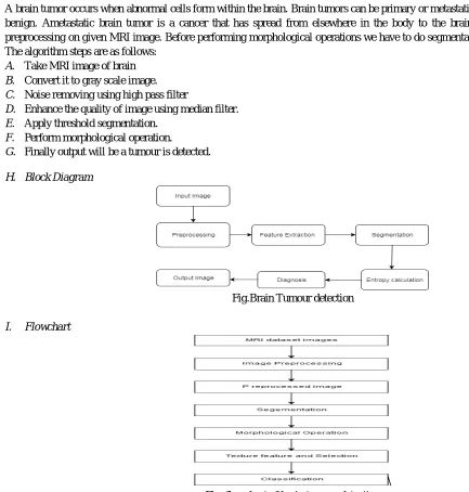
A brain tumor occurs when abnormal cells form within the brain. Brain tumors can be primary or metastatic, and either malignant or benign. Ametastatic brain tumor is a cancer that has spread from elsewhere in the body to the brain. In this project we are preprocessing on given MRI image. Before performing morphological operations we have to do segmentation of given MRI image. The algorithm steps are as follows:
A. Take MRI image of brain
B. Convert it to gray scale image.
C. Noise removing using high pass filter
D. Enhance the quality of image using median filter.
E. Apply threshold segmentation.
F. Perform morphological operation.
G. Finally output will be a tumour is detected.
H. Block Diagram
Fig.Brain Tumour detection
I. Flowchart
[image:2.612.36.469.263.717.2]II. LITERATURE SURVEY
An intelligent Highway is an innovative concept for smart roads of future smart cities. It is a Intracranial Neoplasm or Brain Tumour is abnormal growth of cells in the brain. Brain is the most complicated part of our body. The symptoms of a Tumour may be frequent headaches and migraines. Over the years it may even lead to vision loss. At this moment science is scarce about the origins and factors leading to this abnormal growth. Tumours are classified on two bases: whether they are cancerous or not and their place of origin. The noncancerous form of the Tumour is referred to as Bengn. These are easily distinguishable and have a slow growth rate. Cancerous Tumours are called Malignant. These are very aggressive and can be life threatening as these is hard to detect.When it comes to detecting a Tumour, doctors can opt for either an X-ray or an MRI. MRI’s are appropriate when all other test fail to provide sufficient information. An MRI scan uses the properties of magnetism and radio waves to produce accurate images. Neurosurgeons most commonly prescribe MRI’s as it provides them with sufficient information to detect even the smallest abnormalities. However, as MRI uses magnetic waves, so it is unsuitable for patients with pacemakers and metal implants. Now once we have the scanned image of the brain, it is important to accurately detect the Tumour, its size, and its location. All this information is necessary for the Neurosurgeon to complete his diagnosis. This is where Computerized Image Processing comes to help. With the se of different segmentation techniques and feature extraction method, we can accurately detect the Tumour [1]. In this project, proposed system divides the input image into number of slices and pre-processing takes place in parallel. This software also runs on Multi-Core environment for processing and extraction of each and every image slices separately[2]. In this paper, we have investigated the different Entropy functions for Tumour segmentation and its detection from various MRI images. The different threshold values are obtained depend on the particular definition of the entropy. The threshold values are dependent on the different entropy function which in turn affects the segmented results. the segmented results depend on the Shannon and Non-Shannon behavior at different instance of parametric selections. The texture analysis of medical images is also performed in order to get a better accuracy. The best result are obtained from HavrdaCharvat Entropy that is better than the other Entropy functions used in sense of detecting Tumours and which will help in earlier detection of the Tumours and will provide the pro treatment to the patients and thus, they can be cured [3]. In this paper we propose brain Tumour detection, Image processing for detection of Tumour, only MRI images are not able to identify the Tumourous region in this paper we are using K-Means segmentation with preprocessing of image. Which contains denoising by Median filter and skull masking is usedfor more detailed information of Tumour region[4].
III. PROPOSED WORK
A. Brain Tumour
A brain tumor occurs when abnormal cells form within the brain. There are two main types of tumors: malignant or cancerous tumors and benign tumors. Cancerous tumors can be divided into primary tumors that start within the brain, and secondary tumors that have spread from somewhere else, known as brain metastasis tumors. All types of brain tumors may produce symptoms that vary depending on the part of the brain involved. These symptoms may include headaches, seizures, problem with vision, vomiting, and mental changes. The headache is classically worse in the morning and goes away with vomiting. More specific problems may include difficulty in walking, speaking, and with sensation. As the disease progresses unconsciousness may occurs.
1) Diagnosis nervous:A brain scan is a picture of the internal structures inside the brain.
2) MRI(Magnetic Resonance Imaging):
3) A functional MRI (fMRI) provides information about the location of specific areas of the brain that are responsible for muscle movement and speech. During the fMRI examination, the patient is asked to do certain tasks that cause changes in the brain and can be seen on the fMRI image. This test is used to help plan surgery, so the surgeon can avoid damaging the functional parts of the brain while removing the tumor
4) Magnetic resonance spectroscopy (MRS) is a test using MRI that provides information on the chemical composition of the brain. It can help tell the difference between dead tissue caused by previous radiation treatments and new tumor cells in the brain.
B. Gray scale Imaging
International Journal for Research in Applied Science & Engineering Technology (IJRASET
)
ISSN: 2321-9653; IC Value: 45.98; SJ Impact Factor:6.887 Volume 5 Issue XI November 2017- Available at www.ijraset.com
429
©IJRASET (UGC Approved Journal): All Rights are ReservedC. High Pass Filter
A high pass filter is the basis for most sharpening methods. An image is sharpened when contrast is enhanced between adjoining areas with little variation in brightness or darkness. A high pass filter tends to retain the high frequency information within an image while reducing the low frequency information. The kernel of the high pass filter is designed to increase the brightness of the center pixel relative to neighboring pixels.
D. Median filter:
Such noise reduction is a typical pre-processing step to improve the results of later processing (for example, edge detection on an image). Median filtering is very widely used in digital image processing because, under certain conditions, it preserves edges while removing noise.
E. Threshold Segmentation:
The simplest method of image segmentation is called the throes holding method. This method is based on a clip-level (or a threshold value) to turn a gray-scale image into a binary image. The key of this method is to select the threshold value.
F. Morphological operation:
Morphological image processing is a collection of nonlinear operations related to the shape or morphology of features in an image.
G. Advantages
1) The image could be split progressively according to our demanded resolution because the number of splitting level is determined by us.
2) We could split image using criteria we decide, such as mean or variance of segment pixel value.
3) In addition, The merging criteria could be different to the splitting criteria.
4) Uses single MRI image.
H. Application:
5) Morphological operations have proved very helpful extraction and filtering techniques where operators like open, spur, delite, erode and close have proved to be helpful in extracting the brain tumour from MRI brain images.
IV. CONCLUSION
This project, various techniques that are being used to detect the brain Tumour from MRI images of brain are evaluated. The proposed technique has the capability to produce effective results even in case of high density of the noise. The proposed project will detect the presence of brain Tumour with increased accuracy.
REFERENCES
[1] A Survey on Brain Tumour Detection Using Image Processing Techniques. LuxitKapoor Amity School of Engineering and Technology Amity University, Noida
[2] Brain Tumour Detection and Segmentation In MRI Images AbhijithSivarajan S1, Kamalakar V. Thakare2, Shailesh Kathole3, Pramod B. Khamkar4, Danny J. Pereira5 Department of Computer Engineering, Govt. College of Engineering and Research, Avasari (Kd), Pune,
[3] An efficient Brain Tumour Detection from MRI Images using Entropy Measures DevendraSomwanshiAshutosh Kumar Pratima Sharma Deepika Joshi Poornima University, Research Scholar, JNU.