Update on Malaria Diagnostics and Test
Utilization
Blaine A. Mathison,aBobbi S. Prittb
Parasitology and Fecal Testing Laboratory, Infectious Disease Division, ARUP Laboratories, Salt Lake City, Utah, USAa; Division of Clinical Microbiology, Mayo Clinic, Rochester, Minnesota, USAb
ABSTRACT Malaria is a potentially life-threatening disease requiring rapid diagnosis and treatment. Although microscopic examination of thick and thin blood films re-mains the gold standard for laboratory diagnosis, rapid antigen tests and nucleic acid amplification methods may also play a useful role in detection of acute infec-tion. This review discusses the advantages and disadvantages of the commonly used diagnostic methods and provides important practice points for optimal malaria test utilization.
KEYWORDS diagnostics, malaria, utilization
CAUSAL AGENTS
M
alaria is a potentially life-threatening disease caused by apicomplexan parasites inthe genusPlasmodium (1–3). Human infection occurs throughout much of the
tropics and subtropics and is caused primarily by four species:P. falciparum,P. vivax,P.
ovale, andP.malariae.In parts of Southeast Asia, the zoonotic parasiteP. knowlesialso causes a high proportion of locally acquired cases (1). Another nonhuman primate
Plasmodiumspecies,P. cynomolgi, was also reported as a rare cause of human malaria
in southeast Asia (4). Plasmodium simiumandP. brasilianumwere documented from
human patients in South America, although there is increasing evidence that these
species actually represent P. vivax and P. malariae, respectively, which adapted to
nonhuman primates after being introduced to South America (4).Plasmodium
falcipa-rum is responsible for the majority of malaria-related deaths, with far fewer deaths
associated withP. vivaxand, rarely, other species (1, 2).
MALARIA TODAY
Malaria remains an important cause of morbidity and mortality worldwide (3, 5). The World Health Organization (WHO) estimates that 212 million cases occurred during 2015 (range, 148 million to 304 million), with 429,000 deaths (range 235,000 to 639,000) (3). The majority of infections occur in Africa (90%), followed by the southeast Asia region (6%), and the eastern Mediterranean region (2%) (3). While there has been a 29% decrease in malaria mortality rates since 2010, significant work is still needed to meet the WHO global targets of reducing malaria case incidence and mortality
rates byⱖ90% by 2030 (6).
In settings in which malaria is not endemic, such as the United States, infection is seen primarily among individuals who have traveled to, or emigrated from, regions with ongoing malaria transmission (i.e., “imported malaria”) (7). The Centers for Disease Control and Prevention (CDC) received reports of 1,727 malaria cases in 2013 with 10 associated fatalities; this is a 2% increase from the number of cases reported in 2012 and the highest number of malaria-associated deaths since 2001, underscoring the need for considering malaria in the clinical diagnosis and maintaining expertise in
malaria diagnosis in the United States (7).Plasmodium falciparumwas identified as the
causative agent in 60.8% of cases, followed byP. vivax(14.1%),P. ovale(3.7%), andP.
Accepted manuscript posted online12
April 2017
CitationMathison BA, Pritt BS. 2017. Update
on malaria diagnostics and test utilization. J
Clin Microbiol 55:2009 –2017.https://doi.org/
10.1128/JCM.02562-16.
EditorColleen Suzanne Kraft, Emory University
Copyright© 2017 American Society for
Microbiology.All Rights Reserved.
Address correspondence to Bobbi S. Pritt, [email protected].
crossm
on May 16, 2020 by guest
http://jcm.asm.org/
malariae(2.5%), and the causes of the remaining cases were undetermined (16.6%) or mixed (2.3%).
DIAGNOSIS
The clinical features of malaria are nonspecific and overlap significantly with those of other febrile illnesses. Therefore, the WHO recommends that all patients have a parasite-specific laboratory test performed to confirm the clinical impression (8). Lab-oratory testing options vary based on the geographic and clinical setting. While a variety of options may be available in high-income countries, resource-poor settings of malaria endemicity often have limited options, and test results may not be reliable due to a lack of staff training and quality assurance measures. Given the limitations in these settings, this update focuses on testing options that are widely available in middle- to high-income settings and provides guidance on how these different methods may be employed for routine patient care.
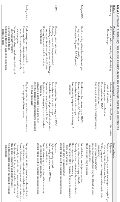
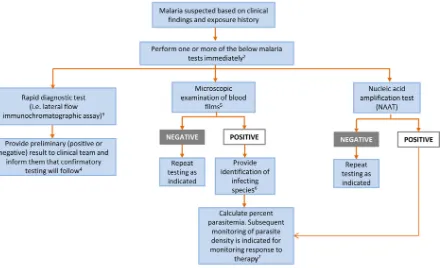
Regardless of the method used, testing should be available and performed on a STAT basis 24 hours/day, 7 days/week due to the potentially life-threatening nature of the infection. The most commonly used methods for laboratory diagnosis of malaria are microscopic examination of stained blood films and detection of parasite antigen or nucleic acid (2, 9). Of these, microscopic examination of thick and thin blood films remains the gold standard for malaria diagnosis. Rapid antigen detection methods and molecular amplification tests are also increasingly employed for malaria diagnosis and are useful adjunctive tests. Tests for detection of antiplasmodial antibodies are com-mercially available but are not recommended for diagnosis of acute disease. The details of these methodologies and their clinical utility are discussed below and summarized in Table 1, while Fig. 1 provides a laboratory testing algorithm. It is important to note that examination of a single set of blood films may be insufficient for malaria diagnosis, particularly with low levels of parasitemia. The Clinical and Laboratory Standards Institute (CLSI) recommends that repeat blood films be obtained and examined every 6 to 8 h for up to 3 days (if clinically indicated) until malaria is definitively excluded from the differential diagnosis (10). Similarly, the CDC recommends that blood smears in nonimmune individuals be repeated every 12 to 24 h for a total of 3 evaluations before ruling out malaria (11). To our knowledge, no similar guidance exists for use of other methodologies.
Inherent to any laboratory test is the need for a strong quality assurance program, including measures for adequate staff training, competency assessment, quality con-trol, and proficiency testing. Proficiency testing programs are available commercially for both blood film microscopy and antigen detection methods. In the absence of com-mercial testing programs, laboratories should use an alternative assessment of profi-ciency.
Morphological diagnosis. Morphological diagnosis may be accomplished using both light- and fluorescence-based microscopic methods (2, 9, 10). While fluorescent stains such as acridine orange may decrease screening time, they are nonspecific and may be difficult to interpret. Therefore, the discussion below focuses on morphological diagnosis using light microscopy. Regardless of the method, proper collection, smear preparation, and staining are crucial for an accurate and reliable morphological diag-nosis.
(i) Blood collection. Blood specimens should be collected without delay, ideally before antimalarial treatment is initiated. Capillary blood obtained via finger stick and venous blood obtained via venipuncture are both adequate for malaria diagnosis. The finger stick method is generally considered preferable, as anticoagulants in blood collected by venipuncture might alter parasite morphology. However, blood is more commonly obtained via venipuncture in settings in which malaria is not endemic; when this method is used, the preferred anticoagulant is EDTA. Blood films should be made as soon as possible after collection to avoid prolonged exposure to EDTA.
(ii) Smear preparation.Both thick and thin blood films should be made whenever possible, since the thick film provides the greatest sensitivity for malaria screening
on May 16, 2020 by guest
http://jcm.asm.org/
TABLE 1 Comparison of microscopy, rapid antigen detection, nucleic acid amplification methods, and serologic tests Method Practical use Advantage(s) Disadvantages Microscopy Gold standard for detecting and identifying Plasmodium spp. Allows for detection and identification to the species level of all species Allows for parasite quantification More sensitive than RDTs a Relatively inexpensive Can be used for monitoring treatment success Subjective Delays in processing can result in changes in morphology that may hinder reliable identification Challenging to train morphologists and maintain their competency Less sensitive than NAATs b Species-level identification may be difficult at lower parasitemia rates Mixed infections may be missed Antigen RDTs Rapid screening while other methodologies (e.g., microscopy) are pending Presumptive diagnosis of P. falciparum c Rapid (faster than microscopy or NAATs) Presumptive diagnosis of P. falciparum Less subjective than microscopy Low complexity; requires minimal training of personnel Not reliable for non-falciparum species c Less sensitive than microscopy or NAATs Generally requires confirmation by other methods More expensive than microscopy May not detect HRPII-negative strains of P. falciparum from South America Does not allow for quantification Should not be used to evaluate treatment success NAATs Detecting mixed infections d Detecting cases with low parasitemia d Identifying species when parasite morphology is inadequate for microscopic identification Resolving discrepant results from other methodologies More sensitive than microscopy and RDTs d Less subjective than microscopy Allows detection and species-level identification Superior for detecting mixed infections d Requires less training of personnel than does microscopy Allows for quantification (real-time PCR) Provides detection of polymorphisms associated with drug resistance d Expensive High-complexity method Not usually performed on a STAT basis Limited availability Quantification may not correlate with microscopy-determined percent parasitemia Should not be used to evaluate treatment success Serologic tests Epidemiologic surveys and research Evaluating febrile patients with recent travel to areas of endemicity who are repeatedly smear negative Supporting the diagnosis of suspected tropical splenomegaly syndrome Screening blood donors Evaluating donors in suspected transfusion-associated cases May be positive in cases where parasites are not seen on peripheral blood smears Not appropriate for detection of acute disease due to time it takes for antibodies to reach detectable levels Cannot differentiate between past and current infections (especially in patients native to areas of endemicity) Not always reliable for species-level identification May be time-consuming and labor intensive Not often available except at specialized reference laboratories a RDT, rapid diagnostic test. b NAAT, nucleic acid amplification test. c Depends on the kit used; applicable to the BinaxNOW Malaria test. d Method dependent.
on May 16, 2020 by guest
http://jcm.asm.org/
[image:3.585.72.474.55.728.2]while the thin film provides the best detection of morphology for parasite species identification. The thick film consists of 1 to 2 drops of blood spread into a circle of 1.5 to 2.0 cm in diameter. The final film is approximately 20 to 30 cell layers thick and, when dry, should be of a thickness through which newsprint can just barely be read. The erythrocytes are lysed when placed into the relatively hypotonic stain solution, releas-ing intraerythrocytic stages (i.e., trophozoites, gametocytes, intact schizonts) and thus allowing a large volume of blood to be examined in a single film. In comparison, the thin film is made using a single drop of blood that is spread in a layer such that the thickness decreases progressively toward the feathered edge. The thin film is fixed in absolute methanol and allowed to dry completely prior to staining and therefore the erythrocytes remain intact. In the feathered edge, the cells should be in a monolayer with little to no overlap. Thick and thin films can be made on the same slide or separate slides (11, 12). If thin and thick films are made on the same slide, it is very important not to allow the thick film to be exposed to methanol or methanol vapors. Otherwise, the thick film may become inadvertently fixed and possibly rendered inadequate for reading (11, 12).
It is important to let both the thick and thin smears dry completely before fixing and/or staining. Thick films take longer to dry than thin films, and this could result in a delay of the diagnosis. A scratch method was developed as an alternative method for
FIG 1Malaria laboratory testing algorithm.1Algorithm includes only commonly used/available laboratory methods. Serology should not be used
for detection of acute malaria.2Malaria can be a rapidly fatal disease, particularly when due toP. falciparum, and less commonlyP. vivaxandP.
knowlesi, and testing must be performed on a STAT basis. If testing is not available at the local laboratory, then arrangements must be made with
another nearby laboratory that can provide immediate testing. A single negative test does not rule out malaria. Perform testing every 6 to 8 h for up to 3 days if clinically indicated. Other laboratory tests (e.g., complete blood count with differential, electrolyte panel, blood glucose, bilirubin, urinalysis, blood cultures) may be indicated to assess the severity of malaria and evaluate other potential causes of the patient’s illness.
3Rapid screening tests such as lateral flow immunochromatographic assays generally provide sensitive detection of high levels ofP. falciparum
andP. vivaxinfection (i.e., 2,000 parasites/l) but lack sufficient sensitivity for detecting low levels of parasitemia (i.e.,ⱕ200 parasites/l) and other
Plasmodiumspecies.4Confirmatory testing may be performed by microscopic examination of blood films or NAAT.5Examination of both thick and
thin blood films is the gold standard for malaria diagnosis.6If necessary, refer for confirmation of species identification by blood film microscopy
or NAAT at a reference laboratory.7Percent parasitemia is calculated using microscopic examination of thick or thin blood films. (Used with
permission of Mayo Foundation for Medical Education and Research. All rights reserved.)
on May 16, 2020 by guest
http://jcm.asm.org/
[image:4.585.50.497.101.369.2]making thick films that allows for improved adherence and faster turnaround times (13). The process is similar to that of making a normal thick film, but the edge of a glass microscope slide is used for spreading the blood, while simultaneously firm pressure is applied to create small scratches in the underlying slide. The scratches allow for improved adherence of the blood film to the slide without affecting the smear morphology. The smear can then be stained as soon as it is dry, generally within 20 to 30 min of smear preparation (13).
(iii) Staining.Thick and thin films for malaria diagnosis are typically stained with Giemsa, Wright, or combined Wright-Giemsa stain (10, 12). The Wright-Giemsa stain is commonly used in the United States, as it can be used in automated hematology systems. However, the pH of this method (approximately 6.8) does not adequately highlight cytoplasmic inclusions (e.g., Schüffner’s stippling and Maurer’s clefts), which are useful for determining the infecting species. Therefore, the recommended stain for species identification is Giemsa with a pH at or around 7.2. Field stains may be helpful for rapid diagnosis of malaria but are not recommended for routine diagnosis in settings in which malaria is not endemic. Various recipes and meth-odologies for staining are available (9–12, 14). The CDC provides additional guid-ance for preparing and examining blood films when there is a clinical suspicion of
Ebola virus disease (seehttps://www.cdc.gov/vhf/ebola/healthcare-us/laboratories/
safe-specimen-management.html).
(iv) Microscopic examination.The thick film should be examined first for parasites, and the thin film used to identify organisms to the species level. Both the thick and thin
films should be examined at⫻100 magnification with oil immersion for a minimum of
100 fields, and up to at least 300 fields for immunologically naive patients (i.e., those
without previousPlasmodiumexposure), as they may present with symptoms at a lower
level of parasitemia. The region of the thin film that provides the best parasite morphology is within the feathered edge, where erythrocytes have minimal overlap and maintain their central pallor. Parasite morphology may be significantly distorted outside this region, which may lead to erroneous species identification.
When examined by knowledgeable microscopists under optimal conditions, the thick film has a reported detection threshold of 10 to 50 parasites/microliter of blood
(approximately 0.001% parasitemia, assuming an erythrocyte count of 5⫻106cells/l)
(15). The sensitivity is generally lower under field conditions, with reported detection
thresholds of 100 to 500 parasites/l of blood (15).
(v) Identification.Because the differentPlasmodiumspecies are clinically managed differently, it is important to identify the causal agent to the species level and rule out
morphologically similar organisms, such as Babesia. The morphological specifics are
beyond the scope of this work; however, we recommend several publications that may assist the technologist in making an accurate diagnosis (2, 4, 9, 11, 14, 16, 17). The treatise by Coatney et al. (4) is available for free from the CDC on both the DPDx
website (11) and as a CD-ROM (seehttp://www.cdc.gov/dpdx/CDProducts.html).
If species-level identification cannot be performed by the local laboratory, the specimen should be forwarded to a reference laboratory (commercial lab, county or state public health lab, or the CDC) for further study. In addition, the CDC’s Division of
Parasitic Diseases and Malaria offers a telediagnostic service (http://www.cdc.gov/dpdx/
contact.html) that can provide a rapid diagnosis, usually to the species level (11). A cost-savings analysis has showed that the CDC’s telediagnostic service is one-twentieth the cost of normal specimen submissions to the CDC and can provide results within 1 to 2 h during normal business hours of operation (18, 19). In a study evaluating data between October 2005 and September 2010, the CDC’s telediagnostic service was able
to confidently obtain a species-level identification in 70% of the cases (n⫽1192) (18).
While awaiting confirmatory identification, the clinical team should be notified of the potential diagnosis, and treatment begun if indicated. Given the potentially
life-threatening nature ofP. falciparuminfection, it is important to communicate with the
clinical team whenP. falciparumcannot be excluded from the differential diagnosis so
on May 16, 2020 by guest
http://jcm.asm.org/
that appropriate treatment can be started while awaiting the definitive species iden-tification.
(vi) Quantification. The quantification of malaria parasites can be used to make clinical management decisions as well as for monitoring response to treatment. Quan-tification can be performed using the thick or thin film (1, 4, 14). For quanQuan-tification using the thin film, the number of infected erythrocytes among 500 to 2,000 erythro-cytes viewed in successive fields is counted and expressed as percent parasitemia
according to the equation % parasitemia⫽ (parasitized red blood cells [RBCs]/total
RBCs)⫻100.
As with species determination, percent parasitemia should be calculated based on observations from the feathered edge monolayer, where there is little to no overlap of cells. When parasitized cells are counted, it is important to count multiply infected RBCs only once. Gametocytes are not counted, as they are a dead-end stage in the human host. Also, since some drugs are not gametocidal, the presence of gametocytes cannot be used to accurately monitor the effectiveness of treatment. For quantification using the thick film, the numbers of parasites and white blood cells (WBCs) are counted in successive fields until 500 parasites or 1,000 WBCs have been tallied (whichever comes first). The results are expressed as the number of parasites per microliter of blood using a predetermined WBC count (if the WBC count is not known, the results can be
calculated with an assumption of 8,000 WBCs/l of blood) according to the equation
parasites/ml of blood⫽(parasites/WBCs)⫻WBC count (or 8,000 cells)/ml of blood.
(vii) Monitoring response to therapy. Examination of serial blood smears with parasite quantification is recommended for monitoring response to therapy, looking for decrease in percent parasitemia and eventual parasite clearance (1). The frequency of monitoring is generally based on the clinical severity of the patient. Daily (or more frequent) testing is recommended initially for patients with severe malaria, with the caveat that the degree of parasitemia may rise during the initial 12 to 24 h since commonly used antimalaria drugs do not inhibit release of merozoites from circulating schizonts (20). Increasing parasite loads after 36 to 48 h are indicative of treatment failure due to parasite resistance (20). It may take 6 days or longer for parasite forms (not including gametocytes) to become undetectable on blood films. Therefore, repeat testing is generally recommended, at minimum, on days 7 and 28 after illness onset (20).
Antigen detection.Several antigen rapid detection tests (RDTs) are commercially available and are increasingly used for malaria diagnosis worldwide (21, 22). These methods are lateral flow assays (cassette, dipstick, or card formats) consisting of a nitrocellulose membrane with bound parasite antigens. Commonly used antigens are
P. falciparumhistidine-rich protein-II (HRPII),Plasmodiumspp. lactate dehydrogenase, and Plasmodium spp. aldolase. Depending on the format and number of antigens
present, assays may detect to the Plasmodiumgenus level only, or they may detect
specific species (e.g.,P. falciparum,P. vivax).
The WHO, in conjunction with the CDC, the Foundation for Innovative New Diag-nostics, and the Special Programme for Research and Training in Tropical Diseases, completed extensive comparisons of commercially available RDTs in 6 separate rounds and found significant disparities in performance characteristics among available
meth-ods (21). While many RDTs were capable of detectingP. falciparumat a high level of
parasitemia (2,000 or 5,000 parasites/l of blood), the sensitivities were often
signifi-cantly lower for non-falciparumspecies or for low levels of parasitemia (200 parasites/
l). Clinicians and laboratorians must therefore be familiar with the RDT used in their
facilities and its associated performance characteristics.
At this time, the BinaxNOW Malaria test (Alere, Waltham, MA) is the only test cleared
by the Food and Drug Administration (FDA) for in vitrodiagnosis of malaria in the
United States. This test targets P. falciparum-specific HRPII and an unspecified
pan-malaria antigen common toP. falciparum,P. vivax,P. malariae, andP. ovale.According
to the package insert, the BinaxNOW test has sensitivities for the detection of P.
on May 16, 2020 by guest
http://jcm.asm.org/
falciparum andP. vivax of 100% and 81.6%, respectively, using blood obtained by venous draw (23). The sensitivity of the BinaxNOW test has also been shown to vary by the degree of parasitemia; WHO product testing using wild-type (clinical) samples
revealed sensitivities of 100% and 85% for detection of high levels ofP. falciparumand
P. vivax, respectively, and only 85% and 10% for detection of low levels ofP. falciparum
and P. vivax, respectively (21). The BinaxNOW test has also been shown to be an insensitive method for detecting other species, with reported sensitivities less than 30% forP. ovale(Western Africa) andP. knowlesi(24, 25).
Several other RDTs that were evaluated in the WHO trials obtained superior
sensi-tivity rates for detection of high and lowP. falciparumandP. vivaxdensities. The reader
is referred to the free online results of the WHO trials for a full list of the assays evaluated (21). It is important to note that some of the tests used in the WHO evaluation program have been updated or discontinued since the original data were generated, and not all commercially available RDTs were represented. New RDTs continue to be developed and may provide additional advantages over existing tests.
Given the significantly lower sensitivities for non-falciparumspecies and for
detec-tion of low levels of parasitemia (all species), the BinaxNOW Malaria test should be used for malaria diagnosis only in conjunction with other laboratory tests (e.g., blood film examination) and the clinical findings (23). This holds true for use of many other
commercial malaria RDTs as well, particularly for non-falciparuminfections. A common
use of RDTs in settings in which malaria is not endemic is for preliminary malaria diagnosis when experienced microscopists are not available (e.g., night shift, small laboratories); confirmatory blood film examination is generally performed the following day. In settings of malaria endemicity, RDTs are often used alone without confirmatory blood smear examination, given the lack of resources and reliable microscopy. While not optimal, this strategy is superior to the use of clinical diagnosis alone and is
generally sufficient for identifying clinically significant infections with P. falciparum
when a suitably sensitive RDT is used.
Some other important caveats to RDT use bear mentioning. First, strains of P.
falciparumlacking thepfhrp2gene were described in Peru and, more recently, in Africa, and therefore tests targeting the HRP2 antigen may produce false-negative results (26). Furthermore, RDT sensitivity for detecting malaria in pregnant women may be de-creased, possibly due to sequestration of antigens in the placental circulation (27). Finally, antigens may remain in the bloodstream after successful treatment and there-fore RDTs should not be used to evaluate the efficacy of antimalarial therapy (11).
Nucleic acid detection. Multiple methods have been described for detection of
Plasmodium spp. nucleic acid detection, including DNA/RNA hybridization, conven-tional and real-time PCR, loop-mediated isothermal amplification (LAMP), and nucleic acid sequence-based amplification (NASBA) (2, 28). The preferred specimen type is whole blood (1 to 5 ml) collected in EDTA (2, 22, 23). Another suitable method is collection of fresh blood (e.g., from a finger prick) onto specially designed filter paper. Specimens collected in this manner are used primarily for research and epidemiologic studies and can be archived for future DNA extraction and amplification testing (29). No tests are currently cleared or approved by the FDA, but multiple laboratory-developed tests have been described and several PCR-based kits are commercially available outside the United States. The 18S small subunit rRNA gene is the most commonly used target for amplification and detection (28). Many real-time PCR assays employ different primers and probes to detect each species, using nested, seminested, and single-tube multiplex reactions. These assays take advantage of polymorphisms in the 18S rRNA gene that allow for specific detection of each species without the need for subsequent sequencing. Assays targeting a single conserved region of the 18S rRNA gene can also be used, with species differentiation performed using melting temperature analysis (28). Although there is significant variation in the performance characteristics of reported tests, nucleic acid amplification tests (NAATs) generally provide superior
sensitivity over other methods, with reported detection thresholds of⬍10 parasites/l.
on May 16, 2020 by guest
http://jcm.asm.org/
Authors at the CDC have described a nested conventional PCR with a detection
threshold of at least 6 parasites/l blood, while a highly cited real-time multiplex PCR
using TaqMan probes has reported sensitivities of 0.7, 1.5, and 4.0 parasites/l forP.
falciparum,P. ovale, andP. vivax, respectively (28). Other assays report sensitivities as
low as 0.002 parasites/l. The reader is referred to a review on this topic for a list of
assays described as of 2013 (28). New assays, and refinements to existing assays, continue to be described in this rapidly evolving field, and some have been adapted for use in resource-limited settings.
The advantages and disadvantages of NAATs for diagnosis of acute malaria are listed in Table 1. Of these, important disadvantages include their high cost and high com-plexity, which hinder the widespread implementation of molecular amplification meth-ods, particularly in low-resource settings. Also, NAAT results are not usually available in a time frame that is conducive for acute patient management. Unless testing can be performed within several hours of specimen collection, the laboratory must have an alternative method (e.g., RDT or blood film examination) for rapid laboratory detection of malaria. Regardless of the method used, blood film examination is still indicated for positive patients to calculate the percentage of parasitemia. For these reasons, the primary roles of NAATs are for confirmation of the infecting species (particularly when the parasite morphology is suboptimal), detection of low levels of parasitemia, and enhanced detection/confirmation of mixed infections. Of note, nucleic acid may remain in the bloodstream after successful treatment and therefore NAATs should not be used to evaluate the efficacy of antimalarial therapy (11). The CDC’s Division of Parasitic Diseases and Malaria provides malaria PCR testing at no cost in select situations upon consultation, and testing is also available at select reference laboratories. The CDC also offers molecular characterization using PCR and gene sequencing to detect known mutations associated with resistance to select antimalarials. Testing for drug resistance is recommended by the CDC for all cases diagnosed in the United States; however, the CDC’s resistance testing is currently for epidemiologic purposes only and is not used for clinical management of patients (1).
As with other methodologies, regular proficiency testing should be performed for NAATs. Unfortunately, there are currently no commercial options available and so testing laboratories may have to rely on in-house proficiency testing programs.
Antibody detection.Serologic testing is generally not recommended for routine diagnosis of malaria except for a few scenarios, such as febrile patients with travel to areas of endemicity who are repeatedly smear negative (especially if immunologically naive) and diagnosis of cases of suspected topical splenomegaly syndrome. Serologic testing is also the primary modality for screening blood donors and is commonly used for evaluating donors in cases of suspected transfusion-transmitted malaria. Most available tests are either immunofluorescence assays or enzyme immunoassays and may be performed on serum or plasma derived from blood samples collected in EDTA (11, 30).
REPORTING CRITERIA
Malaria is nationally reportable in the United States. All laboratory-confirmed cases are to be reported to the local state or territorial health department. Epidemiological and clinical data on confirmed malaria cases in the United States are transmitted to the
CDC via the National Malaria Surveillance System (NMSS) (see https://www.cdc.gov/
malaria/report.html).
SUMMARY
Malaria is a potentially life-threatening disease that requires rapid diagnosis and treatment. Testing should be available continuously throughout the day and night on a STAT basis; laboratories that cannot provide definitive testing (i.e., blood film exam-ination or molecular amplification methods) must provide options for rapid examina-tion elsewhere or offer preliminary testing (e.g., RDT) with confirmatory testing shortly afterward (ideally within 8 h). Microscopic examination of thick and thin blood films,
on May 16, 2020 by guest
http://jcm.asm.org/
particularly using Giemsa stain (pH 7.2), remains the gold standard for laboratory detection of malaria and identification of the infecting species, although molecular amplification methods are also suitable. Regardless of the method, all positive results should be accompanied by calculation of percent parasitemia using blood film exam-ination. All laboratory-confirmed cases should be reported to the local state or terri-torial health department.
REFERENCES
1. Centers for Disease Control and Prevention. 2017. Malaria.https://www
.cdc.gov/malaria/. Accessed 2 April 2017.
2. Pritt BS. 2015.PlasmodiumandBabesia, p 2338 –2356.In Jorgensen JH,
Pfaller MA, Carroll KC, Funke G, Landry ML, Richter SS, Warnock DW (ed), Manual of clinical microbiology, 11th ed, vol 2. ASM Press, Washington, DC. 3. World Health Organization. 2016. World malaria report 2016. World Health
Organization, Geneva, Switzerland. http://www.who.int/malaria/
publications/world-malaria-report-2016/report/en/Accessed 2 April 2017. 4. Coatney GR, Collins WE, Contacos PG. 1971. The primate malarias. U.S.
National Institute of Allergy and Infectious Diseases, Bethesda, MD. 5. Centers for Disease Control and Prevention. 2016. CDC health
informa-tion for internainforma-tional travel 2016. Oxford University Press, New York, NY. 6. World Health Organization. 2015. Global technical strategy for malaria
2016-2030. World Health Organization, Geneva, Switzerland.http://www
.who.int/malaria/publications/atoz/9789241564991/en/. Accessed 2 April 2017.
7. Cullen KA, Mace KE, Arguin PM. 2016. Malaria surveillance—United
States, 2013. MMWR Surveill Summ 65:1–22.https://doi.org/10.15585/
mmwr.ss6502a1.
8. World Health Organization. 2015. Guidelines for the treatment of
ma-laria, 3rd ed. World Health Organization, Geneva, Switzerland.http://
www.who.int/malaria/publications/atoz/9789241549127/en/. Accessed 18 March 2017.
9. Ash LR, Orihel TC. 2007. Ash and Orihel’s atlas of human parasitology, 5th ed. American Society for Clinical Pathology Press, Chicago, IL. 10. National Committee for Clinical Laboratory Standards. 2000. Laboratory
diagnosis of blood-borne parasitic diseases, vol 20. Approved guideline M15-A. National Committee for Clinical Laboratory Standards, Wayne, PA.
11. Centers for Disease Prevention and Control. 2013. DPDx.http://www.cdc
.gov/dpdx/malaria. Accessed 19 December 2016.
12. Garcia LS, Isenberg HD. 2007. Clinical microbiology procedures hand-book, 2nd ed. ASM Press, Washington, DC.
13. Norgan AP, Arguello HE, Sloan LM, Fernholz EC, Pritt BS. 2013. A method for reducing the sloughing of thick blood films for malaria diagnosis.
Malar J 12:231.https://doi.org/10.1186/1475-2875-12-231.
14. World Health Organization. 2000. Bench aids for the diagnosis of malaria infections. World Health Organization, Geneva, Switzerland.
15. Ochola LB, Vounatsou P, Smith T, Mabaso ML, Newton CR. 2006. The reliability of diagnostic techniques in the diagnosis and management of malaria in the absence of a gold standard. Lancet Infect Dis 6:582–588.
https://doi.org/10.1016/S1473-3099(06)70579-5.
16. Pritt BS. 2014. Parasitology benchtop reference guide: an illustrated guide for commonly encountered parasites. College of American Pathol-ogists, Northfield, IL.
17. Swierczynski G, Gobbo M. 2008. Atlas of human malaria. Az Color, Sirmione, Italy.
18. Mathison BA, Bishop H, Johnston SP, Xayavong MV, Arguin PM, Da Silva AJ. 2010. Trends in malaria diagnosis: combining the use of telediagno-sis, microscopy and PCR in the identification of Plasmodium spp. 59th Annu Meet Am Soc Trop Med Hyg, Atlanta, GA, 3 to 7 November 2010. 19. Rhoads DD, Mathison BA, Bishop HS, da Silva AJ, Pantanowitz L. 2016.
Review of telemicrobiology. Arch Pathol Lab Med 140:362–370.https://
doi.org/10.5858/arpa.2015-0116-RA.
20. Trampuz A, Jereb M, Muzlovic I, Prabhu RM. 2003. Clinical review: severe
malaria. Crit Care 7:315–323.https://doi.org/10.1186/cc2183.
21. World Health Organization. 2017. WHO-FIND malaria RDT evaluation
pro-gramme. http://www.who.int/malaria/areas/diagnosis/rapid-diagnostic
-tests/rdt-evaluation-programme/en/. Accessed 18 March 2017.
22. Wilson ML. 2012. Malaria rapid diagnostic tests. Clin Infect Dis 54:
1637–1641.https://doi.org/10.1093/cid/cis228.
23. Alere. 2016. BinaxNOW Malaria Test Kit,http://www.alere.com/en/home/
product-details/binaxnow-malaria.html. Accessed 2 April 2017. 24. Foster D, Cox-Singh J, Mohamad DS, Krishna S, Chin PP, Singh B. 2014.
Evaluation of three rapid diagnostic tests for the detection of human
infections with Plasmodium knowlesi. Malar J 13:60.https://doi.org/10
.1186/1475-2875-13-60.
25. Bigaillon C, Fontan E, Cavallo JD, Hernandez E, Spiegel A. 2005. Ineffec-tiveness of the Binax NOW malaria test for diagnosis of Plasmodium
ovale malaria. J Clin Microbiol 43:1011.https://doi.org/10.1128/JCM.43
.2.1011.2005.
26. World Health Organization. 2016. False-negative RDT results and
impli-cations of new reports ofP. falciparumhistidine-rich protein 2/3 gene
deletions. World Health Organization, Geneva, Switzerland.http://www
.who.int/malaria/publications/atoz/information-note-hrp2-based-rdt/
en/. Accessed 18 March 2017.
27. Fried M, Muehlenbachs A, Duffy PE. 2012. Diagnosing malaria in
pregnancy: an update. Expert Rev Anti Infect Ther 10:1177–1187.https://
doi.org/10.1586/eri.12.98.
28. Vasoo S, Pritt BS. 2013. Molecular diagnostics and parasitic disease. Clin
Lab Med 33:461–503.https://doi.org/10.1016/j.cll.2013.03.008.
29. Strom GE, Tellevik MG, Hanevik K, Langeland N, Blomberg B. 2014. Comparison of four methods for extracting DNA from dried blood on filter paper for PCR targeting the mitochondrial Plasmodium genome.
Trans R Soc Trop Med Hyg 108:488 – 494.https://doi.org/10.1093/trstmh/
tru084.
30. Wilkins P, Nutman TB. 2015. Immunological and molecular approaches
for the diagnosis of parasitic infections.InJorgensen JH, Pfaller MA,
Carroll KC, Funke G, Landry ML, Richter SS, Warnock DW (ed), Manual of clinical microbiology, 11th ed, vol 2. ASM Press, Washington, DC.