INVFSTIGATIOOS OF
NONHISTONE CHROMa>OMAL PROfEINS
Thesis by
Sarah Carlisle Roberts El.gin
In Partial Fulfillment of the Requirements For the Degree of
Doctor of Philosopbiv
California Institute of Technology Pasadena, California
1972
i i
To
Robert,
iil
ACKNOWLEDGMENTS
I woulci like to thank my first teachers who encouraged and directed rrry explorations in chemistry and biochemistry: 1"'.:r. George Birrell of South Salem High School, and Dr. John E. Quinlan and
Dr. Neal Cornell of Pomona College. I would like to thank my cornmi ttee members for their vad ous contributions to my education: Dr. Bonner, for teaching me to ask important questions, Dr. Dreyer, for teaching me to ask the questions
I
want most answered, and Dr.N.
Davidson, for keeping the process somewhat down to earth. Dr. Dreyer, Dr. Hood, and many members of their research groups, particularly John Smart, Terry Laico, Erikki R.ouslahti, and Jim Prahl deserve thanks for assistance a11d advtce concernine techniques of protein chemistry. Dr. Strumwasser directed my tentative explorations of neurobiology, a field in which it is hoped research similar to that presented here will be possi0le in the future. Many colleaeues deserve thanks for assistance, helpful rUscussions, and good companionship: David McConnell, Stan Froehner, Lois Toevs, John Mayfield, John Smart, Mike Dahmus, Ron Kono:)ka, ann others. Most important, I would like to thank my research advisor, Dr. Ja.mes Bonner, for continuing advice, freedom, criticism, and encour;1gement, all in appropriate measure,without whose enthusiasm science would be much less fun.iv
Abstract
The major nonhistone chromosomal proteins (NHC proteins) are a
group of 14-20 acidic proteins associated with DNA in eukaryotic
chroma.tin. In comparisons by SDS gel electrophoresis (molecular
weight sieving) one observes a high degree of homology among the NHC
protein fractions of different tissues from a given species.
Tissue-specific protein bands are also observed. The appearance of a new
NHC protein, A, in the NHC proteins of rat liver stimulated to divide
by partial hepatectomy and of rat ascites cells suggests that this
protein may play a role in preparing the cell for division. The NHC
proteins of the same tissue from different species are also very
similar. Quantitative but not qualitative changes in the NHC proteins
of rat uterus are observed on stimulation (.!!l vivo) with estrogen.
These observations suggest that the major NHC proteins play a general
role in chromatin structure and the regulation of genome expression;
several may be enzymes of nucleic acid and histone metabolism and/or
structural proteins analogous to histones. One such enzyme, a
protease which readily and preferentially degrades histones, can be
extracted from chromatin with 0.7 N NaCl.
Although the NHC proteins readily aggregate, they can be
separated from histone and fractionated by ion exchange chromatography
on Sephadex SE C-25 resin in 10 M urea-25~ formic acid (pH 2.5). Following further purification, four fractions of NHC protein are
obtained; two of these are single purified proteins, and the other two
v
ratio of acidic to basic amino acids from 2.7 to 1.2 and isoelectric points from apparently less than
3.7
to 8.0. These isolated fractions appear more soluble and easier to work with than any whole NHCvi
TABIE OF CCNTENTS
CHAPTER TITLE PAGE
Acknowledgments iii
Abstract iv
1 INTRODUCTION TO NONHISTWE CHROMC60MAL 1
PRorEINS
The Reason Why 2
Possible Roles of the NHC Proteins 3
Preparations 0 /
Heterogeneity 11.
Interaction with Histones and DNA 12
Enzyme Activities 16
References 21
2 LIMITED HETEROOENEITY OF THE MAJCR 27
NONHISTWE CHROMC60MAL PRorEINS
Introduction 28
Methods 29
Results
JO
Discussion )3
References 34
3 A NEW CHROMC6 OMAL PRorEIN IN RAPIDLY Jh
DIVIDING RAT LIVER CEU.S
Introduction J7
Methods 37
Results 40
Discussion 54
vii
TABLE OF CCNTmTS
CHAPTER
TITLE
PAGE
4
PARTIAL FRACTICNATICN AND CHEMICAL
58CHARACTERIZATIW OF THE MAJCR NONHISTWE
CHROMOOOMAL PRO!'EINS
Introduction
59
Methods 60
Results 72
Disaussion
9
8
References 106
5
COLIABCRATIVE STUDIES CN THE BIOLOOICAL
109FUNCTICN OF THE NWHISTCNE CHROMOOOMAL
PRO!'EINS
I
HISTOOE PROTEASE: A CHROMCSOMAL ENZYME
110Introduction 111
Methods 115
Results 119
Discussion 119
References 123
II
QUANTITATIVE CHANGES IN RAT UTERINE
125NONHISTCNE CHROMOSOMAL PRO!'EINS 00
STIMULATION WITH ESTROOEN
Introduction 126
Methods 127
Results 13?
Discussion 1.35
1
CHAPTER 1
2
The Reason Why
The chromatin of the eukaryotic cell is a complex of DNA, RNA
histone, and nonllitone chromosomal protein (NHC protein). Chromatin
can be isolated for in vitro study by chemically gentle techniques
based on differential centrifugation (Bonner ,!1 al., 1968a). That
such isolated chromatin is a good model for chromatin as it exists
in tlY2, has been established by experiments which show that its properties as a template for DNA-dependent RNA polymerase are not
altered by isolation. To the extent that can be determined by
present analytical techniques, the same regions of the genome are
transcribed in, vitro as
in
~ (Marushige and Bonner, 1966; Pauland Gilmour, 1966, 1968; Bekhor et al., 1969; Smith et al.,1969;
Tan and Miyagi, 1970). The role of the various components of
chromatin in directing the observed limited, tissue-specific
trans-cription of the genome is a problem of central interest in biology
today. The structure and function of the histones have been studied
extensively. The relevant literature on this subject has been
reviewed in Stellwagen and Cole (1969), Hearst and Botchan (1970),
and Elgin et al., (1971). The conclusion emerging from these studies
is that the histones are structural proteins of chromatin as well as
nonspecific repressors of template activity. There are only a small
number of different histones, eight to twelve. They act in concert,
no one species playing a key role in template repression, etc.,
although histone I has particular effects on chromatin solubility
(Smart and Bonner, 1971). Experiments in which chromatin is
3
that the tissue specificity of transcription, specificity in binding
hormone receptor complexes, etc., is not a property of the histones
but of the NHC protein component (which in these experiments also
contains chromosomal RNA) (Gilmour and Paul, 1969, 1970; Bekhor et al.,
1969; Spelsberg et al., 197la,b).
In the past there has been very little work done on the NHC
proteins, chiefly because of technical difficulties in their
isolation and fractionation; they tend to aggregate with nucleic
acid, histones, and one another. Because of the above results,
however, it is apparent that we must learn more of the chemistry and
biology of NHC proteins if we are to understand the mechanisms and
specificity of controlled gene transcription on eukaryotes. For
these reasons this thesis research was undertaken.
Possible Roles of the NHC Proteins
The nonhistone chromosomal proteins (NHC proteins) most
probably include polymerases, nucleases and other enzymes involved
in the metabolism of chroma.tin; specific repressor or activator
molecules, both those analogous to RNA polymerase sigma factor and
those analogous to the lac repressor (if such exist in eukaryotic
cells); structural proteins analogous to histones; and some nuclear
membrane components. That this protein fraction f'unctions in part in
a regulatory fashion has been inferred from findings that the
quantity of NHC protein is related to the type and physiological state
of the starting tissue (Dingman and Sporn, 1964; Bonner et al., 1968b),
4
metaphase chromosomes (Sadgopal and Bonner, 1970), and from its high meta~ bolic activity (Holoubek and Crocker, 1968). Recently, evidence has been presented for a sigma-like, species-specific enhancement of template activity by a nuclear acidic protein fraction (Teng~
f!!.,
1971). A regulatory protein factor in transcription of ribosomal genes in a eukaryotic system has also been reported (Crippa, 1970).It seems possible that some NHC proteins will be found to be
components of the nuclear membrane. A number of studies in prokaryotes, both electron microscope observations and biochemical experiments, have suggested that the bacterial chromosome is associated with the cell mei'.brane; the DNA replicase is thought to be attached to the DNA, ~nd
may also be part of the membrane complex, although recent experiments with pol A cells suggest not (Stratling and Knippers, 1971). In several cases, it appears that viral DNA's must be associated with the host cell membrane for normal replication to occur (Knippers and Sinsheimer, 1968; Hallick et al., 1969). Similarly, there is considerable evidence from electron microscope observations and biochemical studies that the DNA of eukaryotes is firmly
associated with the inner nuclear membrane, possible at the annuli (Comings and Okada, 1970a, b, c; Ormerod and Lehmann, 1971;
Zentgraf et !!l,., 1971), and that DNA synthesis occurs at the membrane (Comings and Kakefuda, 1968; Mi.zung et al., 1971; Yoshida et al., 1971). It is also known that isolated chrom~tin
5
contamination from biologically significant association, one must
seriously consider the idea that some of the NHC proteins observed
will prove to be inner nuclear membrane proteins.
There is some evidence concerning a structural role of NHC
proteins. It seems likely that the protein elements of the
s.ynaptonemal complex, etc., will proV$. to be NHC proteins (Comings and
Okada, 1971). Proteins anal~gous to the gene 32 protein of T4,
necessary for the stabilization of single stranded DNA during
replication and recombination, are now being isolated from eukaryotes.
Since such proteins are required in stoicheometric rather than
catalytic amounts, they might be observed by the techniques used in
Chapter 2 (Alberts and Frey, 1970; Alberts et al. , 1971) • A
residual NHC protein structure has also been observed in extracted
preparations of metaphase chromosomes (Maio and Schildkraut, 1967).
In
addition to problems of isolation, a further difficulty inthe study of NHC proteins is in defining what is a chromos oma.l
protein. Generally proteins prepared from nuclei, or preferably
chromatin, which can be shown to coprecipitate with histones and DNA
from low concentrations of NaCl are considered chromosomal. Johns
and Forrester (1969) have shown that in 0.14 M NaCl, calf teymus
deoxyribonucleoprotein adsorbs acidic protein from cytoplasm or
nuclear sap; approximately two-thirds of such protein is apparently
removed by 0.35 M NaCl. However, if chromatin prepared according
to·Bonner ~al., (1968a) is extracted with 0.3 N NaCl, only 10~ of
the protein, principally nonhistone, is removed (Smart, 1970). The
6
weight polypeptides, but otherwise resembles the total standard
preparations (Elgin, unpublished observation). Presumably all such
NHC proteins are in dynamic equilibrium with the chroma.tin complex
in the nucleus, so the question of the degree of association with
DNA in :tlY.2, is somewhat academic as long as cytoplasmic contamination
is ruled out.
Preparations
Several very different procedures have been used to isolate
NHC proteins. Unfortunately, most involve conditions of extreme
pH or high concentrations of denaturing reagents, ma.de necessary by
the tendency of NHC proteins to aggregate.
Benjamin and Gellhorn (1968) have described a preparation of
nuclear acidic proteins from rat and mouse liver. Purified nuclei
are extracted with 0.15 M NaCl - 0.01 M EDTA-Na2 and with 0.1 M HCl
to remove ribonucleoprotein and histones, respectively. The acidic
nonhistone protein and DNA are then extracted with 4 M CsCl at pH
11.6 (lysine buffer) and separated by equilibrium density
centrifuga-tion. The recovered protein is dialyzed and concentrated in 0.01 M
lysine-4M deionized urea-0.002 M EDTA-Na2-0.005 M e-mercaptoethanol (pH 11.6). The resulting solution of acidic proteins can be
examined for chemical characteristics and heterogeneity by gel
electrophoresis. Some of the proteins appear to be phosphoproteins.
At pH 11 the protein sediments as a single band of sedimentation
coefficient 2.?S. Unfortunately, the high pH necessary to extract
7
Holoubek and Crocker (1968) have prepared an acidic protein
fraction from Ehrlich ascites cell nuclei and have studied the
synthesis of this fraction relative to RNA synthesis. Nuclei are
extracted to remove acid-soluble proteins and RNA, after which a
0.5
M hot perchloric acid extract removes the DNA together withclosely associated acidic proteins. Some protein remains associated
with this DNA on CsCl density gradient centrif'ugation. Labeling
experiments with radioisotopes suggest that the acidic protein
fraction represents a mixture of proteins labeled independentzy.
This observation has been confirmed. with another NHC protein
preparation (see Chapter
4).
The proteins are synthesized in theconventional fashion and appear to have a higher specific activity
in situations wherein DNA-like RNA is being made in the cell.
Wang (1967) has done a considerable amount of work on the
isolation and fractionation of "chromosomal" acidic proteins. Rat
liver nuclei are isolated, washed with Mg-Tris buffer to remove
soluble proteins, and the chromatin then solubilized with 1 M NaCl.
The salt solution extract is then diazyzed to 0.14 M NaCl,
precipitating the DNA together with such proteins as are again bound
to it. The proteins left in the supernatant are considered to be
chromatin acidic proteins. These proteins can be further fractionated
by ammonium sulfate precipitation and by acid precipitation at pH
5
.
7
and pH 4.8. The protein fractions are very heterogeneous asindicated. by starch gel electrophoresis; the ammonium sulfate
fraction includes a protein with isoelectric point greater than
8
in solutions of low salt concentration, and coprecipitate with histone. Each fraction can be :f'urther purified by DEAE cellulose chromatography at pH 8.2.
In
all cases there is a significant run-off peak (suggesting histone contamination), a protein peak which elutes at approximately 0.2-0.J N NaCl, and a peak which elutes at 0.5-0.6 M NaCl (Wang and Johns, 1968). I f chroma.tin (purified according to the method of Bonner et al., (1968a)) is precipitated from 0.15 N NaCl and centrifuged. at ca. 12,000 g, the proteins of the supernatant include the whole population of nonhistonechromosomal protein and histone in small amounts; this is because centrifugation under these conditions does not pellet all of the chromatin. Wang's preparation no doubt includes these as well as other miclear proteins. A phosphoprotein fraction can be prepared from "chromatin acidic proteins" by fractionation with calcium-phosphate gel (Langan, 1967; Kleinsmith and Allfrey, 1969). Unfor-tunately, these proteins rapidly aggregate and are difficult to handle (Gershey and Kleinsmith, 1969a).
A different means of preparing nonhistone chromosomal protein has been developed by Marushige et al. (1968). Chromatin isolated according to Bonner
.!.!:..
al. (1968a) is acid extracted to remove histones and the residue solubilized in l~ sodium dodecyl sulfate9
In the presence of O.l~ Sll3 proteins prepared by this procedure
have an S value of 2.7 and an average molecular weight of 14,JQO. In
the absence of detergent the protein aggregates to higher molecular
weight forms; doubtless the standard protein preparation still has
detergent associated with it. The NHC proteins generated by this
isolation procedure can be examined directly by Sll3 gel electrophoresis
(Shapiro.!!!:, al., 1967). Thus the isolation procedure is a very
useful one for determining the heterogeneity of the nonhistone
chromosomal proteins and for comparing the NHC protein populations of
various tissues. Although the acid extraction of chromatin may
appear to be a somewhat drastic treatment, Sll3 gel electrophoresis of
native chromatin in O.l~ Sll3 indicates the presence of the same
population of NHC proteins as prepared by this method (see Chapter 2).
Shirey and Huang (1969) have used Sll3 to prepare a total chromosomal
protein fraction; however, solubility problems are again encountered
as the detergent is removed.
A method of fractionating chromosomal proteins by selective
coprecipitation with polyethylene sulfonate (PES) has been developed
(Krauze et al., 1969). Chromatin is dissociated in 5 M urea-2 N NaCl
and the DNA removed by ultracentrif'ugation. If a 20-fold dilution is
made in the presence of PES, pH 7, a PES-histone complex precipitates.
The nonhistone proteins are precipitated from the supernatant with
PES at pH 4. This fraction appears similar to Wang's on disc gel
electrophoresis (MacGillivray
!1
!J:..,
1969). Unfortunately thefractionation is incomplete and degradation and aggregation problems
10
It is also possible to fractionate chromosomal proteins by
dissociating chromatin in 5 M urea-2 M NaCl, removing nucleic acid
by centrifugation, and separating histones from NHC proteins by ion
exchange chromatography, gel electrophoresis, etc. Unfortunately,
the methods described to date are unsatisfactory; the yield of NHC
protein is low, cross contamination with histones occurs, the proteins
aggregate, etc. (Cameron et al., 1969; Elgin, unpublished
observation). Shaw and Huang (1970) have described the dissociation
of chromatin in 7 M urea-3 M NaCl; they also achieve a fairly good
separation of chromosomal proteins and DNA by shearing chromatin in
3 M NaCl and fractionating it on a Bio-Gel A-50 column. To separate
the NHC proteins from histones, these authors utilize either the
method of Marushige et
!1·
(1968) discussed above or disc gelelectrophoresis at pH 2.7 (Panyim and Chalkley, 1969).
Separate fractions of histone and NHC protein can be obtained by
eluting 2 M NaCl-5 M urea solubilized chroma.tin from hydro:xyapatite
with increasing phosphate. The NHC protein recovery is only ca.
50%,
but the fraction appears representative of total NHC protein by SDS
gel electrophoresis. One is forced to conclude, however, that NHC
proteins prepared by this method tend to aggregate, since the next
fractionation procedure employed by the authors in studying the NHC
proteins is gel filtration in 0.1~
s:r:s
(MacGillivray et al. ' 1971).AlJfrey•s group has employed a phenol extraction procedure to
obtain a nuclear acidic protein fraction that probably includes the
NHC proteins as a subset. Purified nuclei are extracted with 0.14 M
11
soluble proteins, histones, and lipids, respectively. The nuclear
acidic proteins are then extracted with an equal volume of phenol.
These proteins can be dialyzed into 0.1 M Tris-HCl (pH 8.4) containing
8.6 M urea-0.01 M EDTA-0.14 M 2-mercaptoethanol without aggregation
(Shelton and Allfrey, 1970, Teng et al., 1971). Although the authors
report that this fraction looks like NHC proteins by SDS gel
electrophoresis analysis, only about half the expected protein is
recovered. and the gels do not resemble those produced by direct
analysis of chromatin by others (e.g., Elgin and Bonner, 1970).
Finally, a method for dissociating chromatin in 25~ formic
acid-8 M urea-0.2 M NaCl (pH 2.5) and fractionating the chromosomal
proteins by ion exchange chromatography has been developed. by Elgin
and Bonner. Details of this procedure and its advantages and
disadvantages are discussed in Chapter 4.
None of the present methods of preparing NHC proteins are ideal,
particularly if one wishes to consider questions of biological
activity. One must compromise between the necessity of using solvents
to prevent aggregation and the desirability of using non-denaturing
solvents.
In
:f'urther discussion, only acidic proteins prepared fromchromatin will be referred to as NHC proteins; nuclear acidic
proteins, such as the preparations of Wang and of Allfrey discussed
above, probably include these as a subset.
Heterogeneity
There is remarkable agreement among different laboratories
on the question of heterogeneity of the major NHC proteins. In a
comparison of the NHC proteins of different tissues of a given
/ /
12
organism, or of homologous tissues of different vertebrates, one
generally observes that the banding pattern in SOS gels (molecular
weight sieving) is highly conserved. Apparent tissue-specific bands
are also observed. This aspect of the study of NHC proteins is
discussed in Chapter 2. Subsequent to the publication of Chapter 2
(Elgin and Bonner, 1970), several studies have appeared which confirm
the conclusion of limited heterogeneity of the NHC proteins.
MacGillivray et al. (1971) have compared the NHC proteins of mouse
kidney, liver, and spleen, and the NHC proteins of kidney, liver,
and brain of several organisms; in all cases they find very similar
SOO electrophoresis patterns. Shaw and Huang (1970) observe similar
patterns in urea gels, pH 2.7, for the NHC proteins of pig cerebellum
and pig pituitary. Considerable overlap but somewhat greater tissue
specificity has been described for nuclear acidic phosphoproteins
(Platz et al., 1970) and for phenol-extracted nuclear acidic proteins
(Teng et al., 1971; Shelton and Neelin, 1971). The results of these
studies are consistent with the view that the nonhistone chromosomal
protein fraction includes enzymes of chromatin metabolism and/or
structural proteins common to all tissues as well as proteins that
are tissue-specific. Further chemical and functional studies of these
proteins are required to confirm this idea.
Interaction with Histones and DNA
One of the important reasons for interest in the NHC proteins
is the possibility that they might be part of a mechanism which
13
although such specificity caildalso be dependent on the associated
chromosomal RNA (Bekhor et al., 1969; Huang and Huang, 1969). A
number of studies have suggested that nonhistone chromosomal
proteins may be involved in gene activation.
In
order to demonstratebiologically significant derepression in an
iu
vitro RNA synthesizingsystem, one must show that the observed effects cannot be explained
by a simple coprecipitation of the histone; that the nonhistone
protein is associated with the DNA; that it occurs at the normal
salt concentrations of the nucleus, approximately 0.2 M (Langendorf
et al., 1961, 1966); that the effect is on the chromatin and not on
exogenous RNA polymerase; that proteases have not been added; and
that the derepression is specific. Such experiments have not been
done. It has been observed that nonhistone chromosomal proteins can
coprecipitate with histones (Ma.rushige ~al., 1968; Wang and Johns,
1968) and so can reduce histone-nucleic acid association
(Dastugue ~al., 1970). Such coprecipitation is most pronounced at
low salt concentrations, such as those frequently used for RNA
polymerase assay systems. A nonhistone phosphoprotein fraction has
been isolated from rat liver nuclei by Langan (1967). This fraction
also forms insoluble complexes with histones in low salt concentration.
Early studies indicated that if histones are selectively
removed from chroma.tin leaving most of the NHC proteins, the resulting
material has the template activity of DNA (Marushige and Bonner,
1966). When DNA and Sll3-prepared nonhistone chromosomal proteins
are mixed in solutions of high salt concentration and associated by
14
DNA and the reconstituted material is as effective a template for
RNA synthesis as is deproteinized DNA (Marushige !!!:, al., 1968).
Early experiments suggesting that NHC protein has some repressor
capability were not interpretable since they involved acid-extraction
of chromatin (Paul and Gilmour, 1968). Acid extraction, in addition
to removing histones, can damage the remaining structure (probably
by depurination of the DNA), resulting in changes in template
activity and melting profile (Bannai and Terayama, 1969). However,
Seligy and Neelin (1970) have removed histones from chicken
erythrocyte chromatin with acid under conditions such that there is
no significant damage to the DNA and find that the residual NHC
proteins inhibit transcription by 20 to 25 percent (compared to
deproteinized DNA). There are also reports that the chromatin of
embryos shows restricted template activity not due to the presence
of histones (e.g., Johnson and Hnil.ica,1970). These suggestions of
repressor activity by NHC proteins stem from experiments with
special systems.
Gilmour and Paul (1969) have shown that a reconstituted template
(salt-urea dialysis) consisting of DNA, histone, and NHC protein
has the template activity of chromatin, while a reconstituted
template consisting of DNA and histone has no template activity.
The ability of acidic protein to inhibit the restriction of DNA
template activity by histones is not a unique property of the NHC
proteins; in the experiments of Gilmour and Paul (1969) bovine
serum albumin will do as well or better. However, the NHC protein
15
specific in terms of RNA sequences produced
(hybridization-competition criteria) (Gilmour and Paul, 1970). Similar results
have been obtained by Spelsberg !:!::, al., (1971a). It has been shown
that such NHC protein fractions contain RNA (Cameron!:!::, al., 1969).
This RNA may be identical with that shown to play a role in specific
reconstitution of chromatin (Bekhor ,!i al., 1969; Huang and Huang,
1969). Wang has shown that his acidic chromosomal protein fraction
can significantly reduce the amount of inhibition by exogenous
histone of an l!:!, vitro RNA synthesis system (DNA template). The
degree of derepression depends upon the histone used for inhibition.
Apparently the order of addition of components has no effect, but
the other problems have not yet been investigated (Wang, 1968a).
The preparation also reverses histone repression of DNA synthesis,
apparently by competitive binding of the histone (Wang, 1969). Teng
and Hamilton (1969) have demonstrated that the addition of
non-histone protein to RNA synthesizing mixtures reverses inhibition by
added histone (calf endometrium chroma.tin template). In this system
the order of addition of protein components has a definite effect,
but some mitigation of histone inhibition is achieved even if the
histone is added prior to the NHC protein. Acidic nuclear proteins
from other tissu~s are also effective. Complexes of nuclear
phosphoprotein and histone cause some inhibition of DNA-dependent
RNA synthesis in vitro, yet are not as effective as histones alone
(Lan~an and Smith, 1966; Langan, 1967). Spelsberg and Hnilica (1969)
have reported that acidic nuclear proteins, including the
16
of DNA-dependent RNA synthesis ~vitro only if they interact
directly prior to the addition of DNA template. They obtain similar results with a nuclear acidic protein fraction prepared according to Wang (1967). The observations are consistent with the ideas that NHC proteins do not in themselves repress template activity and that they do interact with histones and/or DNA to mitigate the repression by histones. To date neither a specific derepression or repression by NHC proteins per se has been demonstrated.
Enzyme Activities
A number of enzyme activities have been found in preparations of NHC proteins or acidic nuclear proteins. Several of these appear to be involved in the alteration or metabolism of histones. Among the best established is a neutral protease that preferentially attacks other histones; this enzyme is discussed in detail in
Chapter
5.
A protein phosphokinase in a phosphoprotein preparation from acidic nuclear proteins (analogous to Wang, 1967) has been demonstrated (Langan, 1968a.; Kleinsmith and Allfrey, 1969).Unfortunately, it appears that histones tend to be good substrates for kinases (possibly because of their relatively open conformation), and that histone kinase activity is widely distributed in the cell. However, it has been shown that the histone kinase(s) phosphorylate histones at specific sites, and do not phosphorylate NHC proteins
17
during spermatogenesis (Marushige, et al., 1969). Thus histone
kinase must be a nuclear if not chromosomal enzyme. Kinases which
phosphorylate nuclear phosphoproteins are also found in the nucleus
and/or associated with chromatin (Gershey and Kleinsmith, 1969b;
Takeda et al., 1971). The work of Com9 et al., (1966), Park and Kim
(1970) and Burdon and Garven (1971) suggests that a methylase
producing c -N-methyl-lysine in histones is a chromosomal protein
(see also Sekeris et al., 1967). Gallwitz (1968, 1970, 197la) has
isolated several histone-specific acetylases which are chromosomal
enzymes, i.e., are tightly bound to chromatin in the cell. The
distribution of these enzymes is organ specific (Gallwitz, 197lb).
For a more complete discussion of histone metabolism and the possible
significance of modifications such as phosphorylation, methylation,
and acetylation, see Elgin~ al., 1971.
A number of enzymes concerned with nucleic acid metabolism are
associated with chromatin. The presence of aggregate RNA polymerase
(EC 2.7.7.6) in chromatin is well established (Weiss, 1960; Huang
et al., 1960).
In
addition, soluble RNA polymerase not associatedwith the chromatin has been prepared by several laboratories
(Liao et~., 1968; Seifart and Sekeris, 1969). It has been suggested
that only half of the total RNA polymerase is firmly associated with
nuclear chromatin and/or participating in active RNA synthesis
(Liao et al., 1968). It now appears that there.are at least two,
possibly more, different DNA-dependent RNA polymerase activities in
the eukaryotic nucleus (Roeder and Rutter, 1970; Kedinger et al., 1970;
18
been purified from a Wang preparation of acidic chromosomal proteins of rat liver and calf thymus. The chromatin acidic proteins show a higher specific activity than do other fractions such as the nuclear sap (Patel, et al., 1967). The enzyme is active with templates from a variety of sources, but the greatest activity is shown with
homologous rat liver nuclear DNA (Howk and Wang, 1969a). At low enzyme/DNA ratios the preferred template is native DNA; however, at higher enzyme/DNA ratios, the preferred template is heat denatured DNA. The alteration does not seem to be the consequence of endo-nuclease activity (Howk and Wang, 1970b). The preparation may contain two polymerases, one with a preference for each type of template (Howk and Wang, 1969b). The reaction product consists of small, heterogeneous DNA fragments (Howk and Wang, 1970a).
Unfortunately, there is no evidence that these enzymes are chromosomal proteins in the strict definition, although Loeb (1970) and others
have also reported DNA polymerase activity associated with chromatin. These preparations may also include some terminal transferase
activity; such enzyme activity has been reported in a similar
preparation from calf thymus (Wang, 1968b). Cytidine triphosphate
polymerase activity has also been reported to be associated with
isolated chromatin (Duda and Cherry, 1971). A neutral
deoxyribo-nuclease (possibly EC 3.1.4.5) has been observed in the isolated
nuclei of mammalian cells and remains associated with chromatin
prepared by the method of Paul and Gilmour (1966), i.e., precipitated
from 0.2 M sodium phosphate buffer. However, preparations of
19
free of the enzyme. The enzyme is of the deoxyribonuclease-1 type, the products being 3'-hydroxyl terminated fragments of DNA (Swingel
!:!::, al., 1967). O'Connor (1969) has isolated a deoxyribonuclease from
rat liver acidic chromosomal proteins (Wang method) which functions predominantly as an endonuclease and may be identical with that reported above. A comparison of the template preference of DNA polymerase and deoxyribonuclease isolated from the acidic protein preparation and from the nuclear sap shows that enzymes with
difference preferences for template are present in different amounts
in the two fractions. Consequently, O'Connor (1969) has suggested that some of the enzymes under discussion may be chromosomal enzymes in the sense of being largely bound to chromatin in ~' while others are not. There have also been numerous reports of NAD
nucleosidase (3.2.2.5) activity associated with chromatin (e.g., Bock
.!i
!l•t
1968).Present research confirms the idea that enzymes concerned with nucleic acid metabolism are isolated with chromatin. These most likely function in repair and replication of the DNA and synthesis of RNA. It appears that the neutral protease may play a major role in changes of chromosome structure, such as the replacement of histones with protamines in spermatogenesis. Other enzymes modify
20
This introductory review of the methods of isolating NHC
proteins and their possible biological role is in part taken from
Chapter IV, ''Nonhistone Chromosomal Proteins," of the review "The
Biology and Chemistry of Chromosomal Proteins" by Sarah C.R. Elgin,
Stanley
c.
Froehner, John E. Smart, and James Bonner, in Advancesin.
Cell and Molecular Biology ,l,l (ed. by E.J. DuPraw, Academic Press,
21
References
Alberts, B.M., and Frey, L. (1970), Nature 227, 1313.
Alberts, B.M., Herrick, G., Sigal, N., and Frey,
L.
(1971),Fed. Proc.
JQ.,
1036 Abs.Bannai,
s.,
and Terayam.a, H. (1969), J. Biochem. (Tokyo) 66, 289.Bekhor,
I.,
Kung, G.M., and Bonner, J. (1969), J. Mol. Biol.12_, 351.
Benjamin, W., and Gellhorn, A. (1968), Proc. Natl. Acad. Sci.
u .s.
.22,, 262.Bock, K.W., Gang,
v.,
Berr, H.P., Kronau, R., and Grunicke, H.(1968), Eur. J. Biochem. ~' 357.
Bonner, J., Chalkley, G.R., Dahrnus, M., Fambrough, D., Fujimura,
F., Huang, R.C., Huberman, J., Jensen, R., Marushige, K.,
Ohlenbusch, H., Olivera, B.N., and Widholm, J. (1968a).
Methods Enzymol. 12b, 3.
Bonner, J., Dahrnus,
M.E.,
Fambrough, D., Huang, R.c., Ma.rushige,K., and Tuan, D.Y.H. (1968b), Science 12.2,, 47.
Burdon, R.H., and Garven,
E.V.
(1971), Biochem. Biopbivs. Acta232, 371.
Cameron, A., Gilmour,
R.s.,
Hill,E.,
Ingles, C.J., Kranze, R.J.,MacGillevray, A.J., Paul, J., and Threlfall, G. (1969).
Personal communication.
Chesterton, C.J., and Butterworth,
P.H.W.
(1971), FEBS Letters12, 301.
Comb, D.G., Sarkar, N., and Pinzino, C.J. (1966), J. Biol. Chem.
241, 1857.
Comings, D.E., and Kakefud11, 'r.(19(,8), J. Mol. Biol.
Jl,
225.22
Comings, D.E., and Okada, T.A. (1970b), Exptl. Cell Res. .§1, 62. Comings, D.E., and Okada, T.A. (1970c), Ex:ptl. Cell Res. .§1, 471. Comings, D.E., and Okada, T.A. (1971), Ex:pt. Cell Res. §.2., 104. Crippa, M. (1970), Nature 227, 1138.
Dastugue, B., Tichonicky, L. , Hanoune, J., and Kruh, J. (1970)' FESS Letters ~' 133.
Dingman,
c.w.,
and Sporn, M.B. (1964), J. Biol. Chem. ~' 3483. Duda, C.T., and Cherry, J.H. (1971), J. Biol. Chem. 246, 2489. Elgin, S.C.R., and Bonner, J. (1970), Biochemistry2,
444o.Elgin, s.c.R., Froehner, s.c., Smart, J.E., and Bonner, J. (1971) in Advances in Cell and Molecular Biology ,l, 1 (ed. by E.J. DuPraw. Academic Press, New York).
Frenster, J.H. (1965), Nature 206, 680.
Gallwitz, D. (1968), Biochem. Biophys. Res. Commun. Jg,, 117. Gallwitz, D. (1970), Biochem. Biophys. Res. Commun. 40, 236. Gallwitz, D. (197la), Fed. Proc.
JQ.,
1083 Abs.Gallwitz, D. (197lb), FESS Letters
JJ.,
306.Gershey, E.L., and Kleinsmith, L.J. (1969a), Bioch1.m Biophys. Acta 194, 331.
Gershey, E.L., and Kleinsmith, L.J. (1969b), Biochim Biophys. Acta 194, 517.
Gilmour, R.S. and Paul, J. (1969), J. Mol. Biol. 40, 137. Gilmour, R.S., and Paul, J. (1970), FEBS Letters
2,
242.Hallick, L., Boyce, R.P., and Echols, H. (1969), Nature 223, 1239. Hearst, J., and Botchan, M. (1970), Ann. Rev. Biochem. ~ 151. Holoubek,
v.,
and Crocker, T.T. (1968), Biochim. Biophys. Acta23
Howk, R., and Wang, T.Y. (1969a), Arch. Biochem. Biophys.
JJl,
238.Howk, R., and Wang, T.Y. (1969b), 158th ACS Meeting, #2l(abstract).
Howk, R., and Wang, T.Y. (1970a) Arch. Biochem. Biophys. 136, 422.
Howk, R.' and Wang, T.Y. (1970b), Europ. J. Biochem.
JJ.,
455.Huang, R.C., and Huang, P.C. (1969), J. Mol. Biol.
J.2.,
365.Huang, R.C., Ma.heshwari, N., and Bonner, J. (1960), Biochem.
Biophys. Res. Commun.
1,
689.Jackson, V., Earnhardt, J., and Chalkley, R. (1968), Biochem.
Biophys. Res. Commun.
JJ.,
253.Johns, E.W., and Forrester,
s.
(1969), Europ. J. Biochem. ~' 547. Johnson, A.W., and Hnilica, L.S. (1970), Biochem. Biophys. Acta224, 518.
Kedinger,
c.,
Gniazdowski, J.L., Mandel, J.L. Jr., Gissinger, F.,and Chambon, P. (1970) , Biochem. Biophys. Res. Commun.
1§.,
165.Kleinsmith, L.J., and Allfrey, V.G. (1969), Biochim. Biophys. Acta
m,
123.Knippers, R. and Sinsheimer, R.L., (1968), J. Mol. Biol.
1!,
17.Krauze, R.J., MacGillivray, A.J., and Paul, J. (1969), Biochem. J.
114, 40P.
Langan, T.A. (1967) in "Regulation of Nucleic Acid and Protein
Biosynthesis," Proc. Internat. Symp. Lunteren, Netherlands
(V.V. Konigsberger and L. Bosch, eds.). p. 233. (Elsevier,
Amsterdam).
Langan, T.A. (1968a), Science 162, 579.
Langan, T .A. (1968b), in "Regulatory Mechanisms of Protein
24
Langan, T.A. (1969), J. Biol. Chem. 244, 5763.
Langan, T.A., and Smith, L.K. (1966), Fed. Proc. ~' 778.
Langan, T.A., and Smith, L.K. (1967), Fed. Proc. 26, 603.
Langendorf, H., Siebert, G., Lorenz, I., Hannover, R., and
Beyer, R. (1961), Biochem Zeit,
J.:12,
273.Langendorf, H., Siebert, G., Lorenz, I., and Hannover, R., (1966),
Nature 209, 11)0.
Liao,
s.,
Sagher, D., and Fang, S.M. (1968), Nature 220, 1336.Loeb, L.A. (1970), Nature 226, 448.
Ma.cGillivray, A.J., Ingles, C.J., and Krauze, R.J. (1969),
Abstracts 6th FEBS Meeting ~2)0.
MacGillivray, A.J., Carrol, D., and Paul, J. (1971), Fb13S Letters
JJ.,
204.Maio, J., and Schildkraut, C.L. (1967), J. Mol. Biol. 24, 29.
Marushige, K., and Bonner, J. (1966), J. Mol. Biol.
1.2.,
160.Ma.rushige, K., Brutlag, D., and Bonner, J. (1968), Biochemistry
z,
3149.Marushige, K., Ling, V., and Dixon, G.H. (1969), J. Biol. Chem.
244, 5953.
Mizung,
N.s.,
Stoops, C.E., and Sinha, A.A. (1971), Nature NewBiol. 229, 22.
O'Conner, P.J. (1969), Biochem. Biop~ys. Res. Commun.
J.2,
805.Or.mered, M.G., and Lehmann, A.R. (1971), Biochim. Bioplzys. Acta,
228, 331.
Paik, W.K., and Kim,
s.
(1970), J. Biol. Chem. 245, 6010.25
Patel, G., How:k, R., and Wang, T.Y. (1967), Nature 215, 1488.
Paul, J., and Gilmour, R.s. (1966), J, l·~ol. Biol. 16, 242.
Pau1, J.', and Gilmour, R,S. (1968), J. l~ol. Biol. 1±,, 305.
Platz, R.D. , Kish, V.H., and K1einsmith, L.J. (1970). FEES
Letters 12, J8.
Roeder, R.G., and Rutter, J.J. (1970), Biochemistry
2,
254J.Sadgopal, A., and Bonner, J, (1970), Biochim. Biophys. Acta.
207, 227.
Seif~rt, K.H., and Sekeris, C,E. (1969), Xurop. J. Biochem.
2.,
408.Sekeris, C.E., Sekeris, K.S., and Gnllwitz, D. (1967), H-S. Z.
Physiol. Chem. 148, 1660.
Seligy,
v.z.,
and Neelin, J,N. (1970), Biochim. Biophys. Acta213, 380,
Shapiro, A.L., Vinuela, 8., and ~aizel, J,V. (1967), Biochem.
Biophys. Res. Commun. 28, 815.
Shaw, L.t-1,J., and Huang, R.C. (1970), Biochemistry
2,
45JO.Shelton, K,R., and Neelin, J, M. (1971), Biochemistry 10, ;~J42.
Shelton, K.R. and Allfrey, V.G. (1970), Nature 77-8, l)?.
Shirey, T., and Huang, R,G, (1969). Biochemistry§., 41J8.
Smart, J.E. (1970). Ph.D. Thesis, Calif. Inst. Tech.
Smart, J.E., and Bonner, J. (1971), J, Nol. Biol. .2§., 675.
Sm..ith, K.E., Church, R.B., and i·~cCarthy, B.J. (1969), Biochemistry
§., 4271.
26
Spelsberg, T.C., Hnilica,
L.s.
and Ansevin,A.T.
(197la),Biochim. Biophys. Acta 228, 550.
Spelsberg, T
.c.,
Steggles, A.W., and O'Nalley, B.W. (197lb),J. Biol. Chem. 246, 4188.
Stellwagon,
R.H.,
and Cole,R.D.
(1968), Ann. Rev. Biochem.J.5i,
951.Stratling, '>I. and Knippers, R. (1971), Europ. J. Biochem. 20, 3JO.
Swingle, K.F., Cole, L.J., and Bailey, J.S. (1967), Biochim.
Biophys. Acta 149, 467.
Takeda, M., Y.<imamura, H., .!lnd Ohga, Y. (1971), Biochem. Bi ophys.
Res. Co"ll..~un. 42, 103.
Tan, C.H. and Hiyagi, M. (1970), J. Nol. Biol. 2Q., 641.
Teng,
c.s.,
a!1d Hamilton, T.H. (1969), Proc. Natl. Aca.d. Sci. U.S.fil,
465.Teng,
c.s.,
Teng, C.T., and Allfrey, V.G. (1971), J. Biol. Chem.246, 3597.
':la!1g, T.Y. (1967), J. Biol. Chem. 242, 1220,
'fane.;, T. Y. (1 Of.Ra) , Exptl. Cell Res. .5,1, 2~R.
tfang, T.Y. (l96Rb), Arch. 9ioche.-r.. Biophys. 127, ?35.
W:>.ng, T.Y. (1?69), Exptl. Cell Res. :iJ..., 4£:-7.
1fang, T.Y. and Johns, E.\rl. (1Q6R), Arch. Biochem. Biophys. 124, 176.
Weiss, S,B. (1960), Proc. Natl. Acad. Sci. U.S. 46, 1020.
Yoshida,
s.,
.Modak, F.J., and Yagi, K. (1971)., Biochem. Biophys.Res. Commun. ,il, 1409.
Zentp;raf, H., Deumling, B., Jarasch, E.D., :md Franke, H.W. (1971),
J. Biol. Cehm, 246, 2986,
27
Chapter 2
LIMITED HEl'ERCXHl.'NETIY OF THE MAJOR
2?.
IKl'pri11tcd from Biochl'111i>try, ( 1970) 9, 4440.I
Copyright I (>70 hy thl· A111'-·rir.in Chemical Society .ind rl·prinll·d by pn111issio11 of the ,opyright ow111.:r.
Limited Heterogeneity of the
Major Nonhistone Chromosomal Proteins·
Sarah C. R. Elgin and James Bonner
ABSTRACT: There has been much discussion but little detailed work on the chemistry and biology of nonhistone chromo-somal protein. The principal hindrances in their study have been the tendency of the proteins to aggregate and the difficulty in dissociating them from histone and DNA. For this study purified chromatin was used as starting material. The histones were extracted with 0.4 N H,S04, and the remaining nonhistone chromosomal proteins then solu-bilized by 1 % sodium dodecyl sulfate (SOS) in O.OS M Tris
(pH 8).
DNA was next removed by ultracentrifugation. The nonhistone chromosomal proteins were then examined by SOS gel electrophoresis (molecular weight sieving). Our preparations of rat liver nonhistone chromosomal protein
Isolated interphase chromatin is composed of DNA, RNA, histones, and nonhistone chromosomal proteins (NHC proteins). 1 Little is known about the latter; their isolation and fractionation have been severely hampered by the tendency of NHC proteins to aggregate with DNA, histones, and one another. Chromatin possesses several enzymatic activities which may be associated with NHC proteins. These include RNA polymerase (Weiss, 1960; Huang et al., 1960) and a neutral protease that preferentially degrades deoxyribonucleo-histone (Furlan and Jericijo, 1967; Furlan et al., 1968).
Several general findings suggest that the NHC proteins play
• From the Division of Bioloi)I, California Institute of Technology, Pasadena, California. Recelo•d May 4, 1970. This work was supported in part by U. S. Public Health Service Grant No. GM-ll762 and in part by a predoctoral National Science Foundation fellowship to S. C. R. E. A preliminary statement of these results is appearing in Elain ttal. (1970).
l Abbreviations used are: NHC proteins, nonhistone chromosomal
proteins; SOS, sodium dodecyl sulfate; PPO, 2,5-diphenylouzole; POPOP, p-bis[2-(5-phenyloxazoiyl)lbenzcne; DOC, sodium deoxy-cholato.
B I 0 C' II l! M I ST R Y, v 0 I.. 9, N 0. 2 2, I 9 7 0
include 13 major polypeptide bands of molecular weight ca.
5000 to ca. 100,000. Homologous peptides are found in chicken liver nonhistone chromosomal protein, while an additional high molecular weight band is found in prepara-tions from chicken erythrocyte. Rat kidney nonhistonc chrom-osomal protein lacks two and possesses one additional band rdative to the rat liver protein fractions. Pea bud nonhistone chromosomal proteins include half of these same bands. The striking similarity of the nonhistonc chromosomal proteins of different organs and creatures suggests that some of them are common enzymes, such as those of nucleic acid metabolism, and/or common structural proteins (analogous to histoncs). Some of the apparent differences may be organ and species specific.
29
NONHISTONE CHROMOSOMAL PROTEINS
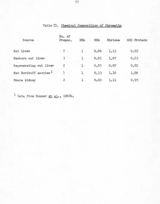
TABLE 1: Chemical Composition of Chromatin. No. of
Source Prepns DNA
Rat liver 7
Rat liver nuclei 2
Rat kidney 4
Chicken liver 3
Chicken erythrocyte 5
Pea bud 2
chromatin has been found to include lipids (Jackson et al.,
1968). Thus at present it may be supposed that the NHC
protein fraction includes DNA and RNA polymerases,
nucleases, and other enzymes involved in the metabolism
of chromatin; structural proteins perhaps analogous to,
but more acidic than, histones; nuclear membrane compo-nents; and possibly specific n:pressor or activator proteins
if they exist in eukaryotes.
For study of the structures and functions of the NHC
proteins it is necessary to dissociate them from DNA and from each other. Ideally, one would wish to have a method that would (I) extract and solubilize all NHC proteins, and (2) maintain them in their native states. Unfortunately, none
of the present methods (acid or base, salt, urea, or detergent
extraction or a combination of these) achieves both goals. We have chosen, therefore, to examine initially only the chemistry
of the NHC proteins. Acid-extracted (and therefore
histone-free) chromatin is treated with 1 % SOS, in which all NHC
proteins are solubilized. We then examine the NHC protein fraction by SOS gel electrophoresis to determine its hetero-geneity as well as the similarities and differences between the NHC proteins of selected tissues.
Methods
Preparation of Chromatin. Chromatin from rat liver, rat
kidney, and chicken liver was prepared essentially as
previ-ously described (Bonner et al., 1968a). Frozen tissue was
ground (Waring Blendor) in saline-EDTA (0.075 M NaCl plus 0.024 M EDT A, pH 8). The homogenate was filtered
through two layers of Miracloth (Chicopee Mills, Inc.) and
the pellet collected by centrifugation at l 500g for 10 min. The
pellet was washed once in saline-EDT A and four times in O.ot M Tris buffer (pH 8), being collected the last two times by centrifugation at 12,000g for 10 min. The. gelatinous crude
chromatin was further purified by centrifugation through I. 7
M sucrose (buffered with 0.01 M Tris, pH 8) for 2.5-4 hr at
50,000g. This purified chromatin was washed once, resus-pended, and dialyzed overnight against 0.01 M Tris (pH 8). The chromatin was then sheared in a Virtis homogenizer at 30 V for 90 sec and centrifuged at 12,000g for 30 min; the
super-natant, referred to as purified chromatin, or nucleohistone,
was used as the starting material for the preparation of NHC
protein.
Pea bud chrom11tin was prepared by the similar method
described by Bonner ti al. ( 1968a) with the following
alter-ations: the grinding medium w11s 0.25 M sucrose, 0.05 M
RNA 0.04 ± 0.01 0.06 0.06 ± 0.03 0.03 ± 0.01 0.02 ± 0.01 0.05
Histone 1.15±0.10 1.40 0.95±0.12 1.17 ± 0.10 1.08 ± .16 1.10
NHC Protein 0.95±0.11 1.19 0.70±0.15 0.88 ± 0.16 0.54 ± 0.14 0.41
Tris buffer (pH 8), and 0.01 M MgCl,. Crude chromatin was resuspended in 0.01 M Tris buffer (pH 8) and centrifuged through 1. 7 M sucrose for 2.5 hr to yield purified chromatin, which was resuspended, dialyzed, and sheared as above.
Chicken erythrocyte chromatin was prepared as follows.
Fresh chicken blood was centrifuged at 500g for 10 min and the supernatant and top l11yer of white cells were rcmovcd.
The erythrocytes were washed three times in saline (0.85 %
NaCl) and then lysed by dilution in an equal volume of O.ot
M CaCI,. The nuclei were then purified once by centrifugation at 750g through 0.33 M sucrose, 0.0033 M CaCI,, and 0.005
M Tris buffer (pH 7.9). The pellet was washed once in
saline-EDTA and then repeatedly with 0.01 M Tris (pH 8); the chromatin was purified and sheared as above.
That chromatin prepared by the method of Bonner et al.
(1968a) is highly purified and free from gross contamination by ribonucleoprotein particles is shown by (a) the low RNA
content of the preparations (see Table I) and (b) the absence
of basic proteins other than histones, as shown by disc gel
electrophoresis of acid extracts of chromatin (Bonner et al.,
1968a). Electron micrographs of typical pea bud chromatin show that it contains little granular matter (Griffith, 1970).
For several experiments rat liver or chicken liver chromatin
was prepared from nuclei purified by the method or Douncc
et al. (Dounce and kkowicL, 1969; Umana and Douncc, 1964). Fresh rat livers were minced and homogenized in 0.44 M sucrose, pH adjusted to S.8 with 0.1 N citric acid. The homogenate was filtered through gauze, rchomogcnized, and
diluted with one volume of 0.44 M sucrose. The pellet was collected by centrifugation at ca. 800g and washed twice with 0.44 M sucrose; it was then resuspended in 2.2 M sucrose and centrifuged at 58,500g for 90 min. The nuclei were resus-pended in saline-EDT A. Such nuclei exhibited nearly
com-plete morphological integrity with little debris under a
phase-contrast microscope as described by Chauveau et al. (1956).
The nuclei were next homogenized in 0.01 M Tris (pH 8), and the pellet collected by centrifugation at 12,000g; this step was repeated once. The final suspension was purified by
centrifu-gation through I. 7 M sucrose and sheared to nucleohistone
as detailed above.
Rat liver and rat kidney were from mah: Sprague-Dawley
rats, approximately 200g. Frozen tissues were obtained from
Pel-Freeze Biologicals, Rogers, Ark. Chicken liver and blood were from adult male White Leghorns.
Preparation of NHC Protein. The NHC proteins were
pre-pared following the procedure of Murushige l't al. (1968) with alterations as noted. Histoncs were cxtrnctcd from the
nucleohistone with 0.4 N H,SO, at 4° for 30 min. The pellet
was washed once with 0.4 N H,SO, and briefly with 0.01 M
Tris (pH 8). Over 95 % of the acid-soluble protein is removed
by this treatment (Fambrough and Bonner, 1966). The
pellet was dissolved by gentle homogenization in 1 %
SDS-0.05 M Tris (pH 8). stirred overnight at 37°, and dialyzed to
0.1 % SDS-0.01 M Tris (pH 8) at 37°. The DNA was removed
by centrifugation at 36,000 rpm for 18 hr at 25 ° in a Spinco
SW-50 rotor. The top two-thirds of the supernatant were
taken as the NHC protein preparation and analyzed by SDS
gel electrophoresis following dialysis against buffer Ill (see
below). This buffer dissociates most proteins into their
individual polypeptide chains (Shapiro et al., 1967).
Preparation of Labeled NHC Proteins. To obtain labeled NHC proteins, a ra~ was given intraperitoneally 0.055 mg
of algal protein hydrolysate-"C (0.1 mCi, uniformly labeled,
New England Nuclear Corp.) 24 hr before killing. The
liver was frozen in Dry Ice and processed, and NHC protein
was prepared from the sheared chromatin as detailed above.
Disc Gel Electrophoresis. SDS disc gel electrophoresis was
carried out according to the method of Shapiro et al. (1967) (final gel composition is 5% acrylamide, 0.133 N,N'-bis-methyleneacrylamide, 0.1 % SDS, 0.1 M sodium phosphate buffer (pH 7.1), 0.05 3 N,N,N',N'-tetramethylenediamine, and 0.075 % ammonium persulfate). Purified acrylamide
(Bio-Rad Laboratories) was used. The gels were 6 cm in length and were run at 47 V for 75 min. Gels were routinely
stained in 0.25 % coomassie brilliant blue R-250 (Mann
Research Laboratories) in 5: 5: 1 water-methanol-acetic
acid and destained sideways electrophoretically in 17 : I : 2
water-methanol-acetic acid. Gels were photographed using
an orange filter with Kodak TriX-10 4 X 5 film; the pictures
were printed on Dupont Varilour-VL-RW-SW paper. All
gels photographed together were run at the same time.
Human 'Y-globulin (Mann Research Laboratories) was used
as a molecular weight marker.
In order to detect low molecular weight proteins, the method
of Laico et al. (1970) was occasionally employed. In this case
11-cm long disc gels (same gel composition as above) are run
at 40 V for approximately 6 hr. The gels are fixed in 20 %
sulfosalicylic acid (three changes, 24-hr total), stained for 5 hr in 0.25 % coomassie brilliant blue, and photographed after
4-hr destaining in 103 acetic acid.
Samples other than NHC proteins were prepared for SDS
gel electrophoresis by dialysis against buffer I, 12 hr, room
temperature; buffer I, 12 hr, 37°; buffer II, 12 hr, room
temperature; buffer III, 4-12 hr, room temperature. (Buffer I is 13 SDS-1 % tl-mercaptoethanol in 0.01 M sodium
phos-phate buffer, pH 7.1; buffer II is 0.1 % SDS-0.1 %
tl-mercap-toethanol in 0.01 M sodium phosphate buffer, pH 7.1; buffer
Ill is 0.1 % SDS-0.1 % ,S-mercaptoethanol-10% glycerol in 0.01 M sodium phosphate buffer, pH 7.1.)
Acrylamide disc gel electrophoresis of acid-extracted
his-tones dialyzed against 8 M urea-0.01 M Tris (pH 8) was also performed by the method of Bonner et al. (1968a) at pH 4.3 in the presence of urea (15 % acrylamide gel).
To obtain NHC protein fractions for amino acid analysis,
identical samples were electrophoresed in SDS in an
eight-slot, vertical slab gel electrophoresis unit (E-C Apparatus
Corp., Model EC470) by the usual method. Samples were
run at 110-150 V for ca. 2.5 hr or until a bromophenol blue
marker (Matheson, Coleman & Bell) had traveled 9 cm.
B 1 0 C H EM 1 ST R Y, V 0 L. 9, NO. 2 2, 1 9 7 0
ELGIN AND BONNER
One strip was removed, stained, and destained as usual (first paragraph, this section) and the desired bands were cut out using this guide. The gel was broken up (by forcing it through a fine stainless steel mesh) and put in a short column; the protein was eluted with one column volume of running buffer (0.1 % SDS in 0.1 M sodium phosphate buffer, pH 7.1). This protein solution was dialyzed extensively against water at 37°, lyophilized, and analyzed for amino acid composition with a Beckman Model 120B instrument. A gel blank had to be subtracted from these values. Cytochrome c (horse heart, Mann Research Laboratories) was analyzed by this procedure as a control; the mole per cent amino acid compo-sition after gel electrophoresis of cytochrome c differed by less than 2 3 from the standard before gel ele;:trophoresis. Portions of protein samples used for amino acid analysis were dialyzed to buffer Ill and reelectrophoresed. Some higher molecular weight material, presumably aggregates, was observed.
Genual Methods. Chromatin samples were analyzed as follows. Histones were extracted with 0.4 N H,SO, and their
concentration was determined by ultraviolet absorption at 230mµ using t = 4.15 (I.fem g)(R. H.Jensen, 1966, unpublished data). The pellet was dissolved in 1.0 N NaOH and the NHC pN>tein content was determined by the method of Lowry
et al. (1951) using bovine serum albumin (Sigma) as a stan-dard. Using fresh chromatin samples RNA was separated from DNA by the modified Schmidt-Tannhauser procedure ofTs'o and Sato (1959). RNA was determined by the orcinol method (Dische and Schwarz, 1955) after hydrolysis in 0.3 M KOH; yeast RNA (Sigma) was used as a standard. DNA was determined from the ultraviolet spectrum of the nucleohistone,
assuming that all absorption at 260 mµ is due to nucleic acids and making an empirical correction for scattering (Marushige
and Bonner, 1966). The absorptivity of DNA contained in
chromatin is 22 (I.fem g) at 260 mµ (Tuan, 1967); for RNA contained in chromatin it is assumed to be 25 (I.fem g) at 260 mµ. Radioactivity of samples was determined as follows:
aliquots were dried on Bac-T-Flex membrane filters (Schlei-cher & Schuell Co.) using a vacuum oven. This technique minimizes quenching differences between the samples due to different solvent systems. Samples were counted on a Beckman liquid scintillation system LS-2008 in toluene scintillation
fluid (22.6 g of PPO plus 0.75 g of POPOP in eight pints of toluene; fluors from New England Nuclear Corp.).
Deoxy-ribonuclease I was obtained from Worthington Biochemicals; ribonuclease A from bovine pancreas was from Sigma.
Results
Chemical Composition of Chromatin. All the chromatins
used in the preparation of NHC proteins were analyzed for composition by the methods described. The results, given in
Table I, are in approximate agreement with values in the literature (Dingman and Sporn, 1964; Bonner et al., 1968a;
Smart, 1970). The preparations are reasonably reproducible
as shown by their standard deviations. The larger protein content of rat liver chromatin prepared from purified nuclei
as compared to that prepared from a crude nuclear pellet possibly results from the reduced exposure of the chromatin to cytoplasmic proteases, such as that with a preference for basic proteins (Paik and Lee, 1970). Alternatively, the increase