i
THE USE OF TWO-DIMENSIONAL
MOTION ANALYSIS AND FUNCTIONAL
PERFORMANCE TESTS FOR ASSESSMENT
OF KNEE INJURY RISK BEHAVIOURS IN
ATHLETES
ALLAN G. MUNRO
School of Health Sciences
University of Salford, Salford, UK
Submitted in Partial Fulfilment of the Requirements of the
ii Contents
Chapter 1
Introduction
1.1 Knee Injuries in Sport 1
1.2 Frequency and causes of knee injuries in men and women 2
1.3 Methods to identify high-risk athletes 2
1.4 2D motion analysis: reliability and validity 3
1.5 Functional Performance Tests: reliability, validity and clinical utility 5 1.6 Causative factors of dynamic valgus and potential interventions 7
1.7 Aims 10
Chapter 2
Literature Review
2.1 Introduction 11
2.1.1 Injuries in Sport 11
2.1.2 Anterior Cruciate Ligament Injuries 13
2.1.3 Patellofemoral Joint Injuries 14
Page Title page
Table of contents II
Tables and figures VIII
Acknowledgements XV
Glossary of terms XVI
iii
2.1.4 Incidence of Anterior Cruciate Ligament and Patellofemoral Joint Injury 14
2.2 Mechanisms of Knee Injury 16
2.2.1 Mechanisms of Anterior Cruciate Ligament Injury 16
2.2.2 Mechanisms of Patellofemoral Joint Injury 19
2.3 Risk Factors for Anterior Cruciate Ligament injuries 21 2.3.1 Extrinsic Risk Factors for Anterior Cruciate Ligament Injury 21 2.3.2 Intrinsic Factors for Anterior Cruciate Ligament Injury 21
2.3.2.1 Anatomical Risk Factors 22
2.3.2.2 Hormonal Risk Factors 25
2.3.2.2 Sagittal Plane Risk Factors 27
2.3.3 Risk Factors for Patellofemoral Joint Injury 31
2.3.3.1 Vastus Medialis Muscle Properties 31
2.3.3.2 The Illiotibial Band 33
2.3.4 Neuromuscular Control 34
2.3.4.1 Dynamic Knee Valgus and Anterior Cruciate Ligament and Patellofemoral Joint Injury Risk
35
2.3.4.2 Differences in Dynamic Valgus between Men and Women 42
2.3.4.3 Muscle Strength 46
2.3.4.4 Muscular fatigue 49
2.3.4.5 Gender differences in muscle strength 50
2.4 Summary 50
2.5 Intervention Studies 53
iv
2.5.2 PFPS Intervention Studies 64
2.5.3 Feedback 65
2.6 Screening for ACL and PFJ Injury Risk 67
2.6.1 Motion Analysis 70
2.6.2 Functional Performance Tests 74
2.6.2.1 Hop for Distance Tests 75
2.6.2.2 The Star Excursion Balance Test (SEBT) 79
2.7 Summary 81
Chapter 3
Reliability and Validity of Two-Dimensional Frontal Plane Projection Angle during common athletic screening tasks
3.1 Aim 82
3.2 Introduction 82
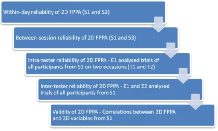
3.3 Methods 85
3.4 Results 96
3.5 Discussion 104
3.6 Conclusion 112
Chapter 4
Reliability of the Hop for Distance and Star Excursion Balance Tests
4.1 Aim 113
4.2 Introduction 113
4.3 Reliability of the Hop for Distance Tests 114
v
4.3.2 Aim 116
4.3.3 Methods 116
4.3.4 Results 119
4.3.5 Discussion 124
4.4 Reliability of the Star Excursion Balance Test 129
4.4.1 Introduction 129
4.4.2 Aim 131
4.4.3 Methods 131
4.4.4 Results 133
4.4.5 Discussion 137
4.5 Conclusion 141
Chapter 5
Factors Contributing to Dynamic Knee Valgus
5.1 Aim 142
5.2 Introduction 142
5.3 Methods 145
5.4 Results 154
5.5 Discussion 157
5.6 Conclusion 164
Chapter 6
The Use of Feedback to Modify Movement Patterns during Common Screening Tasks
vi
6.2 Introduction 165
6.3 Methods 166
6.4 Results 170
6.5 Discussion 173
6.6 Conclusion 178
Chapter 7
Prospective Assessment of Anterior Cruciate Ligament injury Risk in a Women’s Football Player
7.1 Aim 179
7.2 Introduction 179
7.3 Methods 180
7.4 Results 183
7.4.1 Case Study Results 183
7.5 Discussion 186
7.6 Conclusion 189
Chapter 8
Summary, Conclusions and Recommendations for Future Work
8.1 Summary 190
8.2 Conclusions 195
vii
Appendices 198
Appendix 1: Feedback questions lists 199
Appendix 2: Basketball injury report form 201
Appendix 3: Football injury report form 202
Appendix 4: Anterior Cruciate Ligament injured athlete injury report form 203
References 204
Ethical Approval Forms 229
Publications 233
Herrington, L., & Munro, A. (2010). Drop jump landing knee valgus angle; normative data in a physically active population. Physical Therapy in Sport, 11 (2), pp. 56-59
Munro, A.G., & Herrington, L.C. (2010). Between-session reliability of the star excursion balance test. Physical Therapy in Sport, 11 (4), pp. 128-132
Munro, A.G., & Herrington, L.C. (2011). Between-session reliability of four hop tests and the agility T-test. Journal of Strength and Conditioning Research, 25 (5), pp. 1470-1477
Munro, A., Herrington, L., & Carolan, M. (2012). Reliability of 2-Dimensional Video Assessment of Frontal-Plane Dynamic Knee Valgus During Common Athletic Screening Tasks. Journal of Sport Rehabilitation, 21 (1), pp. 7-11
viii
Tables and Figures
Chapter 2
Page
Figure 2.1: The knee joint muscles and direction of action. 12 Figure 2.2: The knee joint ligaments - a) anterior view; b) posterior view. 12 Figure 2.3: Comparison of overall Anterior Cruciate Ligament injury rates per
1000 exposures between men and women across a number of sports and levels of competition.
15
Figure 2.4: Comparison of non-contact Anterior Cruciate Ligament injury rates per 1000 exposures between men and women across a number of sports and levels of competition.
16
Figure 2.5: Dynamic knee valgus during the plant and cut mechanism of ACL injury in Team Handball
17
Figure 2.6: Dynamic knee valgus during the one-legged landing mechanism of ACL injury in Team Handball.
18
Figure 2.7: Dynamic knee valgus
18 Figure 2.8: The effect of changes in patella, tibial or femoral position on the load
bearing surface of the patella – a) neutral position with equal load bearing at both the medial and lateral patella facets; b) increased lateral displacement with resultant increase load bearing of the lateral patella facet; c) increased medial displacement with resultant increase load bearing of the medial patella facet
20
Table 2.1: Summary of section content for intrinsic risk factors for Anterior Cruciate Ligament injury.
22
Figure 2.9: Femoral condyle notch width measures. A - femoral intercondylar notch width; B – femoral bicondylar width: A:B – notch width index
ix
Figure 2.10: ACL impingement on the femoral condyle caused by tibial external rotation and knee valgus
24
Figure 2.11: A free-body diagram of the quadriceps (Q) and hamstring (H) forces acting upon the proximal tibia in the sagittal plane during different degrees of knee flexion (a) with the knee at full extension; (b) with the knee is a moderately flexed position
28
Figure 2.12: Muscles affecting motion of the patella. 31
Figure 2.13: Dynamic valgus of the lower limb 35
Figure 2.14: The influence of femoral rotation on a) position of the patella and b) contact pressures of the patella facets
36
Figure 2.15: The influence of tibial rotation on a) position of the patella and b) contact pressures of the patella facets
41
Table 2.2: Summary of the studies using 2D motion analysis that have shown differences in the dynamic knee valgus between men and women.
44
Table 2.3: Summary of the studies using 3D motion analysis that have shown differences in the dynamic valgus kinematics and kinetics between men and women.
45
Figure 2.16: Summary of the potential risk factors for Anterior Cruciate Ligament and Patellofemoral Joint injury.
52
Table 2.4: Summary of prevention programmes aimed at reducing ACL injury rates
59-60
Table 2.5: Summary of prevention programmes aimed to modify risk factors for ACL injury.
61-63
Table 2.6: Summary of the studies assessing the reliability of the four hop tests. 78
Chapter 3
x
Figure 3.2: The Single Leg Land task. 88
Figure 3.3: The Single Leg Squat task. 89
Figure 3.4: 3D anatomical and rigid marker setup. 89
Figure 3.5: 3D tracking marker setup. 90
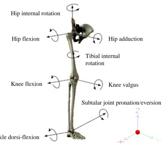
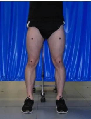
Figure 3.6: Lower extremity segment and joint rotation denotations. 92 Figure 3.7: 2D marker placement for measurement of Frontal Plane Projection
Angle.
93
Figure 3.8: Frontal Plane Projection Angle during drop jump, single leg land and single leg squat tasks.
94
Figure 3.9: A flow diagram showing statistical analyses undertaken. 96 Table 3.1: Mean, standard deviation (SD) for session 1 (S1) and session 2 (S2)
and within-day intraclass correlation coefficient (ICC), 95% confidence intervals (CI) for ICC, standard error of measurement (SEM), and smallest detectable difference (SDD).
97
Table 3.2: Mean and standard deviation (SD) values for sessions 1 (S1) and 3 (S3), between-day intraclass correlation coefficient (ICC), 95% confidence intervals (CI) for ICC, standard error of measurement (SEM), and smallest detectable difference (SDD).
97
Table 3.3: Mean and standard deviation (SD) values for test 1 (T1) and test 2 (T2) and intra-tester intraclass correlation coefficient (ICC), 95% confidence intervals (CI) for ICC, standard error of measurement (SEM), and smallest detectable difference (SDD).
98
Table 3.4: Mean and standard deviation (SD)values for experimenter 1 (E1) and experimenter 2 (E2), inter-tester intraclass correlation coefficient (ICC), 95% confidence intervals (CI) for ICC, standard error of measurement (SEM).
xi
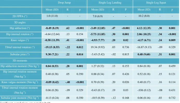
Table 3.5: Means, standard deviations (SD), Pearson’s correlations (r) and p valuesbetween 2D FPPA and 3D variables for the screening tasks.
100
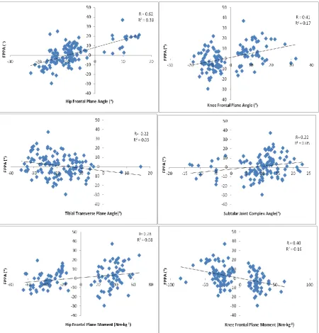
Figure 3.10: Scatterplot illustrating the significant relationships, including R and R2 values, between FPPA hip frontal plane angle (top left), knee frontal plane angle (top right), tibial transverse plane angle (middle left), subtalar joint complex angle (middle right), hip frontal plane moment (bottom left) and knee frontal plane moment (bottom right) during the drop jump task.
101
Figure 3.11: Scatterplot illustrating the significant relationships, including R and R2 values, between FPPA and hip frontal plane angle (top left), hip transverse plane angle (top right), knee frontal plane angle (bottom) in the single leg land task.
102
Figure 3.12: Scatterplot illustrating the significant relationships, including R and R2 values, between FPPA and hip frontal plane angle (top left), hip transverse plane angle (top right), knee frontal plane angle (bottom left) and subtalar joint complex angle (bottom right) in the single leg squat task.
102
Figure 3.13: Ensemble averages for frontal plane angles at the hip and knee during the drop jump (DJ), single leg land (SLL) and single leg squat (SLS) tasks.
103
Figure 3.14: Example force-time graphs for vertical ground reaction forces in the drop jump (blue) and single leg land (red) tasks.
103
Chapter 4
Figure 4.1: The single, triple and crossover hop for distance and timed hop tests 118 Table 4.1: Mean (SD), p-value and effect sizes for hop test scores in men and
women
121
Table 4.2: Week one mean ± standard deviation values for all trials of the four hop tests for men and women (Values are percentage of leg length *
xii 100, except for timed hop).
Table 4.3: Mean, standard deviation (SD), intraclass correlation coefficient (ICC), 95% confidence intervals (CI) for ICC, standard error of measurement (SEM), and smallest detectable difference (SDD) values for the four hop tests (After practice trials. All values presented as percentage of leg length * 100 except timed hop).
123
Table 4.4: Mean, standard deviation (SD), intraclass correlation coefficient (ICC), 95% confidence intervals (CI) for ICC, standard error of measurement (SEM), and smallest detectable difference (SDD) values for the four hop tests (After practice trials. All values presented are raw scores).
123
Table 4.5: Limb symmetry index (LSI) mean, standard deviation (SD), intraclass correlation coefficient (ICC), 95% confidence intervals (CI) for ICC and standard error of measurement (SEM) values for the four hop tests.
124
Table 4.6: Comparison of standard error of measurement scores between studies. 127 Figure 4.2: (A) Anterior reach direction, (B) lateral reach direction and (C)
posterior reach direction of the Star Excursion Balance Test.
132
Table 4.7 Mean (SD) and p-values for SEBT reach direction in men and women 134 Table 4.8: Mean ± standard deviations for normalised maximum excursion
distance (excursion distance/ leg length x 100).
135
Table 4.9: Mean, standard deviation (SD), intraclass correlation coefficient (ICC), 95% confidence intervals (CI) for ICC, standard error of measurement (SEM), and smallest detectable difference (SDD) values for normalised trials 5-7 of all reach directions.
136
Table 4.10: Mean, standard deviation (SD), intraclass correlation coefficient (ICC), 95% confidence intervals (CI) for ICC, standard error of measurement (SEM), and smallest detectable difference (SDD) values for raw trials 5-7 of all reach directions.
xiii
Table 4.11: Limb symmetry index (LSI) mean, standard deviation (SD), intraclass correlation coefficient (ICC), 95% confidence intervals (CI) for ICC and standard error of measurement (SEM) values for all reach directions.
137
Chapter 5
Figure 5.1: Contributing factors to dynamic knee valgus. 142 Figure 5.2: Standing isometric hip abduction strength test. 148
Figure 5.3: Standing isometric clam strength test. 149
Figure 5.4: Seated isometric external rotation strength test. 149
Figure 5.5: Gastrocnemius range of motion test. 151
Figure 5.6: Soleus range of motion test. 151
Figure 5.7: Measurement of navicular drop. 152
Table 5.1: Mean and standard deviation (SD) of all measured variables for recreational men and women.
154
Table 5.2: Pearson’s product correlation coefficients between 2D Frontal Plane Projection Angle in the Drop Jump and Single Leg Land tasks and the independent variables measured.
155
Table 5.3: Means and standard deviations for each measured variable in high FPPA limbs and normal limbs in the drop jump task
155
Table 5.4: Means and standard deviations for each measured variable in high FPPA limbs and normal limbs in the single leg landing task.
156
Table 5.5: Means and standard deviations for each measured variable in high FPPA limbs and the opposing limb within-subjects in the drop jump task.
xiv
Table 5.6: Means and standard deviations for each measured variable in high FPPA limbs and the opposing limb within-subjects in the single leg landing task.
156
Chapter 6
Table 6.1: Frontal plane projection angle (FPPA), ground reaction force (GRF), contact time and jump height means and standard deviations (SD) for baseline and post feedback/repeat test in the feedback and control groups for the drop jump (DJ) task.
170
Table 6.2: Frontal plane projection angle (FPPA), ground reaction force (GRF), contact time and jump height means and standard deviations (SD) for baseline and post feedback/repeat test in the feedback and control groups for the single leg landing (SLL) task.
171
Figure 6.1: Change in FPPA from baseline to post feedback in intervention group partcipantswho exceeded normative values in the drop jump task.
172
Figure 6.2: Change in FPPA from baseline to post feedback in intervention group partcipantswho exceeded normative values in the single leg landing task.
172
Figure 6.3: Example photograph of changes in drop jump technique from pre to post feedback
177
Figure 6.4: Example photograph of changes in single leg landing technique from pre to post feedback
177
Chapter 7
Table 7.1 Participant demographics. 183
Table 7.2 Results of pre-season screening tests for the Anterior Cruciate Ligament injured player, women’s football and women’s basketball players.
xv
Acknowledgments
A huge thank you must go to Dr. Lee Herrington who has been a constant source of support, ideas and inspiration not just for this PhD but also for my clinical practice. You have always been available to answer my questions and challenge my ideas no matter where in the world you have been! I also extend my sincere gratitude to Professor Alison Hammond whose help, guidance and experience in the final stages of bringing this thesis together has been invaluable.
Thank you to Paul Comfort for travelling to help me test all those football and basketball players and providing continuous support reading drafts of papers and my thesis. Thanks also go to Paul Jones, it took a while but we finally got some footballers in the lab!
To those colleagues who I have shared an office with; Kath and Liz who helped me when I first started this journey and more recently Jon, John, Rachel and Tamara, you have all provided entertainment, someone to have a moan and bounce ideas off, and the odd night out! Mike Carolan, cheers for coming to Cardiff and also starting up the business, maybe I can do some real work now! Thank you to Steve Horton and Laura Smith, all your help in the lab has been hugely appreciated. To all those who participated in the research which contributed to this thesis, thank you, this could not have happened without you.
To all my family and friends, I will not be a student much longer, and you might actually know what I do for a living soon! Mum and Dad, you have been amazing for the last 28 years and I would not be here if it wasn’t for your continued love, support and encouragement.
xvi Glossary of terms
2D Two dimensional
3D Three dimensional
ACL Anterior cruciate ligament
ACL- D Anterior cruciate ligament deficient ACL-R Anterior cruciate ligament reconstructed BMI Body mass index
CAI Chronic ankle instability
DJ Drop Jump
EMG Electromyography
FPPA Frontal Plane Projection Angle FPT Functional performance test GRF Ground reaction force ITB Illiotibial band
LCL Lateral collateral ligament LESS Landing Error Scoring System
LSI Limb Symmetry Index
MCL Medial collateral ligament
MVIC Maximal Isometric Voluntary Contraction NMC Neuromuscular control
OA Osteoarthritis
PCL Posterior cruciate ligament PFJ Patellofemoral joint
xvii TFJ Tibiofemoral joint
vGRF Vertical ground reaction force VL Vastus lateralis
VM Vastus medialis
xviii Abstract
Dynamic knee valgus and limb asymmetry have been linked to greater risk of anterior cruciate ligament (ACL) or patellofemoral joint (PFJ) injury. Two-dimensional (2D) frontal plane projection angle (FPPA) is more clinically useful than three-dimensional (3D) motion analysis techniques used to assess dynamic knee valgus in the literature. Further, hop for distance tests and the star excursion balance test (SEBT) offer a clinically useful assessment of limb symmetry.
1. Reliability and validity of 2D FPPA
Within-day and between-session reliability of 2D FPPA during the drop jump (DJ), single leg land (SLL) and single leg squat (SLS) tasks was fair to good. Intra- and inter-tester reliability was excellent. Significant correlations were found between 2D FPPA and 3D measures of dynamic knee valgus. These results indicate that 2D FPPA is a reliable and valid measure of dynamic knee valgus.
2. Reliability of hop for distance tests and the SEBT
Between-session reliability of the hop for distance tests and SEBT was good. Error measurement values were calculated to evaluate future performance.
3. Investigation of factors contributing to 2D FPPA
Significant correlations were found between DJ FPPA and isometric hip abduction, external rotation and combined abduction/external rotation (clam) strength. Clam strength accounted for 20% of the variance in 2D FPPA. No significant correlations were found for SLL FPPA.
4. Use of feedback to modify movement patterns
Augmented feedback was shown to significantly improve landing patterns during the drop DJ and SLL tasks. In the DJ task a significant reduction in FPPA and increase in contact time were found post-feedback. A significant reduction in FPPA and vertical ground reaction forces were found for the SLL task.
5. Prospective assessment of ACL injury risk in women’s sport
1 Chapter 1 Introduction
Knee injuries are among the most common and problematic injuries in both professional and amateur sports people. Much research has been devoted to how these injuries occur, what factors contribute to them, and how this risk might be reduced. A key component of this is the identification of those who are more susceptible to such injuries, without the use of expensive laboratory equipment. This thesis focuses on building upon this area of sports injury expertise, in particular it aims to improve the identification of those athletes who are at greatest risk of injury. To achieve this, a variety of measurement tools for assessing injury risk in the field will be identified and evaluated for their clinical utility to recognise those at greatest risk. This will help clinicians to identify modifiable risk factors and plan preventative training to limit the occurrence of these injuries.
This introduction will provide an overview of the literature pertaining to knee injury risk in the athletic population and the risk factors for these injuries. Following this, methods to identify those who demonstrate high-risk movement patterns for use in the field will be identified and potential intervention strategies to improve these movement patterns will be reviewed.
1.1. Knee Injuries in Sport
Injury to the knee joint complex is one of the most common in sport (Hootman, Dick, & Agel, 2007; Starkey, 2000). In particular, injury to the anterior cruciate ligament (ACL) and patellofemoral joint (PFJ) are responsible for a significant amount of time-loss in sport (Starkey, 2000). ACL injuries can result in inability to return to previous activity levels and both injuries are associated with early onset of knee osteoarthritis (OA) (Lohmander, Englund, Dahl, & Roos, 2007; Lohmander, Ostenberg, Englund, & Roos, 2004; Myklebust, Holm, Maehlum, Engebretsen, & Bahr, 2003b; Utting, Davies, & Newman, 2005). The majority of ACL and PFJ injuries occur through non-contact and overuse mechanisms (Agel, Arendt, & Bershadsky, 2005; Finestone et al., 2008; Mountcastle, Posner, Kragh, & Taylor, 2007; Olsen, Myklebust, Engebretsen, & Bahr, 2004) which are widely regarded as avoidable if injury mechanisms and risk factors can be identified and preventative measures taken.
2
Hewett, 2009; Krosshaug et al., 2007a). Altered neuromuscular control (NMC) of the lower limb during these movements has been suggested as an important component of such injuries (Hewett, Myer, & Ford, 2006b; Ireland, 1999). PFJ injuries are commonly overuse in nature and like ACL injuries, are thought to be the result of poor neuromuscular control during common tasks such as running, jumping and landing (Dierks, Manal, Hamill, & Davis, 2008; Souza & Powers, 2009a). Changes in frontal plane movement at the knee can alter the loads placed on the ACL and PFJ, leading to increased stress and microtrauma which over time can lead to pathology (Berns, Hull, & Patterson, 1992; Farrokhi, Colletti, & Powers, 2011a; Ireland, 1999; Lee, Anzel, Bennett, Pang, & Kim, 1994; Markolf et al., 1995; Powers, 2003). Dynamic knee valgus is a term which has been coined to reflect the numerous factors, including frontal and transverse plane motion at the hip, knee and ankle, which contribute to frontal plane motion of the knee during athletic tasks (Hewett et al., 2005). Moreover, increases in dynamic knee valgus may increase the risk of ACL and PFJ injury (Decker, Torry, Wyland, Sterett, & Richard Steadman, 2003; Hewett et al., 2005; Myer et al., 2010).
1.2. Frequency and causes of knee injuries in men and women
Women are typically at least twice as likely to suffer ACL or PFJ injury as men (Agel et al., 2005; Arendt, Agel, & Dick, 1999; Boling et al., 2010; Deitch, Starkey, Walters, & Moseley, 2006; Messina, Farney, & DeLee, 1999; Myer et al., 2010). This is thought in part to be a result of women frequently demonstrating postures which increase the loads imparted on the ACL and PFJ during athletic tasks, including increased dynamic valgus (Herrington & Munro, 2010; Kernozek, Torry, Van Hoof, Cowley, & Tanner, 2005; Zeller, McCrory, Kibler, & Uhl, 2003). This may be due to a number of factors including, increases in frontal and transverse plane hip and knee joint angles and decreases in hip muscle strength and activation compared to men (Beutler, de la Motte, Marshall, Padua, & Boden, 2009; Decker et al., 2003; Willson, Ireland, & Davis, 2006). Despite higher injury rates in women, there are likely to be common factors which may increase injury risk in both men and women. The identification of risk factors for ACL and PFJ injuries is paramount for injury prevention. Risk factor literature will be reviewed in chapter two.
1.3. Methods to identify high-risk athletes
3
However, due to the financial, spatial and temporal cost of 3D motion analysis it is not practical for most clinical settings or for use in large screening programmes useful to sport. Thus, there is a need for a simpler method of knee injury risk assessment to identify potentially high-risk athletes. Two-dimensional (2D) motion analysis of dynamic knee valgus, and functional performance tests commonly used in knee injury rehabilitation outcome measurement, may have the potential to identify these high-risk athletes.
It is important to ensure that any assessment method used in research or clinical assessment is valid and reliable. The ability of clinical tools to accurately measure the desired variable and also to detect differences within or between participants or test sessions is paramount to its utility in the field. A test which is not reliable will not provide consistent measurements in which the clinician or researcher can be confident, limiting the use of these measurements for comparison between sessions in which they are taken. It is desirable for measurement tools used with physically active participants to be able to detect small differences that may exist between populations or within an individual athlete’s performance. In addition, it is important that the observation or measurement made by a clinician or researcher is actually representative of what they are trying to measure.
1.4. 2D motion analysis: reliability and validity
4
reduced the likelihood of finding an association between LESS scores and injury risk. It may be that the sensitivity of the LESS means that only those with the highest scores are at high risk of injury.
Quantitative 2D analysis has been used to measure frontal plane knee motion in athletic, general and injured populations (Herrington, 2011; Herrington & Munro, 2010; Noyes, Barber-Westin, Fleckenstein, Walsh, & West, 2005; Stensrud, Myklebust, Kristianslund, Bahr, & Krosshaug, 2011; Willson & Davis, 2008b; Willson et al., 2006). Different methods of quantitative 2D analysis have been used, including knee separation distance (Barber-Westin, Galloway, Noyes, Corbett, & Walsh, 2005; Noyes et al., 2005) and frontal plane projection angle (FPPA) (Herrington, 2011; Willson & Davis, 2008b; Willson et al., 2006).
Knee separation distance has been used to quantify frontal plane lower limb motion in several studies. Sigward et al. (2011) recently investigated the relationship between knee separation distance and 3D knee valgus angles. They found that knee separation distance accounted for 52% of the knee valgus angle during a drop jump task, where those with smaller knee separation distances had greater knee valgus angles. However, the use of knee separation distance is limited to use during bilateral tasks only and does not allow for comparison between limbs. Considering that many ACL injuries occur during single leg landings and many individuals exhibit asymmetry between limbs, this limitation is likely to be significant when attempting to predict ACL injury using this method.
5
Although validity of FPPA has been investigated during SLS and cutting manoeuvres, this relationship has not been established in other common screening tasks. Knee valgus motion exhibited during the drop jump (DJ) task has been prospectively linked to both ACL and PFJ injury. Additionally, ACL injury commonly occurs during unilateral landings (Faude, Junge, Kindermann, & Dvorak, 2005) and, whilst not confirmed prospectively, the single leg landing (SLL) task may be useful in identifying those at risk of injury. Investigation of the relationship between 2D FPPA and 3D variables during these tasks is therefore important.
Furthermore, only within-day ICCs for the SLS have been presented to demonstrate reliability of FPPA (Willson et al., 2006). Intra-tester, inter-tester, between-session reliability and measurement error values of 2D FPPA have not been established. Therefore, further investigation of the reliability of 2D FPPA is needed before it can be recommended for use in screening tests. Further discussion and analysis of 2D and 3D motion analysis can be found in chapter three.
1.5. Functional Performance Tests: reliability, validity and clinical utility
6
Each of these unilateral FPTs is able to detect differences in function between injured and uninjured limbs following ACL injury (Barber et al., 1990; Goh & Boyle, 1997; Risberg & Ekeland, 1994). However, the stair hopple test requires that a set of stairs, with at least 11 steps, are available for the test to be undertaken. This is not always available in a clinical environment and limits the convenience of this test for use in the field. The ability of the single leg vertical jump test to detect functional deficits in injured populations only (sensitivity) is questionable (Barber et al., 1990). In this study, over half of the normal population were unable to achieve 90% symmetry between limbs, whilst only 69% achieved 85% symmetry, suggesting this test may not be suitable for detecting lower limb functional limitations in injured populations.
Hop tests, which require the participant to hop as far as possible, are routinely used during rehabilitation from ACL injury. Hop tests can detect deficits between ACL reconstructed or deficient and uninjured limbs (Barber et al., 1990; Goh & Boyle, 1997; Reid et al., 2007). In order to compare and evaluate performance between limbs during the hop tests the limb symmetry index (LSI) is used. LSI gives a percentage value of the distance hopped on the injured limb versus the uninjured limb. An LSI of ≥85% indicates that ‘normal’ limb symmetry exists and function of the injured limb is being restored (Bandy, Rusche, & Tekulve, 1994; Barber et al., 1990). The 85% value was chosen as over 93% of the normal population were able to achieve this score (Barber et al., 1990). However, the validity of this value has not been investigated further and is not always sensitive to deficits in ACL injured participants (Barber et al., 1990; Noyes et al., 1991; Petschnig et al., 1998). This lack of sensitivity may be due to this arbitrary LSI value being too low. If hop tests are able to show functional deficits between limbs in injured populations, it would seem plausible to screen healthy individuals for LSI and investigate whether an abnormal LSI is a predisposing factor to injury and to help determine a minimal required LSI score to reduce injury risk.
7
functional deficits in CAI and ACL-D patients (Herrington et al., 2009; Hertel et al., 2006a). A link between SEBT performance and lower extremity injury occurrence in high school basketball players has also been reported (Plisky, Rauh, Kaminski, & Underwood, 2006). These studies suggest that the SEBT may be sensitive to both post-injury deficits between limbs and the prediction of future injury risk.
As both the hop tests and SEBT are indicated to be the most clinically applicable as well as relevant tests in which to potentially detect limb symmetry differences, deficits in functional performance and risk of injury, these will therefore be reviewed in more detail in chapter two. Considering the factors presented, further investigation and understanding of the potential of 2D video analysis and FPTs to identify athletes at high-risk of ACL or PFJ injury is warranted.
1.6. Causative factors of dynamic valgus and potential interventions
Identification of individuals who exhibit dynamic valgus and are at higher risk of ACL or PFJ injury is important. However, in order for this risk of injury to be reduced through interventions aimed at modifying movement patterns, an understanding of the factors that contribute to demonstration of dynamic valgus is required. Despite the frequent use of the drop jump, single leg drop landing, and single leg squat tasks for clinical screening, little is known about which factors contribute to dynamic knee valgus during these tasks. These contributory factors need to be identified, to enable targeted prevention strategies to reduce injury rates.
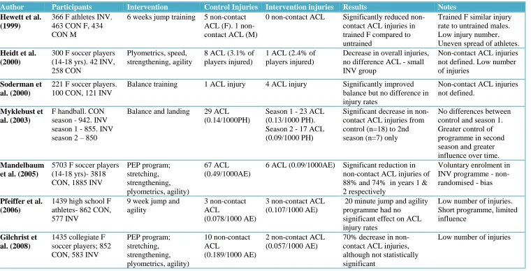
A number of studies have assessed the effect of intervention programmes aimed at modifying the risk factors identified for ACL and PFJ injury. Studies assessing the effectiveness of programmes for ACL injury prevention have used injury rates and changes in lower limb biomechanics as outcome measures. Those assessing PFJ injuries have mainly used changes in pain and function in those already diagnosed with patellofemoral pain syndrome (PFPS), limiting the application of the results to injury prevention strategies. However, the findings of the studies examining changes in biomechanics related to ACL injury could also be applied to the PFJ due to increased stress being brought about by similar movement patterns.
8
demonstrated no difference in injury rates between control and intervention groups (Heidt, Sweeterman, Carlonas, Traub, & Tekulve, 2000; Myklebust et al., 2003a; Pasanen et al., 2008b; Pfeiffer, Shea, Roberts, Grandstrand, & Bond, 2006; Soderman, Werner, Pietila, Engstrom, & Alfredson, 2000). Despite the relatively high incidence of PFPS, only one study has prospectively examined the effect of a multifaceted intervention programme on PFJ injury rates finding no difference between experimental and placebo groups (Brushoj et al., 2008).
Several studies have seen increases in hip and knee flexion angles and decreases in hip internal rotation, knee valgus and internal rotation motion and ground reaction force (GRF) after various training programmes (Barendrecht, Lezeman, Duysens, & Smits-Engelsman, 2011; Cochrane et al., 2010; Irmischer et al., 2004; Lephart et al., 2005; Myer, Ford, McLean, & Hewett, 2006; Pollard, Sigward, Ota, Langford, & Powers, 2006a). These changes are likely to reduce ACL and PFJ stress and therefore help to reduce injury risk. However, these changes in lower limb mechanics are not always evident (Cochrane et al., 2010; Grandstrand, Pfeiffer, Sabick, DeBeliso, & Shea, 2006; Herman et al., 2009; Lephart et al., 2005; Pollard et al., 2006a).
9
Herman et al. (2008) found that a lower limb strength training intervention did not improve hip and knee kinetic and kinematics. However, a second study found that when feedback was introduced the strength training group improved more than a feedback only group (Herman et al., 2009). Recently, there has been an increase in research activity investigating how feedback can influence lower extremity movement patterns. Feedback is a fundamental tool for learning and performing of motor skills and has been shown to improve landing strategies across a number of studies (Cronin, Bressel, & Finn, 2008; Herman et al., 2009; Onate et al., 2005; Onate, Guskiewicz, & Sullivan, 2001). The use of simple verbal feedback decreases GRFs and knee abduction angles and moments during landing tasks (Cowling, Steele, & McNair, 2003; McNair, Prapavessis, & Callender, 2000; Mizner, Kawaguchi, & Chmielewski, 2008; Prapavessis & McNair, 1999).
10 1.7. Aims
The aims of the thesis are therefore to:
1. Review the literature related to Anterior Cruciate Ligament and Patellofemoral Joint injuries, including their occurrence, mechanism and proposed risk factors (chapter 2). 2. Review the literature regarding screening tools to identify potential Anterior Cruciate
Ligament or PFJ injury risk (chapter 2).
3. Establish the reliability and validity of 2D FPPA during the drop jump, single leg landing and single leg squat tasks (chapter 3).
4. Establish the reliability and measurement error of the SEBT and hop for distance tests (chapter 4).
5. Establish what factors contribute to the demonstration of 2D FPPA during screening tasks (chapter 5).
6. Establish whether a simple feedback intervention can modify landing strategies during screening tasks (chapter 6).
11 Chapter 2
Literature Review
2.1. Introduction
This literature review provides the background and rationale for the work conducted in this thesis. The following are therefore discussed:
current trends in sport injury occurrence (2.1.1)
injuries of the knee joint, specifically ACL (2.1.2) and PFJ injuries (2.1.3), their occurrence and comparison between sexes (2.1.4)
mechanisms (2.2) and proposed risk factors (2.3) for ACL and PFJ injuries in relation to knee anatomy
screening tools to identify those at greater risk of ACL and PFJ injuries (2.5) intervention strategies to reduce the risk of ACL and PFJ injuries (2.7)
2.1.1. Injuries in Sport
Physical activity is associated with a potential risk of injury. Increased sports participation leads to an inherent increase in injuries sustained, which results in costs to: the individual, in temporary or long-term disability and loss of earnings; the healthcare system; and the economy.
12
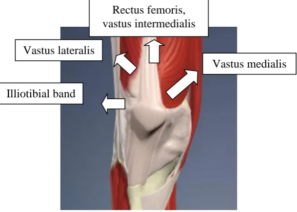
[image:30.595.92.507.278.705.2]The time-loss from training and competition associated with knee injuries is due to their seriousness. The knee joint consists of the tibiofemoral and patellofemoral (PFJ) joints which, due to their relatively shallow articulations, rely primarily on ligamentous and muscular restraints for stability. The anterior cruciate ligament (ACL) and posterior cruciate ligaments (PCL) primarily restrict anterior and posterior translation of the tibia on the femur respectively. The medial collateral ligament (MCL) restrains valgus forces and the lateral collateral ligament (LCL) restrains varus forces applied to the knee. The ligament restraints typically come into play towards the end range of these movements, with the muscles around the knee joint providing added stability. However, the muscles around the knee primarily create, rather than restrict, movement. Thus knee joint stability is heavily influenced by muscular action. Figure 2.1 and 2.2 show the knee joint muscles and ligaments.
Figure 2.1 – The knee joint muscles and direction of action.
A B
Figure 2.2 – The knee joint ligaments - A) anterior view; B) posterior view. ACL – anterior
cruciate ligament; PCL – posterior cruciate ligament; LCL - lateral collateral ligament; MCL – medial collateral ligament (Primal Images, London, UK).
Vastus medialis Vastus lateralis
Illiotibial band
Rectus femoris, vastus intermedialis
MCL
PCL
[image:30.595.159.464.286.504.2]13
No study has evaluated the cost of sports injuries in the United Kingdom. Annual costs of such injuries are $222million in New Zealand (Gianotti & Hume, 2007), and $680million in the United States for people under the age of 24 alone (Burt & Overpeck, 2001). Using current exchange rates and assuming similar participation and injury rates, this equates to between £115-445 million in the UK (exchange rate at 19/07/2013).
2.1.2. Anterior Cruciate Ligament Injuries
ACL injury is catastrophic, resulting in an extended period away from sports participation. For example, only 58% of Norwegian elite handball players returned to the same level of competition after ACL reconstruction (Myklebust et al., 2003b). The remaining 42% either competed at a lower level or did not return to sport at all. Over half of Swedish women football players were unable to return to sport post-ACL injury, and only 15% reported returning to pre-injury activity levels (Lohmander et al., 2004). A recent study in American Football identified that 37% of players who underwent ACL surgery did not return to play (Shah, Andrews, Fleisig, McMichael, & Lemak, 2010).
Most individuals who suffer ACL injury also experience early onset of OA with associated pain and limited function (Fink, Hoser, Hackl, Navarro, & Benedetto, 2001; Lohmander et al., 2007; Lohmander et al., 2004; Myklebust et al., 2003b). Around 40% of ACL patients have signs of early onset OA of the tibiofemoral joint or PFJ six to eleven years post injury (Jarvela, Paakkala, Kannus, & Jarvinen, 2001; Keays, Bullock-Saxton, Keays, Newcombe, & Bullock, 2007; Myklebust et al., 2003b). Studies where follow-up has been conducted after 10-15 years have shown around 75-80% of patients who suffered an ACL injury have radiographic changes in the knee joint complex (Fink et al., 2001; Lohmander et al., 2004; Oiestad et al., 2010; von Porat, Roos, & Roos, 2004). Within-subject comparisons show radiographic changes in only 37% of uninjured knees, suggesting that the ACL injury was the reason for the majority of early onset OA cases (Lohmander et al., 2004).
14
subjects (Jarvela et al., 2001; Lohmander et al., 2004; Myklebust et al., 2003b). Overall, it is evident that ACL injury can lead to detrimental changes to the knee joint complex and/or changes in knee function which may not happen if the injury did not occur.
2.1.3. Patellofemoral Joint Injuries
Retropatellar and peripatellar pain resulting from injury to the PFJ, clinically referred to as patellofemoral pain syndrome (PFPS), is a common pain disorder experienced by athletes (Boling et al., 2010; Loudon et al., 2004; Myer et al., 2010; Natri, Kannus, & Jarvinen, 1998; Starkey, 2000; Taunton et al., 2002; Witvrouw, Lysens, Bellemans, Cambier, & Vanderstraeten, 2000). PFPS results in significant time-loss from training and competition (Starkey, 2000) and causes athletes to limit or cease their sport activities (Blond & Hansen, 1998; Witvrouw et al., 2000). Athletic activity of 74% of PFPS patients is affected in some way, either through taking a break, playing at a lower level or being forced to stop (Blond & Hansen, 1998). In some cases, PFPS patients are forced to change their employment as they cannot meet the physical demands of their job (Blond & Hansen, 1998).
Symptomatic knee OA is more likely to occur in the PFJ than the TFJ and also has a greater impact on daily activities (Duncan et al., 2008; Risberg & Ekeland, 1994). It has been reported that those who experience anterior knee pain during adolescence or early adulthood are more likely to suffer from PFJ OA (Utting et al., 2005). This suggests PFPS can have a large negative impact on an individuals’ short-term athletic activities and, perhaps more importantly long-term, on employment and quality of life.
2.1.4. Incidence of Anterior Cruciate Ligament and Patellofemoral Joint Injury
The incidence of ACL injuries is only 0.1-0.3 per 1000 athlete exposures (Gwinn, Wilckens, McDevitt, Ross, & Kao, 2000; Mihata, Beutler, & Boden, 2006; Myklebust, Maehlum, Holm, & Bahr, 1998). Incidence of PFPS is greater at 1.09 injuries per 1000 exposures (Myer et al., 2010). This seems a small problem in comparison to common injuries, such as ankle ligament and hamstring muscle strains, with incidence rates up to 3.19 per 1000 exposures (Agel et al., 2007; Deitch et al., 2006). However, the consequences of ACL and PFJ injuries, in terms of time-loss, future participation and increased risk of OA, make these among the most serious and problematic injuries in sport.
15
levels (Agel et al., 2005; Arendt et al., 1999; Boling et al., 2010; Deitch et al., 2006; Hewett et al., 1999; Messina et al., 1999; Myklebust et al., 1998; Powell & Barber-Foss, 2000; Taunton et al., 2002). Perhaps most importantly, women consistently suffer a higher rate of non-contact ACL injuries than men (Agel et al., 2005; Hewett et al., 1999; Mountcastle et al., 2007). The findings of previous studies are summarised in Figures 2.3 and 2.4.
16
0 0.05 0.1 0.15 0.2 Soccer (Agel et al., 2005)
Basketball (Agel et al., 2005)
Multi-sport (Hewett et al., 1999)
Multi-sport (Mountcastle et al., 2007)
Injury Rate Per 1000 Exposures
Women Men
Figure 2.4 - Comparison of non-contact Anterior Cruciate Ligament injury rates per 1000 exposures between men and women across a number of sports and levels of competition.
2.2. Mechanisms of Knee Injury
The mechanisms of ACL (2.2.1) and PFJ (2.2.2) injury will now be discussed in detail.
2.2.1. Mechanisms of Anterior Cruciate Ligament Injury
60-70% of ACL injuries occur in non-contact situations (Agel et al., 2005; Faude et al., 2005; Giza, Mithöfer, Farrell, Zarins, & Gill, 2005; Mountcastle et al., 2007; Pasanen, Parkkari, Rossi, & Kannus, 2008c). Non-contact injuries may be avoidable and as these are the most common types of ACL injury, it is important to understand the injury mechanism to help reduce their occurrence.
17
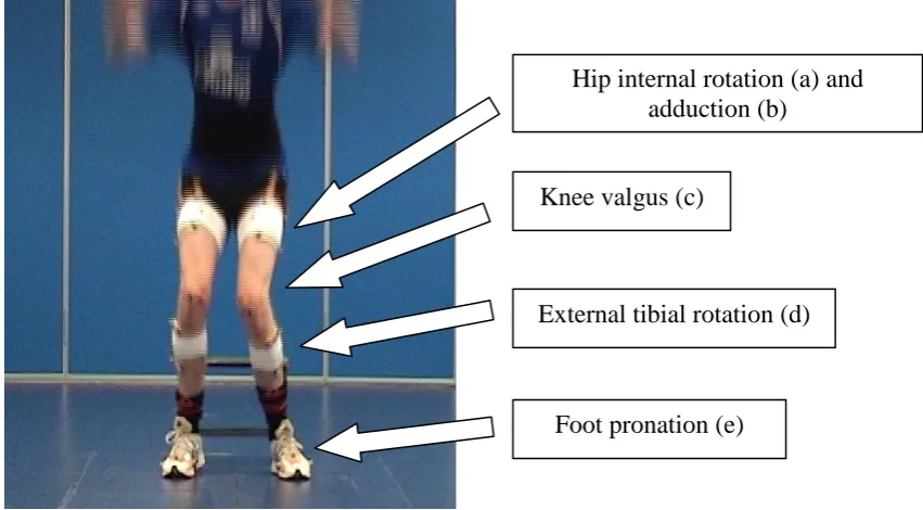
accounted for 16/20 incidents reviewed by Olsen et al. (2004), and 27/39 videos analysed by Krosshaug et al. (2007a). It was also noted that ACL injury occurs during the deceleration phase of these movements (Koga et al., 2010; Krosshaug et al., 2007a). Figures 2.5 and 2.6 show the cutting and landing mechanisms of non-contact ACL injury in Team Handball.
As well as the type of action performed at the time of injury, it is also important to understand the position of the body during these actions. Several studies have estimated lower limb joint angles through video-analysis of injury occurrence by experienced researchers (Boden et al., 2000; Krosshaug et al., 2007a; Olsen et al., 2004). The results show that athletes often land with the hip slightly flexed, adducted and internally rotated, with minimal flexion of the knee, the tibia externally rotated and evidence of a valgus knee collapse. This position can be seen in figures 2.5-2.7 and has been termed dynamic knee valgus or the ‘position of no-return’ (Hewett et al., 2005; Ireland, 1999).
Figure 2.5 – Dynamic knee valgus during the plant and cut mechanism of ACL injury in Team
18
Figure 2.6 – Dynamic knee valgus during the one-legged landing mechanism of ACL injury in
Team Handball (adapted from Olsen et al., 2004)
Figure 2.7 – Dynamic knee valgus (adapted from Hewett et al., 2006)
Most recently, a technique called model-based image-matching, which extracts joint kinematics from video recordings, has been used in an attempt to greater explain ACL injury mechanism (Koga et al., 2010). Difficulties in matching body parts, due to occlusion by other players or clothes, and assessment of axial rotations mean the methodology and joint angles calculated are not 100% accurate. However, they provide the most detailed and accurate description of injury mechanism to date. Despite the limitations of this method it produced
Hip internal rotation and adduction
Knee valgus and external rotation
19
consistent results for knee kinematics during non-contact ACL injury situations (Krosshaug, Slauterbeck, Engebretsen, & Bahr, 2007b). It also confirmed previous findings that the knee flexion angle at initial contact tends to be low (<25º) with knee external rotation (external rotation of the tibia in relation to the femur) and valgus also being evident (Koga et al., 2010). Only knee joint kinematics were observed in this study therefore confirmation of previous findings at the hip is not possible.
Support for the dynamic knee valgus injury mechanism has also come in the form of in-vitro and 3D modelling studies which have explored the strain imparted on the ACL during specific movements at the knee joint. As knee joint stabilisation is achieved through a number of active muscular and passive ligament controls, it would seem plausible that more than one particular excessive movement would be required to bring about enough force to disrupt the ACL. Forces of at least 1500-2000N are required to cause disruption to the ACL (Chandrashekar, Mansouri, Slauterbeck, & Hashemi, 2006; Woo, Hollis, Adams, Lyon, & Takai, 1991). However, tensile properties of the ACL are not uniform throughout the population and forces as low as 1200N may cause ACL injury in women compared to 1700N in men (Chandrashekar et al., 2006). Anterior tibial shear causes the most strain on the ACL, but not with enough force to cause ligament rupture (Berns et al., 1992; McLean, Huang, Su, & Van Den Bogert, 2004b). Even in a ‘worst-case scenario’ sagittal plane injury mechanism computer simulation the resultant force on the ACL never exceeded 900N (McLean et al. 2004). However, anterior tibial shear with combined knee valgus and/or rotational moments cause significantly greater strain on the ACL, increasing the potential for injury (Berns et al., 1992; Markolf et al., 1995; McLean et al., 2004b). This is especially true at angles closer to full knee extension, further supporting the proposed mechanism of ACL injury (Berns et al., 1992; Ireland, 1999).
2.2.2. Mechanisms of Patellofemoral Joint Injury
20
been investigated and has shown that increases in hip adduction, hip internal rotation and tibial external rotation can decrease PFJ contact area and increase PFJ contact pressures (Lee et al., 1994; Lee, Morris, & Csintalan, 2003; Powers, Souza, Draper, & Fredericson, 2010; Salsich & Perman, 2007). Figure 2.8 shows a diagrammatic representation of how changes in patella position, resulting from either patella maltracking or changes in tibial or femoral position, can reduce the load bearing surface of the patella and increase PFJ contact pressures.
Figure 2.8 – The effect of changes in patella, tibial or femoral position on the load bearing
surface of the patella – a) neutral position with equal load bearing at both the medial and
lateral patella facets; b) increased lateral displacement with resultant increased load bearing
of the lateral patella facet; c) increased medial displacement with resultant increased load
bearing of the medial patella facet (adapted from Lee et al., 2004)
Abnormal motion of the patella, femur or tibia can decrease the size of the load bearing surface of the patella, resulting in altered distribution of forces and excessive PFJ stress. Continuous overload of the PFJ in this way can lead to a loss of peripatellar tissue homeostasis, leading to pain (Dye, Staubli, Biedert, & Vaupel, 1999). Patients with PFPS demonstrate greater PFJ stress during walking and squatting as a result of reduction in PFJ contact area (Brechter & Powers, 2002; Farrokhi, Keyak, & Powers, 2011b). Changes in PFJ contact area can cause wear of the articular cartilage (Salsich & Perman, 2007). However, articular cartilage is not an innervated structure and cannot be a source of pain (Biedert, Stauffer, & Friederich, 1992). Therefore, it is thought that the subchondral bone is a source of pain in PFPS (Biedert & Sanchis-Alfonso, 2002; Dye, Vaupel, & Dye, 1998). This is supported by the presence of significantly decreased patella cartilage thickness in PFPS patients, suggesting that by the time symptoms arise, the degenerative process is likely to be well underway (Farrokhi et al., 2011a). The higher incidence of PFJ OA in adults who suffered from anterior knee pain during adolescence also reflects this (Utting et al., 2005). Hence, chronic overloading of the PFJ resulting from changes in lower limb motion causes
b)
a) c)
21
cartilage wear, increasing symptoms and decreasing activity levels (Blond & Hansen, 1998; Fulkerson, 2002).
2.3. Risk Factors for Anterior Cruciate Ligament Injuries
This section reviews the proposed risk factors for non-contact ACL injuries only. If the risk factors for non-contact ACL injuries are better understood, some may be modified and injuries prevented. Extrinsic and intrinsic risk factors linked to ACL injuries include: shoe type, hormonal and anatomical factors and poor NMC (Ireland, 1999). Extrinsic factors will be briefly discussed in section 2.3.1. Intrinsic risk factors will be reviewed in greater detail in section 2.3.2. Neuromuscular control, which is a proposed risk factor for both ACL and PFJ injuries, will be reviewed later in section 2.3.4.
2.3.1. Extrinsic Risk Factors for Anterior Cruciate Ligament Injury
Extrinsic factors are those external to the individual and include; surface type; shoe type; and weather conditions. Injury rates on synthetic surfaces, where the coefficient of friction is greater, are significantly higher than on wooden floors (Olsen, Myklebust, Engebretsen, Holme, & Bahr, 2003; Pasanen et al., 2008c). More cleats on the boots of American football players, which increases torsional resistance between shoe and surface, is associated with greater risk of ACL injury (Lambson, Barhnill, & Higgins, 1996). It has been reported that ACL injuries occur more frequently during periods of lower rainfall when friction between shoe and surface is greater (Orchard, Seward, McGivern, & Hood, 1999; Orchard & Powell, 2003). Increases in friction through these mechanisms mean that the foot is fixed and minimises the rotation available between shoe and surface, which may then transfer to the ankle and knee joints. Thus increased friction may lead to increased risk of sustaining ACL injury risk. Changes in surface and shoe types to decrease friction may be possible, however this may come at the detriment to performance.
2.3.2. Intrinsic Factors for Anterior Cruciate Ligament Injury
22
Table 2.1 – Summary of section content for intrinsic risk factors for Anterior Cruciate
Ligament injury.
Intrinsic risk factors Section Anatomical
Femoral notch width and shape
Joint laxity
2.3.2.1
2.3.2.1a
2.3.2.1b
Hormonal 2.3.2.2 Sagittal plane mechanics 2.3.2.3
2.3.2.1. Anatomical Risk Factors a) Femoral intercondylar notch size:
This is potentially important as the ACL is housed in this notch. Studies investigating femoral intercondylar notch width and its relationship to ACL injury have reported conflicting results (Harner, Paulos, Greenwald, Rosenberg, & Cooley, 1994; Herzog, Silliman, Hutton, Rodkey, & Steadman, 1994; Laprade & Burnett, 1994; Shelbourne, Davis, & Klootwyk, 1998; Souryal, Freeman, & Daniel, 1993; Uhorchak et al., 2003). This conflict is likely due to use of the femoral intercondylar notch width in some studies and the notch width index, i.e. the ratio of the notch width to the femoral bicondylar width in others (Shelbourne et al., 1998). These two measures are demonstrated in figure 2.6. Femoral bicondylar width is influenced by an individual’s height whereas notch width is not. Therefore the notch width index is inherently influenced by the person’s height (Shelbourne et al., 1998). As a result, Shelbourne and colleagues recommended the use of the femoral intercondylar notch width rather than notch width index.
23
A relationship between smaller intercondylar notch width and ACL injury has been shown (Uhorchak et al., 2003). However, the reason for this relationship is unclear with two theories having been proposed; ACL impingement upon the intercondylar notch wall, and smaller ACL size.
Impingement:
24
Figure 2.10 – ACL impingement on the femoral condyle caused by tibial external rotation and
knee valgus (adapted from Olsen et al., 2004).
ACL size:
Shelbourne et al. (1998) hypothesised that notch width alone does not account for differences in injury rates, rather the smaller notches found in women house a smaller ACL, which may be weaker and more susceptible to injury. The basis for this theory followed their study in which they found that patients who undergo ACL reconstruction with the same size ACL graft have similar graft failure rates regardless of notch width and sex (Shelbourne et al., 1998). It has been reported however that femoral notch width is correlated to ACL size in men but not in women (Chandrashekar, Slauterbeck, & Hashemi, 2005). Notwithstanding this, the female ACL has been found to be smaller in length, cross-sectional area and volume and to have lower load resistance than the male ACL (Chandrashekar et al., 2005; Chandrashekar et al., 2006). Therefore, it seems likely that a combination of the difference in ACL properties and smaller intercondylar notch width would contribute to increased injury risk in women.
b) Joint Laxity:
25
knee can result in altered NMC via changes in muscular activity, such as delayed activation of the hamstrings (Shultz, Carcia, & Perrin, 2004), Additionally, participants with greater frontal and trasverse plane knee joint laxity demonstrate greater hip internal rotation, hip adduction and knee valgus angles than those with lower laxity values (Shultz & Schmitz, 2009). Increases in knee joint laxity may therefore lead to greater instability, increased anterior tibial translation and resultant shear force, and increase in dynamic knee valgus therefore increasing ACL strain.
Women tend to exhibit greater knee joint laxity and diminished proprioception compared to men (Myer et al., 2008; Rozzi, Lephart, Gear, & Fu, 1999; Uhorchak et al., 2003) which may increase their injury risk. A combination of smaller intercondylar notch width, high body mass index (BMI) and increased knee joint laxity was able to predict all ACL injuries in women, but none in men (Uhorchak et al., 2003). However, dynamic stability of the knee is affected by both passive and active restraints (Rozzi et al., 1999; Shultz et al., 2004). This further emphasises the complexity of the ACL injury risk paradigm. It would seem that smaller notch widths, structurally weaker ACL’s and increased knee joint laxity in women play a part in explaining some of the disparity in injury rates between men and women. However, each of these anatomical factors cannot be modified, therefore limiting the ability to influence injury rates as a result of their understanding.
2.3.2.2. Hormonal Risk Factors
The different hormonal profile of men and women may contribute to disparity in injury rates. The primary drivers behind this theory are:
a) the changes in hormonal profile during the menstrual cycle
b) differences in neuromuscular characteristics post-puberty (Barber-Westin, Noyes, & Galloway, 2006; Hewett, Myer, & Ford, 2004)
a). Changes in hormonal profile during the menstrual cycle
26
just after onset of menstruation. Arendt et al. (1999) reported injuries were spread evenly between pre and post ovulatory phases with fewest injuries occurring during the ovulatory phase. These differences in injury susceptibility within the menstrual cycle led to the suggestion that use of the oral contraceptive pill may have a protective effect. However, Agel et al. (2006) found that it had no effect on non-contact ACL injury rates.
The effect of hormones on injury risk may not be direct, for example an increase in oestrogen concentration may not automatically increase risk of injury. Rather, changes in ligament properties and NMC have been proposed. ACL laxity progressively increases up to the time of peak oestrogen and progesterone levels (Heitz, Eisenman, Beck, & Walker, 1999), potentially increasing injury risk. However, changes in knee joint laxity and NMC are not evident (Chaudhari et al., 2007; Hertel, Williams, Olmsted-Kramer, Leidy, & Putukian, 2006b). Furthermore, use of the contraceptive pill has no effect on hip and knee angles or moments during several jump landing tasks (Chaudhari et al., 2007). The lack of consensus regarding effects of the menstrual cycle on injury risk may be due to the lack of consistency in terms and phases used to describe the cycle itself.
b). Neuromuscular characteristic differences post-puberty
27
knee as a result. The changes in NMC between men and women post-puberty correlate with, and may be partly responsible for, the divergence in injury rates between the sexes.
The complexity of the female hormonal profile, the effect of the contraceptive pill and different varieties, and individual differences in hormone concentrations and their effects on psychological state, the ACL and neuromuscular system makes this area difficult to study adequately. However, it seems that the change in overall hormonal profile during puberty, which leads to changes in NMC, correlates with higher injury rates in the female athlete. Therefore a greater understanding of the contribution of NMC to injury risk is important.
2.3.2.3 Sagittal Plane Risk Factors
As described earlier it has been reported that movements in the sagittal, frontal and transverse planes contribute to ACL injury. The following section will review the factors that arise in the sagittal plane of movement and how they might influence non-contact ACL injury risk. The frontal and transverse planes of movement will be examined later in section 2.3.4.1.
Anterior tibial shear:
28
cause ACL injury via quadriceps contraction (DeMorat et al., 2004). In addition, disruption of the ACL only occurred in 6 out of 11 cadaveric knees subjected to the 4500N force (DeMorat et al., 2004), suggesting that 4500N quadriceps force does not equate to a 1500-2000N load at the ACL. Furthermore, the synergistic action of the hamstrings and quadriceps muscle groups, joint compression forces, and dissipation of landing forces at the ankle and hip are likely reduce the forces experienced by the ACL (McLean et al., 2004b). Therefore, it is unlikely that anterior shear alone will result in 1500-2000N load required to injure the ACL (Chandrashekar et al., 2006; Woo et al., 1991).
Figure 2.11 – A free-body diagram of the quadriceps (Q) and hamstring (H) forces acting
upon the proximal tibia in the sagittal plane during different degrees of knee flexion (a) with
the knee at full extension; (b) with the knee in a moderately flexed position (adapted from
Hashemi et al., 2011).
Hamstring strength:
Contraction of the hamstring muscle group may help to prevent ACL injury by decreasing anterior shear (Draganich & Vahey, 1990; Li et al., 1999; Renstrom, Arms, Stanwyck, Johnson, & Pope, 1986). When working in isolation the hamstrings can decrease ACL strain throughout knee motion (Renstrom et al., 1986). However, changes in ACL strain and anterior shear when the hamstrings are acting synergistically with the quadriceps are inconsistent.
a b
29
In-vitro studies have shown that antagonistic hamstring contraction can reduce ACL load and anterior shear at knee flexion angles greater than 10º (Draganich & Vahey, 1990; Li et al., 1999). The reduction in ACL load was 30, 43 and 44% at knee flexion angles of 15, 30 and 60° respectively (Li et al., 1999). Other studies have noted that ACL strain is significantly decreased from 30° to 90° of knee flexion but not at angles of 0, 15 and 30° (Pandy & Shelburne, 1997; Renstrom et al., 1986). As demonstrated in figure 2.11, when the knee is close to full extension the angle between the line of action of the hamstrings and the tibia is low, meaning the hamstrings are unable to generate large enough posterior shear forces to counteract anterior shear forces to protect the ACL (Pandy & Shelburne, 1997). It is therefore unclear whether the hamstrings can protect the ACL up to 30° knee flexion, the range in which ACL injury often occurs.
Whilst the hamstrings may decrease ACL strain in-vitro, whether this occurs during dynamic movements is questionable. Quadriceps and hamstring strength and ratio do not predict the amount of anterior tibial shear force exhibited during a drop jump task (Bennett et al., 2008). Evidence has shown that the hamstrings are recruited during running, turning and landing activities (Colby et al., 2000; Gehring, Melnyk, & Gollhofer, 2009) although hamstring electromyography (EMG) activity can be more than 50% lower than the quadriceps during these tasks (Colby et al., 2000). If hamstring muscle activity is low, particularly in comparison to the quadriceps, then increases in hamstring strength are likely to have negligible effects on reducing ACL load.
Increased hamstring torque demonstrated after a jump training intervention has been linked to decreases in vertical ground reaction forces (vGRF) which may decrease injury risk (Hewett, Stroupe, Nance, & Noyes, 1996). However, these decreases in vGRF could also be attributed to increased hip and knee flexion angles which have been seen after similar jump training programmes (Lephart et al., 2005; Myer et al., 2006).
30 Knee flexion angles:
Changes in sagittal plane angles at the knee can alter the load imparted on the ACL. As previously noted ACL strain is often greatest at angles nearer to full extension (Berns et al., 1992; Markolf et al., 1995). The potential for the quadriceps to cause anterior tibial shear, and therefore greater ACL strain, is also greatest at angles close to full extension (Arms et al., 1984; Beynnon et al., 1995; Beynnon et al., 1992; Draganich & Vahey, 1990; Li et al., 1999; Pandy & Shelburne, 1997; Shoemaker et al., 1993). Women often land with 20-25º knee flexion, which on average is 5-10º less than men (Chappell et al., 2005; Decker et al., 2003; Huston, Vibert, Ashton-Miller, & Wojtys, 2001; Malinzak et al., 2001). Additionally, women display decreased flexion angles and absorption of force at the hip which results in increased loads on the knee (Chappell et al., 2005; Decker et al., 2003). The greater sagittal plane loads exhibited by women, coupled with increased quadriceps activation and decreased hamstring activation may all contribute to increased ACL strain and likelihood of injury.
Summary