0095-1137/06/$08.00⫹0 doi:10.1128/JCM.44.3.693–699.2006
Copyright © 2006, American Society for Microbiology. All Rights Reserved.
Identification of Medically Important Yeast Species by Sequence
Analysis of the Internal Transcribed Spacer Regions
Shiang Ning Leaw,
1Hsien Chang Chang,
1Hsiao Fang Sun,
2Richard Barton,
3Jean-Philippe Bouchara,
4and Tsung Chain Chang
5*
Institute of Biomedical Engineering,1Institute of Molecular Medicine,2and Department of Medical Laboratory Science and Biotechnology,5National Cheng Kung University, Tainan, Taiwan, Republic of China; School of Biochemistry and
Microbiology, University of Leeds, Leeds, United Kingdom3; and Host-Parasite-Interaction Study Group (UPRES-EA 3142), Laboratory of Parasitology and Mycology, University Hospital, Angers, France4
Received 18 July 2005/Returned for modification 27 October 2005/Accepted 13 December 2005
Infections caused by yeasts have increased in previous decades due primarily to the increasing population
of immunocompromised patients. In addition, infections caused by less common species such as Pichia,
Rhodotorula,Trichosporon, andSaccharomycesspp. have been widely reported. This study extensively evaluated the feasibility of sequence analysis of the rRNA gene internal transcribed spacer (ITS) regions for the identification of yeasts of clinical relevance. Both the ITS1 and ITS2 regions of 373 strains (86 species), including 299 reference strains and 74 clinical isolates, were amplified by PCR and sequenced. The sequences were compared to reference data available at the GenBank database by using BLAST (basic local alignment search tool) to determine if species identification was possible by ITS sequencing. Since the GenBank database currently lacks ITS sequence entries for some yeasts, the ITS sequences of type (or reference) strains of 15 species were submitted to GenBank to facilitate identification of these species. Strains producing discrepant identifications between the conventional methods and ITS sequence analysis were further analyzed by sequenc-ing of the D1–D2 domain of the large-subunit rRNA gene for species clarification. The rates of correct identification by ITS1 and ITS2 sequence analysis were 96.8% (361/373) and 99.7% (372/373), respectively. Of
the 373 strains tested, only 1 strain (Rhodotorula glutinisBCRC 20576) could not be identified by ITS2 sequence
analysis. In conclusion, identification of medically important yeasts by ITS sequencing, especially using the ITS2 region, is reliable and can be used as an accurate alternative to conventional identification methods.
The incidence of fungal infections has increased in the past few decades. Invasive infections caused by yeasts have become a major cause of morbidity and mortality in patients receiving immunosuppressive chemotherapy for cancer or organ trans-plantation or in immunodeficient patients, such as individuals
with untreated AIDS (4, 13, 49).Candida albicansis the most
common species causing a variety of infections. However, the
incidence rates of non-C. albicans Candida infections have
been increasing in recent years (46, 47). Moreover, outbreaks of systemic infections caused by yeasts in neonatal intensive care units have been described (6, 12, 20, 27, 36, 38). Recently,
infections caused by less common yeast species such asPichia,
Rhodotorula,Trichosporon, andSaccharomycesspp. and other rarely encountered species have been reported (2, 16, 31, 44, 49, 51). More than 100 yeast species have been identified as human pathogens and have been isolated from virtually all body sites (14). Identification of the increasing diversity of pathogens by conventional methods may be difficult and some-times inconclusive (34), especially for unusual yeast species.
The susceptibilities of different species to antifungal agents may be different (32, 52), and reliable identification of yeasts is helpful for treatment with appropriate antifungal agents. Com-mercially available biochemical and enzymatic panels, such as
API ID32C (bioMe´rieux, Marcy-l’Etoile, France) and VITEK
ID-YST (bioMe´rieux Vitek, Hazelwood, Mo.), are convenient
for use. However, the disadvantages of limited databases (33) and misidentification using these kits (7, 24, 30) have been reported.
Molecular approaches have been developed to provide more rapid and accurate identification of fungi compared to tradi-tional phenotypic methods. The internal transcribed spacer 1 and 2 (ITS1 and ITS2) regions of the rRNA gene operon have been used extensively for PCR-based systems for detection and identification of fungal pathogens in a variety of formats. These methods include PCR (24, 26), ITS fragment length polymorphism (3, 4, 46), restriction fragment length polymor-phism (15, 28, 45), DNA probe hybridization (7, 10, 25, 29, 48), and DNA sequencing. Among these molecular methods, ITS sequence analysis has been proven to be an accurate method for species delineation (4, 5, 18, 19, 21, 39, 40). However, until now only a limited number of species or just a specific genus has been evaluated for species identification by ITS sequence analysis (4, 19, 39, 40).
For ITS sequence analysis, several questions remain to be an-swered: (i) is only the ITS1 or ITS2 sequence enough for yeast identification? (ii) is the ITS2 sequence more species specific than ITS1 or vice versa? (iii) can this approach be applied to most species of clinical importance? The aim of this study was to clarify these questions by testing 373 yeast strains from 86 species including type strains, reference strains, and clinical isolates.
* Corresponding author. Mailing address: Department of Medical Laboratory Science and Biotechnology, School of Medicine, National Cheng Kung University, 1 University Road, Tainan 701, Taiwan, Re-public of China. Phone: 886-6-2353535, ext. 5790. Fax: 886-6-2363956. E-mail: [email protected].
693
on May 16, 2020 by guest
http://jcm.asm.org/
Downloaded from
on May 16, 2020 by guest
http://jcm.asm.org/
Downloaded from
on May 16, 2020 by guest
http://jcm.asm.org/
MATERIALS AND METHODS
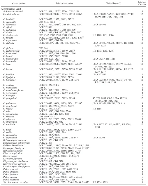
Yeast strains.A total of 373 strains from 86 species, including 41Candidaspp. (207 strains) and 45 non-Candidaspp. (166 strains), were used in this study (Table 1). Among these yeasts, 299 were reference (or type) strains and 74 were clinical isolates. All reference strains were obtained from the Bioresources Col-lection and Research Center (BCRC; Hsinchu, Taiwan) and Centraalbureau voor Schimmelcultures (CBS; Utrecht, The Netherlands). Clinical isolates were obtained from the Mycology Reference Centre, Department of Microbiology, University of Leeds (Leeds, United Kingdom), the Laboratory of Parasitology and Mycology of Angers University Hospital (Angers, France), and the National Cheng Kung University Medical Center (Tainan, Taiwan). Isolates were identi-fied to the species level based on traditional criteria (17) with the API ID32C system (bioMe´rieux Vitek).
DNA preparation. Yeasts were subcultured on Sabouraud dextrose agar (Difco, Detroit, Mich.) and incubated at 28°C for 24 to 48 h. Colonies of these strains were suspended in saline to obtain a turbidity of 0.5 McFarland standard at a 530-nm wavelength. One milliliter of the cell suspension was centrifuged at 5,000⫻gfor 3 min in a microcentrifuge. The genomic DNA was extracted by using the Blood and Tissue Genomic DNA Extraction Miniprep system (Vio-gene, Taipei, Taiwan) in accordance with the manufacturer’s instructions, except that the step of lyticase digestion of yeast cells was omitted. The extracted DNA was stored at⫺20°C for further use.
Amplification and sequencing of the ITS regions.The fungus-specific universal primers ITS1 (5⬘-TCCGTAGGTGAACCTGCGG-3⬘) and ITS2 (5⬘-GCATCG ATGAAGAACGCAGC-3⬘) were used to amplify the ITS1 region, while uni-versal primers ITS3 (5⬘-GCATCGATGAAGAACGCAGC-3⬘) and ITS4 (5⬘-GCATATCAATAAGCGGAGGA-3⬘) were used to amplify the ITS2 region (50). PCR was performed in a total reaction volume of 50l consisting of 10 mM Tris-HCl (pH 8.3), 50 mM KCl, 1.5 mM MgCl2, 0.8 mM deoxynucleoside triphos-phates (0.2 mM each), 1.2 U ofTaqDNA polymerase, 0.4M (each) of the ITS1 region primers (ITS1/ITS2) or the ITS2 region primers (ITS3/ITS4), 2l (1 to 5 ng) of DNA template, and 50l of a mineral oil overlay. PCR was carried out using the following conditions: initial denaturation at 94°C for 3 min; 30 cycles of denaturation (94°C for 1 min), annealing (60°C for 1 min), and extension (72°C for 1 min); and a final extension step at 72°C for 3 min. A negative control was performed with each run by replacing the template DNA with sterile water in the PCR mixture.
All amplicons were purified using the PCR-M Clean Up System (Viogene, Taipei, Taiwan). The DNA fragments were sequenced using an ABI Prism 377 automated DNA sequencer (Applied Biosystems, Taipei, Taiwan) with a BigDye Terminator cycle sequencing kit (version 3.1; Applied Biosystems). All ampli-cons were sequenced on both strands using primers ITS1 and ITS2 for the ITS1 region and primers ITS3 and ITS4 for the ITS2 region. After sequencing, por-tions of the 18S, 5.8S, and 26S rRNA gene sequences of the PCR products were removed to obtain the exact ITS1 and ITS2 sequences. For all yeasts, the sequences of the 3⬘ends of the 18S and 5.8S rRNA genes were GCGGAAGGA TCATTA and GTTTGAGCGTCATTT, respectively, and the sequences of the 5⬘
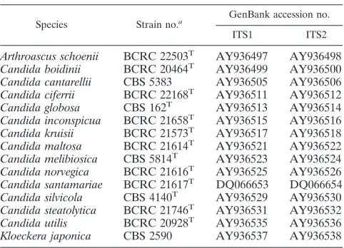
ends of the 5.8S and 26S rRNA genes were AAACTTTCAACAA and GACCTC AAATCAG, respectively. Since the ITS sequences of 15 of the yeast species exam-ined in this study are not currently available in the GenBank database, the ITS sequences of the type strain (or a reference strain) of each of these yeasts were submitted to GenBank (Table 1) to facilitate sequence comparison of strains be-longing to these species.
Identification of yeast by ITS sequencing.A total of 373 strains (86 species) including 41Candidaspecies (207 strains) and 45 non-Candidaspecies (166 strains) were examined. Species were identified by searching databases using the BLAST sequence analysis tool (http://www.ncbi.nlm.nih.gov/BLAST/). The ITS1 or ITS2 sequence was compared using nucleotide-nucleotide BLAST (blastn) with default settings except that sequences were not filtered for low complexity. Species identification was determined from the lowest expect value of the BLAST output. Occasionally, the BLAST search with the query sequence hit sequences from two different species with 100% identity. Under these conditions, the lengths of ITS1 and ITS2 were taken into consideration for species identi-fication, since the lengths of both the ITS1 and ITS2 fragments are important characteristics of a fungal species (3, 4, 30, 46).
For strains producing discrepant identification between the methods based on phenotypic characteristics and ITS sequence analysis, the D1–D2 region of the large-subunit RNA gene was sequenced for species clarification. Primers NL1 (5⬘-GCATATCAATAAGCGGAGGAAAAG-3⬘) and NL4 (5⬘-GGTCCGTGT TTCAAGACGG-3⬘) (22) were used to amplify this region. The procedures for PCR amplification, PCR product purification, and sequencing of the PCR prod-ucts were the same as those described for the ITS regions.
Nucleotide sequence accession numbers.The GenBank accession numbers of the ITS1 and ITS2 regions of type (or reference) strains of 15 species sequenced in this study are given in Table 1.
RESULTS
Amplification of ITS regions.Both the ITS1 and ITS2 regions
were successfully amplified from DNA from all strains by the fungus-specific universal primer pairs ITS1–ITS2 and ITS3–ITS4,
respectively. The lengths of ITS1 ranged from 59 bp (Candida
lipolytica) to 402 bp (C. glabrata), while the lengths of ITS2 ranged
from 69 bp (C. haemulonii) to 261 bp (C. colliculosa) (data not
shown). The ITS fragments of all species studied were less than
300 bp. However, the ITS1 fragments ofC. glabrata(402 bp) and
Saccharomyces cerevisiae(366 bp) were longer than 300 bp. Identification of reference strains by ITS sequence analysis.
A total of 299 reference strains including 41Candidaspecies
(146 strains) and 45 non-Candida species (153 strains) were
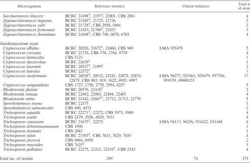
analyzed (Table 2). By ITS1 sequence analysis, 290 strains (97%) were correctly identified to species level. Reference strains producing discrepant identification by phenotypic char-acteristics and ITS sequence analysis are listed in Table 3. Nine
strains (C. intermediaBCRC 22567,C. melibiosicaCBS 6211,
C. silvicola[CBS 4069 and 4141],Pichia ohmeri[BCRC 21592,
22556, and 22557], Cryptococcus albidus CBS 969, and
Rhodotorula glutinisBCRC 20576) were not identified by ITS1
sequencing. In the ITS1 regions,C. intermediaBCRC 22567,C.
melibiosicaCBS 6211, andPichia ohmeri(BCRC 21592, 22556, and 22557) had sequence similarities of only 0.78, 0.65, and 0.76
to 0.89, respectively, with their corresponding type strainsC.
in-termediaBCRC 21250,C. melibiosicaCBS 5814, andPichia
ohm-eriBCRC 22178 (listed in Tables 1 and 2). However, other strains
ofC. intermedia (BCRC 20863, 21250, and 21604) and Pichia ohmeri(BCRC 21349 and 22178) (Table 1) were correctly iden-tified by ITS1 sequence analysis.
A BLAST search revealed thatC. silvicola(CBS 4069 and
4141), in addition to its own species, had an ITS1 sequence and
length (199 bp) identical with those of the type strain ofC.
[image:2.585.43.284.87.261.2]ernobiiCBS 1737 (GenBank accession no. AY585212) (Table 3). The output of the BLAST search of the ITS1 sequence of
TABLE 1. ITS sequences of 15 yeast species submitted to GenBank
Species Strain no.a GenBank accession no.
ITS1 ITS2
Arthroascus schoenii BCRC 22503T AY936497 AY936498
Candida boidinii BCRC 20464T AY936499 AY936500
Candida cantarellii CBS 5383 AY936505 AY936506
Candida ciferrii BCRC 22168T AY936511 AY936512
Candida globosa CBS 162T AY936513 AY936514
Candida inconspicua BCRC 21658T AY936515 AY936516
Candida kruisii BCRC 21573T AY936517 AY936518
Candida maltosa BCRC 21614T AY936521 AY936522
Candida melibiosica CBS 5814T AY936523 AY936524
Candida norvegica BCRC 21616T AY936525 AY936526
Candida santamariae BCRC 21617T DQ066653 DQ066654
Candida silvicola CBS 4140T AY936529 AY936530
Candida steatolytica BCRC 21746T AY936531 AY936532
Candida utilis BCRC 20928T AY936535 AY936536
Kloeckera japonica CBS 2590 AY936537 AY936538
a
T, type strain.
on May 16, 2020 by guest
http://jcm.asm.org/
TABLE 2. Yeast species and strains used in this study
Microorganism Reference strain(s) Clinical isolate(s) Total no.
of strains
Ascomycetous yeast
Arthroascus schoenii BCRC 21401, 22503T, 22504, CBS 2556 4
Candida albicans BCRC 20511, 20512T, 20513, 21538, 22063 LMA 938838, 962507, 499010350, ATTC
66390, RB 1325, 1326, 1331
12
C. boidiniia BCRC 20472, 21432, 21483, 21757 4
C. cantarellii CBS 5445, 5654 2
C. catenulata BCRC 21507, 22316T, CBS 564, 565, 1904 LMA 954976 6
C. ciferrii BCRC 22408 1
C. colliculosa BCRC 21429, 22074T, CBS 158, 6991 4
C. dattila BCRC 22043, CBS 1877, 2803, 2860, 2907 5
C. dubliniensis CBS 2747, 7987, 7988, 8500, 8501 RB 1168, 1271, 1306 8
C. famata BCRC 22304, 22712, CBS 1791, 1792, 1795T 5
C. freyschussii BCRC 21555T, CBS 2161 2
C. glabrata BCRC 20586T, CBS 860, 861, 2175, 7307 LMA 901085, 905756, 945574, RB 1284,
1295, 1324
11
C. globosa CBS 864 1
C. guilliermondii BCRC 20862, 21500T, 21549, 21559 RB 1012, 1055, 1216 7
C. haemulonii BCRC 21572T, CBS 6590, 7801, 7802 4
C. holmii BCRC 21524T, 21999, 22000 3
C. inconspicua CBS 990, 1735, 2833 LMA 90289, RB 1226 5
C. intermedia BCRC 20863, 21250T, 21604, 22567 4
C. kefyr BCRC 20516, 20517, 21269, 21355, 22057T LMA 911323, 938657, 938779, 944459,
947644, RB 1227
11
C. krusei BCRC 20514T, 21321, 21720, 21796, 22342 LMA 911256, 945615, 948501, RB 1222,
1237, 1317
11
C. lambica BCRC 21347, 22067T, 22068, 22071, 22090 LMA 937990 6
C. lipolytica BCRC 20864, 21541, 21542, 21596 4
C. lusitaniae BCRC 20326, 21387T, 21740, CBS 7270 LMA 932648, 947060, 947315, 948764,
RB 1283, 1288, 1294
11
C. maltosa BCRC 21327, 21482 2
C. melibiosica CBS 6211 1
C. membranaefaciens BCRC 21563, 22398T, 22399 3
C. norvegensis BCRC 21851, 22096T, 22097, CBS 1911 4
C. norvegica CBS 2670, 4737 2
C. parapsilosis BCRC 20515T, 20865, 21253, 21544 43, 770, 8053, C4-2, LMA 938558,
961299, RB 1318, 1320
12
C. pelliculosa BCRC 20857, 20858, 21359, 21741, 22583T LMA 892971, RB 766, 778, 913 9
C. pintolopesii BCRC 21439, 22002, 22003, 22239 4
C. rugosa BCRC 21356, 21709T RB 1158 3
C. sake BCRC 21621T, CBS 5690, 5740 3
C. santamariae BCRC 21562, CBS 4261, 4515T 3
C. silvicola CBS 4069, 4141 2
C. sphaerica BCRC 21716, 22153, 22154, 22055, 22604 5
C. steatolytica BCRC 22232, CBS 7652 2
C. tropicalis BCRC 20520T, 20521, 21436, 21437, 21560 LMA 9077, 921810, 945762, RB 1298,
1330
10
C. utilis BCRC 20260, 20325, 20334, 20860, 21357 5
C. valida BCRC 22069T, 21399, 21441 3
C. viswanathii BCRC 21330T, 22554 2
C. zeylanoides BCRC 21743T, 21749, 22396, CBS 947 LMA 91304 5
Debaryomyces marama BCRC 21526, CBS 1958T 2
Debaryomyces polymorphus BCRC 21413 1
Dekkera bruxellensis BCRC 20932, 21414T, 21440, 21517, 21518, 21519 6
Endomyces fibuligera BCRC 20455, 21379, 21380, 21449, 21465, 21511T 6
Hansenula saturnus BCRC 20463, 21360, 21659, 21692, 21765 5
Kloeckera apiculata BCRC 20539, 21362, CBS 312, 314, 2582 5
Kloeckera apis BCRC 22105, 22106, 22112T, CBS 4378 4
Kloeckera japonica CBS 281, 479T 2
Kluyveromyces delphensis BCRC 22017, CBS 2170 2
Kluyveromyces yarrowii BCRC 21747, 22822, CBS 2684, 8242 4
Lodderomyces elongisporus BCRC 21390T, CBS 2606, 5912 3
Pichia carsonii BCRC 21529, 22098T, CBS 4409, 5254 4
Pichia etchellsii BCRC 21479T, CBS 2012, 5519, 5603 4
Pichia farinosa BCRC 21368T, 21682, 21881 3
Pichia ohmeri BCRC 21349, 21592, 22178T, 22556, 22557 5
Pichia spartinae BCRC 22766T, CBS 6059, 6077, 6661 4
Saccharomyces cerevisiae BCRC 20263, 20270, 20271, 20405, 20490, 21447T RB 1254, 1299 8
Continued on following page
on May 16, 2020 by guest
http://jcm.asm.org/
Cryptococcus albidus CBS 969 also showed 100% sequence
identity withCryptococcus albidusCBS 1925 (GenBank
acces-sion no. AB051041) and Cryptococcus adeliensis CBS 8351
(GenBank accession no. AF145328). ITS1 sequencing revealed thatRhodotorula glutinisBCRC 20576, in addition to its own
species, shared an identical sequence with Rhodosporidium
babjevaeCBS 7808 (GenBank accession no. AF444542).
There-fore, these four strains (C. silvicolaCBS 4069 and 4141,
Crypto-coccus albidusCBS 969, andRhodotorula glutinisBCRC 20576) could not be unambiguously identified by ITS1 sequence analysis.
However, other strains of Cryptococcus albidus(BCRC 20527,
21672, and 21860) andRhodotorula glutinis(BCRC 21418) were
correctly identified by ITS1 sequence analysis.
By ITS2 sequencing, 99.7% (298/299) of reference strains were correctly identified. In contrast to the ITS1 region, a
BLAST search of the ITS2 sequences ofCryptococcus albidus
CBS 969 matched only the ITS2 sequence (100% identity) of
Cryptococcus adeliensis CBS 8351 (GenBank accession no. AF145328). Sequence comparison of the D1–D2 region also
revealed that Cryptococcus albidus CBS 969 had 100%
se-quence identify withCryptococcus adeliensis(GenBank
acces-sion no. AF137603). SinceCryptococcus albidusCBS 969 had
identical sequences withCryptococcus adeliensisCBS 8351 at
the ITS1, ITS2, and D1–D2 regions, CBS 969 might be a
misidentification ofCryptococcus adeliensis.
In the ITS2 region, sequence analysis ofRhodotorula glutinis
BCRC 20576, in addition to its own species, revealed 100%
identity withRhodosporidium babjevae JCM 9283 (GenBank
accession no. AB073235). Strain BCRC 20576 also had 100%
sequence identity withRhodosporidium babjevaeA130 (GenBank
accession no. AF485991) and Rhodotorula graminis KCTC
17088 (GenBank accession no. AF459705) in the D1–D2
do-main. Thus, Rhodotorula glutinis BCRC 20576 could not be
differentiated fromRhodosporidium babjevaein both the ITS1
and ITS2 regions and could not be differentiated from
Rho-dosporidium babjevaeandRhodotorula graminisin the D1–D2 domain. In summary, of the 299 reference strains tested, 290 (97%) and 298 (99.7%) strains were correctly identified by sequence analysis of the ITS1 and ITS2 regions, respectively. No strain was misidentified by sequence analysis of either the ITS1 or the ITS2 region.
Identification of clinical isolates by ITS sequence analysis.
A total of 74 clinical isolates, including 15Candidaspecies (61
strains) and 4 non-Candidaspecies (13 strains), were analyzed.
Three isolates (Candida lusitaniae LMA 948764, RB 1283,
andC. pelliculosaLMA 892971) were not identified by ITS1 sequencing, since there were no matching sequences in the
GenBank database. The identification of Candida lusitaniae
LMA 948764 and RB 1283 was correct, as evidenced by
se-quencing of the ITS2 and D1–D2 regions.C. pelliculosaLMA
892971 was a misidentification of Pichia fabianii (GenBank
accession no. AF335967), as confirmed by sequences of the ITS2 and D1–D2 regions (Table 3).
C. dubliniensis RB 1168,C. guilliermondii RB 1055,C. in-TABLE 2—Continued
Microorganism Reference strain(s) Clinical isolate(s) Total no.
of strains
Saccharomyces kluyveri BCRC 21498T, 21977, 22001, CBS 2861 4
Zygosaccharomyces bisporus BCRC 21505T, 21725, 21726 3
Zygosaccharomyces cidri BCRC 21728T, CBS 2950, 5666 3
Zygosaccharomyces fermentati BCRC 21433, 21760T, 22453 3
Zygosaccharomyces florentinus BCRC 21648T, CBS 748, 6078, 6703 4
Basidiomycetous yeast
Cryptococcus albidus BCRC 20526, 21672T, 21860, CBS 969 LMA 935479 5
Cryptococcus curvatus BCRC 21735, CBS 570, 2744, 8770 4
Cryptococcus humicolus CBS 5123 1
Cryptococcus daszewskae BCRC 21639T 1
Cryptococcus laurentii BCRC 20527T, 21997 2
Cryptococcus luteolus BCRC 22372T 1
Cryptococcus neoformans BCRC 20528T, 20532, 22241, 22873, 22874,
22875, CBS 883, 919, 1622, 6955, 6997
LMA 94277, 925461, 935479, 957786, 959159, 49800123
17
Cryptococcus uniguttulatus CBS 1727, 1730, 2770, 2994, 4257 5
Rhodotorula glutinis BCRC 20576, 21418T 2
Rhodotorula minuta BCRC 22482, 22483, 22484, 22485 4
Rhodotorula rubra BCRC 21442, 21667T, 21712, 21713, 21770 5
Sporobolomyces roseus BCRC 22375 1
Sporobolomyces salmonicolor CBS 490, 4474 2
Trichosporon aquatile BCRC 22271T, 22272, CBS 5973, 5988 4
Trichosporon asahii CBS 2479, 2936, 4829, 7631 4
Trichosporon cutaneum BCRC 21675T, 22273 LMA 94117, 94256, 931422, 931440 6
Trichosporon debeurmannianum CBS 1896 1
Trichosporon dermatis CBS 2043 1
Trichosporon inkin BCRC 21503T, CBS 7613, 7629, 7655 4
Trichosporon jirovecii CBS 6864, 6950 2
Trichosporon mucoides CBS 7625T 1
Trichosporon pullulans BCRC 22275, 22313, 22318T, CBS 2543 4
Total no. of strains 299 74 373
aThree strains ofCandida boidinii(BCRC 20472, 21432, and 21483) required assessment of the ITS1 and ITS2 lengths for accurate identification.
on May 16, 2020 by guest
http://jcm.asm.org/
[image:4.585.45.536.80.399.2]conspicuaLMA 90289,C. inconspicuaRB 1226,C. kruseiRB
1237,C. rugosaRB 1158,Cryptococcus albidusLMA 935479,
andTrichosporon cutaneum(LMA 94117, 94256, 931422, and
931440) were misidentifications ofC. albicans,C. parapsilosis,
C. krusei,C. glabrata,Pichia norvegensis,Saccharomyces cerevi-siae,Cryptococcus neoformans, andTrichosporon dermatis, re-spectively, as revealed by sequence analysis of the ITS1, ITS2,
and D1–D2 regions. However, in addition toTrichosporon
der-matis(GenBank accesssion no. AY143555), a BLAST search
of the D1–D2 sequences of Trichosporon cutaneum (LMA
94117, 94256, 931422, and 931440) revealed 100% identity with
an additional sequence of Trichosporon mucoides CBS 7625
(GenBank accession no. AF075515). If the species names of those unidentified or misidentified clinical isolates were cor-rected according to their D1–D2 sequences, the identification rates of clinical isolates by ITS1 and ITS2 sequencing were 95.9% (71/74) and 100% (74/74), respectively. No misidentifi-cation of clinical isolates was caused by sequence analysis of either the ITS1 or the ITS2 region. If reference strains and clinical isolates were taken together, the identification rates were 96.8% (361/373) and 99.7% (372/373), respectively, by sequence analysis of the ITS1 and ITS2 regions.
DISCUSSION
In this study, the feasibility of using ITS sequencing for identification of clinically important yeasts was demonstrated. The whole procedure could be completed within 24 h from isolated colonies. With identification rates of 96.8% (ITS1)
and 99.7% (ITS2), the present approach provides an accurate alternative for species delineation of clinically important yeasts. An important finding of this study was that the ITS2 sequence seems to be more species specific than the ITS1 sequence, and almost all clinically relevant species could be identified by using the ITS2 region alone, providing the ITS2 sequences corresponding to the unknown species are in the GenBank database.
In this study, it was found that the intraspecies sequence divergence of ITS1 is higher than that of ITS2. For example, of
the four reference strains ofC. intermediatested, three were
correctly identified while the remaining strain (BCRC 22567) was not identified by ITS1 sequencing (Tables 2 and 3). In
addition, three reference strains of Pichia ohmeri (BCRC
21592, 22556, and 22557) not identified by ITS1 sequencing were accurately identified by their ITS2 sequences.
Further-more, although all seven clinical isolates ofC. lusitaniaewere
unambiguously identified by ITS2 sequencing, two isolates (LMA 948764 and RB 1283) were not identified by ITS1 se-quence analysis (Tables 2 and 3). It should be noted that the ITS1 sequence may not be sufficiently sensitive for identifying some species.
Cryptococcus albidusCBS 969 might be a misidentification of
Cryptococcus adeliensis, since Cryptococcus albidus CBS 969
had 100% sequence identity withCryptococcus adeliensisCBS
8351 in the ITS1, ITS2, and D1–D2 regions (Table 3). Multiple sequence alignment demonstrated that the nucleotides at
[image:5.585.44.542.89.390.2]po-sitions 203 and 204 in the ITS2 region ofCryptococcus albidus
TABLE 3. List of reference strains and clinical isolates that produced discrepant identification by phenotypic characteristics and ITS sequence analysis
Strain no. Species received as Species identification (% identity with sequences in GenBank) by:
ITS1 sequence ITS2 sequence D1–D2 sequence
Reference strains
BCRC 22567 C. intermedia NIa C. intermedia(100) C. intermedia(99)
CBS 6211 C. melibiosica NI C. melibiosica(100) C. melibiosica(100)
CBS 4069 C. silvicola C. silvicola(100) C. silvicola(100) C. silvicola(99)
C. ernobii(100)
CBS 4141 C. silvicola C. silvicola(100) C. silvicola(100) C. silvicola(99)
C. ernobii(100)
BCRC 21592 Pichia ohmeri NI P. ohmeri(100) P. ohmeri(100)
BCRC 22556 Pichia ohmeri NI P. ohmeri(100) P. ohmeri(100)
BCRC 22557 Pichia ohmeri NI Pichia ohmeri(100) Pichia ohmeri(100)
CBS 969 Cryptococcus albidus Cryptococcus adeliensis(100) Cryptococcus adeliensis(100) Cryptococcus adeliensis(100)
Cryptococcus albidus(100)
BCRC 20576 Rhodotorula glutinis Rhodotorula glutinis(100) Rhodosporidium glutinis(100) Rhodotorula glutinis(100)
Rhodosporidium babjevae(100) Rhodosporidium babjevae(100) Rhodosporidium babjevae(100)
Rhodotorula graminis(100)
Clinical isolates
RB 1168 C. dubliniensis C. albicans(100) C. albicans(100) C. albicans(100)
RB 1055 C. guilliermondii C. parapsilosis(100) C. parapsilosis(100) C. parapsilosis(100)
LMA 90289 C. inconspicua C. krusei(100) C. krusei(99) C. krusei(99)
RB 1226 C. inconspicua C. glabrata(100) C. glabrata(100) C. glabrata(100)
RB 1237 C. krusei Pichia norvegensis(99) P. norvegensis(99) P. norvegensis(99)
LMA 948764 C. lusitaniae NI C. lusitaniae(100) C. lusitaniae(99)
RB 1283 C. lusitaniae NI C. lusitaniae(100) C. lusitaniae(99)
LMA 892971 C. pelliculosa NI Pichia fabianii(100) Pichia fabianii(100)
RB 1158 C. rugosa Saccharomyces cerevisiae(99) S. cerevisiae(99) S. cerevisiae(100)
LMA 935479 Cryptococcus albidus Cryptococcus neoformans(100) Cryptococcus neoformans(100) Cryptococcus neoformans(100) LMA 94117 Trichosporon cutaneum Trichosporon dermatis(100) T. dermatis(100) T. dermatis(100)
T. mucoides(100) LMA 94256 Trichosporon cutaneum Trichosporon dermatis(100) T. dermatis(100) T. dermatis(100)
T. mucoides(100) LMA 931422 Trichosporon cutaneum Trichosporon dermatis(100) T. dermatis(100) T. dermatis(100)
T. mucoides(100) LMA 931440 Trichosporon cutaneum Trichosporon dermatis(100) T. dermatis(100) T. dermatis(100)
T. mucoides(100)
aNI, not identified.
on May 16, 2020 by guest
http://jcm.asm.org/
are C and G (GenBank accession no. AB051026, AB051037, AB051040, and AB051042 to AB051044), respectively, whereas
the two nucleotides are A and C, respectively, in strains of
Crypto-coccus adeliensis (AF145328, AY733078, and AY733079). Therefore, these two positions in the ITS2 regions could be used as a signature sequence to differentiate the two species. Rimek et al. (35) recently published an account of the first case
of meningitis caused by Cryptococcus adeliensis in a patient
with acute myeloid leukemia. Cryptococcus adeliensiscan be
misidentified asCryptococcus albidusdue to the high variability
of phenotypic markers of the latter. Tintelnot and Losert (43) reexamined six isolates from their collection originally
identi-fied as Cryptococcus albidus and found that three of the six
strains in fact turned out to beCryptococcus adeliensis.
Molecular approaches are now being developed to provide a more rapid and objective identification of yeasts compared to traditional phenotypic methods. Ribosomal targets, especially the ITS1, ITS2, and D1–D2 domains of the RNA operon, have shown particular promise for molecular identification. At present, the sequences of the D1–D2 regions of almost all yeasts, including nonpathogenic species, have been determined (11, 22, 23). Analysis of ITS sequences has been carried out mainly for pathogenic yeast species (7, 8, 15, 24, 30, 39). It
should be noted that four clinical isolates ofTrichosporon
cu-taneum(LMA 94117, 94256, 931440, and 931442) were clearly
identified as Trichosporon dermatis by either ITS1 or ITS2
sequencing (Table 3); however, the four isolates could not be
identified asTrichosporon dermatisby sequence analysis in the
D1–D2 domain. A BLAST search revealed that the four
Tri-chosporon cutaneumisolates had 100% sequence identity with
Trichosporon mucoides (GenBank accession no. AF075515) andTrichosporon dermatisin the D1–D2 domain. In this spe-cial case, sequence analysis of both ITS1 and ITS2 is more
specific for identification of Trichosporon dermatis than
se-quence analysis of the D1–D2 region.
An additional advantage of using ITS sequences for yeast identification is length polymorphisms among different species (3, 4, 30, 46). In this study, an interesting example was the fact
that a BLAST search revealed that three C. boidinii strains
(BCRC 20472, 21432, and 21483) had 100% sequence
identi-ties withPichia norvegensisATCC 22977 (GenBank accession
no. AF333096) (data not shown). However, the ITS1 and ITS2
lengths ofC. boidiniiwere 268 and 189 bp, respectively, while
the ITS1 and ITS2 lengths ofPichia norvegensiswere 108 and
142 bp, respectively. For this reason,C. boidiniicould be easily
differentiated fromPichia norvegensis.
The GenBank database currently lacks ITS sequence entries for some yeast species. However, the number of ITS sequences available in public databases has increased rapidly in recent years, and the expanding database may improve the quality and accuracy of fungal identification (19). In this study, the ITS sequences (30 entries) of type (or reference) strains of 15 species were submitted to GenBank (Table 1) to facilitate sequence comparison of these species. As mentioned above, the ITS1 sequence has higher intraspecies divergence than the ITS2 sequence for some species. We also found that different
groups ofC. parapsilosis had divergent ITS1 sequences (data
not shown) as previously reported (37), and this may explain why a relatively lower identification rate was obtained by ITS1
sequence analysis. However, through a multilocus (COX3,
SADH, andSYA1) sequence typing scheme (42) and the fact
that DNA sequence similarities were⬍90% in the ITS1
se-quence,C. orthopsilosisandC. metapsilosiswere proposed to
replace the existing designations of C. parapsilosis groups II
and III, respectively. The speciesC. parapsilosisis retained for
group I isolates. It is anticipated that as more entries of ITS1 sequences become available in public databases, the accuracy of yeast identification based on ITS1 sequences will increase. Phylogenetically closely related species sometimes cannot be identified by sequence analysis of both the ITS and D1–D2
regions (Table 3). In this study, it was found thatRhodotorula
glutinisBCRC 20576 could not be differentiated from Rhodos-poridium babjevaein the ITS1, ITS2, and D1–D2 regions. Re-cently, the intergenic spacer (IGS) region of the fungal rRNA operon was found to have high intraspecies sequence
diver-gence, which might be useful for genotyping of Malassezia
globosa(41) andTrichosporon asahii(40) and for
differentia-tion of varieties ofCryptococcus neoformans(9). However, due
to the high intraspecies divergence in the IGS region, this region may not be suitable for yeast identification.
Many uncommon yeasts are emerging as human pathogens, and their identification may pose a challenge. A rapid and reliable identification method is urgently needed as the incidence of fun-gal infections increases (49). The API ID32C strips or Vitek YBC cards are commonly used for yeast identification. The VITEK system has the advantage of speed (a 15-h incubation) compared to ID32C (a 48- to 72-h incubation). The ID32C kit has a relatively large database (1) for 69 species; however, additional tests are needed for species confirmation of 11 of the 69 spe-cies. With the advanced technology available for sequence analysis and with open access to sequence databases, DNA sequence analysis for microorganism identification is expected to become more popular in the future. In conclusion, the present results clearly demonstrate that sequence analysis of the ITS regions (especially ITS2) can reliably identify yeasts of clinical importance. The method is straightforward and can be completed within 24 h from isolated colonies.
ACKNOWLEDGMENTS
This project was supported by grants (NSC93-2323-B006-007 and NSC93-2314-B006-117) from the National Science Council, Taiwan, Republic of China.
REFERENCES
1.Buchaille, L., A. M. Freydiere, R. Guinet, and Y. Gille.1998. Evaluation of six commercial systems for identification of medically important yeasts. Eur. J. Clin. Microbiol. Infect. Dis.17:479–488.
2.Cassone, M., P. Serra, R. Mondello, A. Girolamo, S. Scafetti, E. Pistella, and M. Venditti.2003. Outbreak ofSaccharomyces cerevisiaesubtypeboulardii
fungemia in patients neighboring those treated with a probiotic preparation of the organism. J. Clin. Microbiol.41:5340–5343.
3.Chang, H. C., S. N. Leaw, A. H. Huang, T. L. Wu, and T. C. Chang.2001. Rapid identification of yeasts in positive blood cultures by a multiplex PCR method. J. Clin. Microbiol.39:3466–3471.
4.Chen, Y.-C., J. D. Eisner, M. M. Kattar, S. L. Rassoulian-Barrett, K. LaFe, S. L. Yarfitz, A. P. Limaye, and B. T. Cookson. 2000. Identification of medically important yeasts using PCR-based detection of DNA sequence polymorphisms in the internal transcribed spacer 2 region of the rRNA genes. J. Clin. Microbiol.38:2302–2310.
5.Chen, Y.-C., J. D. Eisner, M. M. Kattar, S. L. Rassoulian-Barrett, K. Lafe, U. Bui, A. P. Limaye, and B. T. Cookson.2001. Polymorphic internal tran-scribed spacer region 1 DNA sequences identify medically important yeasts. J. Clin. Microbiol.39:4042–4051.
6.Chowdhary, A., K. Becker, W. Fegeler, H. C. Gugnani, L. Kapoor, V. S. Randhawa, and G. Mehta.2003. An outbreak of candidemia due toCandida tropicalisin a neonatal intensive care unit. Mycoses46:269–274.
on May 16, 2020 by guest
http://jcm.asm.org/
7.Coignard, C., S. F. Hurst, L. E. Benjamin, M. E. Brandt, D. W. Warnock, and C. J. Morrison. 2004. Resolution of discrepant results forCandida
species identification by using DNA probes. J. Clin. Microbiol.42:858–861. 8.De Baere, T., G. Claeys, D. Swinne, C. Massonet, G. Verschraegen, A. Muylaert, and M. Vaneechoutte.2002. Identification of cultured isolates of clinically important yeast species using fluorescent fragment length analysis of the amplified internally transcribed rRNA spacer 2 region. BMC Micro-biol.2:21–27.
9.Diaz, M. R., T. Boekhout, B. Theelen, and J. W. Fell. 2000. Molecular sequence analyses of the intergenic spacer (IGS) associated with rDNA of the two varieties of the pathogenic yeast,Cryptococcus neoformans. Syst. Appl. Microbiol.23:535–545.
10.Elie, C. M., T. J. Lott, E. Reiss, and C. J. Morrison.1998. Rapid identifi-cation ofCandidaspecies with species-specific DNA probes. J. Clin. Micro-biol.36:3260–3265.
11.Fell, J. W., T. Boekhout, A. Fonseca, G. Scorzetti, and A. Statzell-Tallman. 2000. Biodiversity and systematics of basidiomycetous yeasts as determined by large-subunit rDNA D1/D2 domain sequence analysis. Int. J. Syst. Evol. Microbiol.50:1351–1371.
12.Fotedar, R., U. Banerjee, and A. R. Chaudhary.2000. Outbreak of systemic candidiasis in low birth weight pre-term infants at a neonatal intensive care unit. J. Mycol. Med.110:100–104.
13.Freydiere, A. M., R. Guinet, and P. Boiron.2001. Yeast identification in the clinical microbiology laboratory: phenotypical methods. Med. Mycol.39:9–33. 14.Fromtling, R. A., J. C. Rhodes, and D. M. Dixon.2003. Taxonomy,
classifi-cation, and morphology of the fungi, p. 1653–1658.InP. R. Murray, E. J. Baron, J. H. Jorgensen, M. A. Pfaller, and R. H. Yolken (ed.), Manual of clinical microbiology, 8th ed. ASM Press, Washington, D.C.
15.Frutos, R. L., M. T. Ferna´ndez-Espinar, and A. Querol.2004. Identification of species of the genusCandidaby analysis of the 5.8S rRNA gene and the two ribosomal internal transcribed spacers. Antonie Leeuwenhoek85:175–185. 16.Han, X. Y., J. J. Tarrand, and E. Escudero.2004. Infections by the yeast
Kodomaea(Pichia)ohmeri: two cases and literature review. Eur. J. Clin. Microbiol. Infect. Dis.23:127–130.
17.Hazen, K. C., and S. A. Howell.2003.Candida,Cryptococcus, and other yeasts of medical importance, p. 1693–1711.InP. R Murray., E. J. Baron, J. H. Jorgensen, M. A., Pfaller, and R. H. Yolken (ed.), Manual of clinical microbiology, 8th ed. ASM Press, Washington, D.C.
18.Henry, T., P. C. Iwen, and S. H. Hinrichs.2000. Identification ofAspergillus
species using internal transcribed spacer regions 1 and 2. J. Clin. Microbiol. 38:1510–1515.
19.Hinrikson, H. P., S. F. Hurst, T. J. Lott, D. W. Warnock, and C. J. Morrison. 2005. Assessment of ribosomal large-subunit D1–D2, internal transcribed spacer 1, and internal transcribed spacer 2 regions as targets for molecular identification of medically importantAspergillusspecies. J. Clin. Microbiol. 43:2092–2103.
20.Huang, Y. C., H. S. Linty, H. L. Peng, and H. Y. Cheng.1999. Outbreak of
Candida parapsilosisfungemia in neonatal intensive care units, clinical im-plications and genotypic analysis. Infection27:97–102.
21.Iwen, P. C., S. H. Hinrichs, and M. E. Rupp.2002. Utilization of the internal transcribed spacer regions as molecular targets to detect and identify human fungal pathogens. Med. Mycol.40:87–109.
22.Kurtzman, C. P., and C. J. Robnett.1997. Identification of clinically impor-tant ascomycetous yeasts based on nucleotide divergence in the 5⬘end of the large-subunit (26S) ribosomal DNA gene. J. Clin. Microbiol.35:1216–1223. 23.Kurtzman, C. P., and C. J. Robnett.1998. Identification and phylogeny of ascomycetous yeasts from analysis of nuclear large subunit (26S) ribosomal DNA partial sequences. Antonie Leeuwenhoek73:331–371.
24.Li, Y. L., S. N. Leaw, J. H. Chen, H. C. Chang, and T. C. Chang.2003. Rapid identification of yeasts commonly found in positive blood cultures by ampli-fication of the internal transcribed spacer regions 1 and 2. Eur. J. Clin. Microbiol. Infect. Dis.22:693–696.
25.Lindsley, M. D., S. F. Hurst, N. J. Iqbal, and C. J. Morrison.2001. Rapid identification of dimorphic and yeast-like fungal pathogens using specific DNA probes. J. Clin. Microbiol.39:3505–3511.
26.Luo, G., and T. G. Mitchell.2002. Rapid identification of pathogenic fungi directly from cultures by using multiplex PCR. J. Clin. Microbiol.40:2860–2865. 27.Lupetti, A., A. Tavanti, P. Davini, E. Ghelardi, V. Corsini, I. Merusi, A. Boldrini, M. Campa, and S. Senesi.2002. Horizontal transmission of Can-dida parapsilosiscandidemia in a neonatal intensive care unit. J. Clin. Mi-crobiol.40:2363–2369.
28.Majoros, L., G. Karods, A. Belak, A. Maraz, L. Asztalos, E. Csanky, Z. Barta, and B. Szabo.2003. Restriction enzyme analysis of ribosomal DNA shows thatCandida inconspicua clinical isolates can be misidentified as
Candida norvegensiswith traditional diagnostic procedures. J. Clin. Micro-biol.41:5250–5253.
29.Martin, C., D. Roberts, M. Van Der Weide, R. Rossau, G. Jannes, T. Smith, and M. Maher.2000. Development of a PCR-based line probe assay for identification of fungal pathogens. J. Clin. Microbiol.38:3735–3742.
30.Massonet, C., J. V. Eldere, M. Vaneechoutte, T. De Baere, J. Verhaegen, and K. Lagrou.2004. Comparison of VITEK 2 with ITS2-fragment length poly-morphism analysis for identification of yeast species. J. Clin. Microbiol. 42:2209–2211.
31.Petrocheilou-Pschou, V., H. Prifti, E. Kostis, C. Papadimitriou, M. A. Dimopoulos, and S. Stamatelopoulos.2001.Rhodotorulasepticemia: case report and minireview. Clin. Microbiol. Infect.7:100–102.
32.Pfaller, M. A., D. J. Diekema, S. A. Messer, R. J. Hollis, and R. N. Jones. 2003. In vitro activities of caspofungin compared with those of fluconazole and itraconazole against 3,959 clinical isolates ofCandidaspp., including 157 fluconazole-resistant isolates. Antimicrob. Agents Chemother.47:1068–1071. 33.Ramani, R., S. Gromadzki, D. H. Pincus, I. F. Salkin, and V. Chaturvedi.
1998. Efficacy of API 20C and ID 32C systems for identification of common and rare clinical yeast isolates. J. Clin. Microbiol.36:3396–3398. 34.Reiss, E., K. Tanaka, G. Bruker, V. Chazalet, D. Coleman, J. P. Debeaupuis,
R. Hanazawa, J. P. Latge, J. Lortholary, K. Makimura, C. J. Morrison, S. Y. Murayama, S. Naoe, S. Paris, J. Sarfati, K. Shibuya, D. Sullivan, K. Uchida, and H. Yamaguchi.1998. Molecular diagnosis and epidemiology of fungal infections. Med. Mycol.36(Suppl.):249–257.
35.Rimek, D., G. Haase, A. Lu¨ck, J. Casper, and A. Podbielski.2004. First report of a case of meningitis caused byCryptococcus adeliensisin a patient with acute myeloid leukemia. J. Clin. Microbiol.42:481–483.
36.Roilides, E., E. Farmaki, J. Evdoridou, A. Francesconi, M. Kasai, J. Filioti, M. Tsivitanidou, D. Sofianou, G. Kremenopoulos, and T. J. Walsh.2003.
Candida tropicalisin a neonatal intensive care unit: epidemiologic and mo-lecular analysis of an outbreak of infection with an uncommon neonatal pathogen. J. Clin. Microbiol.41:735–741.
37.Roy, B., and S. A. Meyer.1998. Confirmation of the distinct genotype groups within the form speciesCandida parapsilosis. J. Clin. Microbiol.36:216–218. 38.Shin, J. H., H. Kook, D. H. Shin, T. J. Hwang, M. Kim, S. P. Suh, and D. W. Ryang.2000. Nosocomial cluster ofCandida lipolyticafungemia in paediatric patients. Eur. J. Clin. Microbiol. Infect. Dis.19:344–349.
39.Sugita, T., A. Nishikawa, R. Ikeda, and T. Shinoda.1999. Identification of medically relevantTrichosporonspecies based on sequences of internal tran-scribed spacer regions and construction of a database forTrichosporon iden-tification. J. Clin. Microbiol.37:1985–1993.
40.Sugita, T., M. Nakajima, R. Ikeda, T. Matsushima, and T. Shinoda.2002. Sequence analysis of the ribosomal DNA intergenic spacer 1 regions of
Trichosporonspecies. J. Clin. Microbiol.40:1826–1830.
41.Sugita, T., M. Kodama, M. Saito, T. Ito, Y. Kato, R. Tsuboi, and A. Nishikawa. 2003. Sequence diversity of the intergenic spacer region of the rRNA gene of
Malassezia globosacolonizing the skin of patients with atopic dermatitis and healthy individuals. J. Clin. Microbiol.41:3022–3027.
42.Tavanti, A., A. D. Davidson, N. A. R. Gow, M. C. J. Maiden, and F. C. Odds. 2005.Candida orthopsilosisandCandida metapsilosisspp. nov. to replace
Candida parapsilosisgroups II and III. J. Clin. Microbiol.43:284–292. 43.Tintelnot, K., and H. Losert.2005. Isolation ofCryptococcus adeliensisfrom
clinical samples and the environment in Germany. J. Clin. Microbiol.43: 1007.
44.Toscano, C. M., and W. R. Jarvis.1999. Emerging issues in nosocomial fungal infections. Curr. Infect. Dis. Rep.1:347–361.
45.Trost, A., B. Graf, J. Eucker, O. Sezer, K. Possinger, U. B. Gobel, and T. Adam.2004. Identification of clinically relevant yeasts by PCR/RFLP. J. Microbiol. Methods56:201–211.
46.Turenne, C. Y., S. E. Sanche, D. J. Hoban, J. A. Karlowsky, and A. M. Kabani.1999. Rapid identification of fungi by using the ITS2 genetic region and an automated fluorescent capillary electrophoresis system. J. Clin. Mi-crobiol.37:1846–1851.
47.Viscoli, C., C. Girmenia, A. Marinus, L. Collette, P. Martino, B. Vandercan, C. Doyer, B. Lebeau, D. Spence, V. Krcmery, B. De Pauw, F. Meunier, and the Invasive Fungal Infection Group of the EORTC.1999. Candidemia in cancer patients: a prospective multicenter surveillance study by the Invasive Fungal Infection Group (IFIG) of the European Organization for Research and Treatment of Cancer(EORTC). Clin. Infect. Dis.28:1071–1079. 48.Wahyuningsih, R., H. J. Freisleben, H. G. Sonntag, and P. Schnitzler.2000.
Simple and rapid detection ofCandida albicansDNA in serum by PCR for diagnosis of invasive candidiasis. J. Clin. Microbiol.38:3016–3021. 49.Walsh, T. J., A. Groll, J. Hiemenz, R. Fleming, E. Roilides, and E. Anaissie.
2004. Infections due to emerging and uncommon medically important fungal pathogens. Clin. Microbiol. Infect.10(Suppl. 1):48–66.
50.White, T. J., T. Bruns, S. Lee, and J. Taylor.1990. Amplification and direct sequencing of fungal ribosomal RNA genes for phylogenetics, p. 315–322.In
M. A. Innis, D. H. Gelfand, J. J. Sninsky, and T. J. White (ed.), PCR protocols: a guide to methods and applications. Academic Press, San Diego, Calif. 51.Yang, R., J. Ao, W. Wang, K. Song, R. Li, and D. Wang.2003. Disseminated
trichosporonosis in China. Mycoses46:519–523.
52.Zaas, A. K., M. Boyce, W. Schell, B. A. Lodge, J. L. Miller, and J. R. Perfect. 2003. Risk of fungemia due toRhodotorulaand antifungal susceptibility testing ofRhotodorulaisolates. J. Clin. Microbiol.41:5233–5235.
on May 16, 2020 by guest
http://jcm.asm.org/
0095-1137/07/$08.00⫹0 doi:10.1128/JCM.00078-07
ERRATUM
Identification of Medically Important Yeast Species by Sequence
Analysis of the Internal Transcribed Spacer Regions
Shiang Ning Leaw, Hsien Chang Chang, Hsiao Fang Sun, Richard Barton,
Jean-Philippe Bouchara, and Tsung Chain Chang
Institute of Biomedical Engineering, Institute of Molecular Medicine, and Department of Medical Laboratory Science and Biotechnology; National Cheng Kung University, Tainan, Taiwan, Republic of China; School of Biochemistry
and Microbiology, University of Leeds, Leeds, United Kingdom; and Host-Parasite-Interaction Study Group (UPRES-EA 3142). Laboratory of Parasitology and Mycology, University Hospital, Angers, France
Volume 44, no. 3, p. 693–699, 2006. Page 694, column 1, third paragraph, lines 2 and 3: “ITS2 (5⬘-GCATCGATGAAGAACG
CAGC-3⬘)” should read “ITS2 (5⬘-GCTGCGTTCTTCATCGATGC-3⬘).”
Page 694, column 1, third paragraph, lines 4 and 5: “ITS4 (5⬘-GCATATCAATAAGCGGAGGA-3⬘)” should read “ITS4
(5⬘-TCCTCCGCTTATTGATATGC-3⬘).”
Page 696, Table 2, column 1, line 9: “Cryptococcus humicolus” should read “Cryptococcus daszewskae.”
Page 696, Table 2, column 1, line 10: “Cryptococcus daszewskae” should read “Cryptococcus humicolus.”
![CAMAC bulletin: A publication of the ESONE Committee Issue #14 December 1975 [last pub of series]](data:image/gif;base64,R0lGODlhAQABAIAAAP///wAAACH5BAEAAAAALAAAAAABAAEAAAICRAEAOw==)