Copyright © 1999, American Society for Microbiology. All Rights Reserved.
Large-Scale Survey of
Campylobacter
Species in Human
Gastroenteritis by PCR and PCR–Enzyme-Linked
Immunosorbent Assay
A. J. LAWSON, J. M. J. LOGAN, G. L. O’NEILL, M. DESAI,ANDJ. STANLEY* Molecular Biology Unit, Virus Reference Division, Central Public
Health Laboratory, London NW9 5HT, United Kingdom
Received 4 June 1999/Returned for modification 17 August 1999/Accepted 13 September 1999
A PCR-based study of the incidence of enteropathogenic campylobacter infection in humans was done on the basis of a detection and identification algorithm consisting of screening PCRs and species identification by PCR-enzyme-linked immunosorbent assay. This was applied to DNA extracted from 3,738 fecal samples from patients with sporadic cases of acute gastroenteritis, submitted by seven regional Public Health Laboratories
in England and Wales over a 2-year period. The sending laboratories had cultured “Campylobacterspp.” from
464 samples. The PCR methodologies detected 492Campylobacter-positive samples, and the combination of
culture and PCR yielded 543Campylobacter-positive samples. There was identity (overlap) for 413 samples, but
79 PCR-positive samples were culture negative, and 51 culture-positive samples were PCR negative. While there was
no statistically significant difference between PCR and culture in detection of C. jejuni-C. coli (PCR, 478
samples; culture, 461 samples), PCR provided unique data about mixed infections and non-C. jejuniand
non-C. colicampylobacters. Mixed infections withC. jejuniandC. coliwere found in 19 samples, and mixed infection withC. jejuniandC. upsaliensiswas found in one sample; this was not apparent from culture. Eleven cases of
gastroenteritis were attributed toC. upsaliensisby PCR, three cases were attributed toC. hyointestinalis, and one
case was attributed toC. lari. This represents the highest incidence of C. hyointestinalis yet reported from
human gastroenteritis, while the low incidence ofC. larisuggests that it is less important in this context.
Gastroenteritis due toCampylobacter jejuniandC. coliis the principal cause of acute bacterial diarrhea (21). Several other species such as C. upsaliensis, C. hyointestinalis, and C. fetus have also been shown to be enteropathogenic for humans, but their significance remains unclear because the procedure for the isolation ofC. jejuniandC. coliuses selective media that may inhibit their growth (1, 9, 15). Furthermore, clinical lab-oratories usually do not identify campylobacter isolates to the species level, since they have relatively fastidious growth re-quirements and lack easily distinguishable biochemical charac-teristics (9). Thus, non-C. jejuniand non-C. colicampylobacters may be underreported in human gastrointestinal illness (2, 15).
The target genes used for PCR identification of Campy-lobacter species from cultured isolates have included 16S rRNA (7, 17), 23S rRNA (4),flaA(flagellin) (29), GTP-bind-ing protein (27), ceuE (iron transport protein) (8), and hip (hippuricase) (16). Flagellin gene and 16S rRNA gene (rDNA) PCR assays have been applied to foodstuffs (7, 30). TheflaA (20),hip(16), and 16S rDNA (14) PCRs have been applied directly to small numbers of fecal samples without culture of an isolate. A recent study of 493 fecal samples (28) was based on enrichment culture and 16S rDNA PCR for detection ofC. jejuni,C. coli, andC. lari.
To our knowledge, the present study represents the largest PCR-based survey ofCampylobactergastroenteritis yet under-taken and demonstrates the utility of this approach for inves-tigation of the incidence and epidemiology of the full spectrum of enteropathogenic campylobacters.
MATERIALS AND METHODS
Study design.Clinical samples were collected over a 2-year period by seven Public Health Laboratories (PHLs) in England and Wales, termed PHLs A, B, C, D, E, F, and G. The samples were from patients with acute gastroenteritis submitted from general practices and outpatient departments or collected by environmental health officers. Repeat, follow-up samples and samples from inpatients were not examined.
DNA was extracted from aliquots of fecal samples sent to the Central Public Health Laboratory not later than 10 days after initial receipt and culture at the collaborating laboratory. Samples were simply cultured on a selective medium,
and no attempt was made to quantify theCampylobactercells present; the lag
between specimen culture and DNA extraction precluded direct quantitative comparison. Culture data collected by the collaborating laboratories were with-held until completion of blind PCR assays with DNA extracted from the corre-sponding fecal samples at the Central Public Health Laboratory. PCR data were then compared with the results of conventional selective culture performed by the contributing laboratories.
Bacterial reference strains.A large range of type and reference strains (19
Campylobacter, 12Helicobacter, and 4Arcobacterstrains, and 11 strains of other enteropathogenic species) were used as controls as described previously (16).
Bacteriological investigation of clinical samples.Fecal samples were examined forCampylobacterspp.,Clostridium difficile,Escherichia coliO:157,Salmonella
spp.,Shigellaspp., and ova, cysts, and parasites by standard methods. With the
exceptions of laboratories A and F, campylobacters were cultured on Campy-lobacter Blood Free Selective Agar Base (Oxoid CM739) with Charcoal Ce-foperazone Desoxycholate Agar supplement (Oxoid SR155). Laboratory A used cefoperazone amphotericin B teicoplanin supplement (Oxoid SR174), while laboratory F used a cefoperazone and amphotericin B supplement (Prolab, Neston, United Kingdom). All plates were incubated for 48 h at 41 to 42°C
(except Laboratory A [37°C]) under microaerobic conditions (5% O2, 5% CO2,
2% H2, and 88% N2, by volume). Isolates were identified to the genus level by
morphology and Gram staining. Laboratory A further identified isolates by hippurate hydrolysis, indoxyl acetate hydrolysis, and urease production.
Extraction of nucleic acid from feces.Approximately 200 mg of each clinical fecal sample was homogenized in 2 ml of brucella broth (Life Technologies Ltd.,
Paisley, United Kingdom). Nucleic acid was extracted from a 100-l aliquot of
the fecal suspension as described previously by Lawson et al. (13). DNA extracts
were stored at⫺20°C prior to screening by PCR.
PCR screening assays.Nucleic acid extracts were screened in batches of 96 samples (including positive and negative controls). Two 16S rDNA PCR assays were used to screen all fecal extracts. The first assay, termed pathgroup, was a
newly designed assay specific for theC. jejuni,C. coli,C. lari,C. upsaliensis, and
* Corresponding author. Mailing address: Molecular Biology Unit, Virus Reference Division, Central Public Health Laboratory, 61 Colin-dale Ave., London NW9 5HT, United Kingdom. Phone: 0208 2004400, ext. 3090. Fax: 0208 2001569. E-mail: sevenwoods@hotmail.com.
3860
on May 15, 2020 by guest
http://jcm.asm.org/
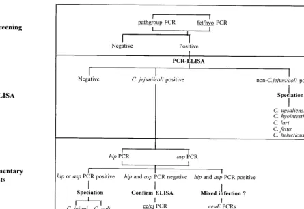
C. helveticusgroup. The second assay, termed fet/hyo, was a duplex assay specific forC. fetusandC. hyointestinalis(17). The screening step is shown in the algorithm presented in Fig. 1.
Each 2.5-l nucleic acid extract obtained from a fecal sample was amplified in
a 25-l reaction volume in a 96-well-format microtiter plate as described
previ-ously (14). Amplification conditions were denaturation at 94°C for 1 min, an-nealing at either 66°C for pathgroup or 65°C for fet/hyo for 1 min, and extension at 72°C for 1 min for 30 cycles in a RoboCycler thermocycler with a hot top assembly (Stratagene, La Jolla, Calif.). The products were analyzed by 96-well-format electrophoresis (on a 1% [wt/vol] agarose gel). The gels were stained with SYBR green I (Flowgen Instruments Ltd., Lichfield, United Kingdom).
Species identification.Samples positive by the screening PCR assays were identified by PCR-enzyme-linked immunosorbent assay (PCR-ELISA) (18) by
using capture probes specific forC. jejuni-C. coli,C. upsaliensis,C. hyointestinalis,
C. lari,C. fetus, andC. helveticus. Those samples identified asC. jejuni-C. coliby PCR-ELISA were further examined by supplementary PCR assays specific for
thehipgene ofC. jejuniand the aspartokinase (asp) gene ofC. coli(16). In cases
in which these two PCRs proved negative, a more sensitive PCR (cc/cj) for the multicopy 16S rRNA gene (16) was applied to confirm the PCR-ELISA data. In
cases of potential mixed culture, two sets ofceuEgene primers (8) capable of
distinguishingC. jejunifromC. coliwere also used. The procedure used for
species identification is summarized in the algorithm (Fig. 1).
Statistical analysis. The results of Campylobacter detection by the PCR screening assay and PCR-ELISA were compared with those obtained by culture on selective agar at contributing laboratories by McNemar’s test (22).
RESULTS
Design and application of primers. Phylogenetic analysis
was conducted with Megalign (Lasergene package; DNA-STAR, Inc., Madison, Wis.). Sequences of 16S rDNA from Helicobacter pylori, Bacteroides ureolyticus, and 15 species of Campylobacter were recovered from the GenBank database and were aligned by the Clustal method (23). PCR primer pairs were designed from the alignment with the aid of the program Oligo (version 4.0; National Biosciences, Plymouth, Mass.). A set of pathgroup primers which inclusively detected C. jejuni,
C. coli,C. upsaliensis,C. lari, andC. helveticuswere designed in this manner. The forward pathgroup primer was 5⬘-ACA TGC AAG TCG AAC GAT GAA GC-3⬘and the reverse pathgroup primer was 5⬘-TAT AGA TTT GCT CCA CCT CGC GG-3⬘. These yielded an amplicon of 1,195 bp from DNA prepared from reference strains of the five species mentioned above but not from the remaining type strains ofCampylobacterand the other enteropathogenic species listed in Materials and Meth-ods. The pathgroup primers were also tested with a set of 200 fecal samples which included 18 cc/cj-positive samples (14) known to containC. jejuniorC. coli. With an annealing tem-perature of 66°C, pathgroup primers detected all 18 previous cc/cj-positive samples. Nonspecific mismatch products could be eliminated by raising the annealing temperature to 68°C, but at the expense of some loss of sensitivity. The lower annealing temperature (66°C) was retained for screening purposes in the full survey. For this and for the fet/hyo assay, any mismatch products were eliminated by the subsequent PCR-ELISA (cf. Fig. 1).
In the full survey of 3,738 samples, the pathgroup PCR was positive for 720 samples, while the fet/hyo duplex PCR was positive for 29 samples.
PCR-ELISA data.The 749 samples positive by the screening
assays described above were subjected to PCR-ELISA accord-ing to the algorithm presented in Fig. 1. This identified campy-lobacters in 492 samples, as follows: C. jejuni-C. coliin 477 samples,C. jejuni-C. coliandC. upsaliensisin 1 sample,C. up-saliensisin 10 samples,C. hyointestinalis in 3 samples, and C. lariin 1 sample. The remaining 257 screening PCR-positive samples were PCR-ELISA negative. In this survey neither C. fetusnorC. helveticuswas detected. A breakdown, by send-FIG. 1. Algorithm forCampylobacterdetection and species identification (speciation) by PCR and PCR-ELISA.
on May 15, 2020 by guest
http://jcm.asm.org/
[image:2.612.96.541.70.376.2]ing laboratory, for detection ofC. jejuni-C. coli and non-C. jejuni–non-C. colicampylobacters is given in Table 1.
Identification ofC. jejuniandC. coli.PCR-ELISA detected
478C. jejuni-C. coli-positive samples. These were subjected to supplementary PCRs, as outlined in Fig. 1 and Materials and Methods. With the hip primers, the campylobacters in 408 samples were positively identified asC. jejuni. With theC. coli -specific aspprimers, the campylobacters in 16 samples were positively identified asC. coli. A further 19 samples were pos-itive for bothhipandasp, indicative of a mixed infection. These 19 samples were therefore investigated with different sets of ceuEprimers, one specific for theC. jejunisequence and one specific for theC. colisequence. All 19 were positive by assays with both sets of primers, confirming mixed infections. From the remaining 35 samples positive forC. jejuni-C. coliby PCR-ELISA, nohiporaspamplicon was obtained. We were there-fore unable to distinguish the two species in these samples but confirmed the PCR-ELISA result by the equivalent simple cc/cj PCR assay (Fig. 1).
Comparison of PCR-based and culture-based detection.The
combination of results from the PCR screening, PCR-ELISA, and supplementary PCR assays allowed each sample to be assigned an overall molecular identification. This was then compared with the culture data: “Campylobacter spp.” were cultured by the seven contributing laboratories from 464 of the 3,738 samples. Of these, the campylobacters in 413 samples (410 with C. jejuni-C. coli, 2 with C. upsaliensis, and 1 with C. hyointestinalis) had been identified by PCR-ELISA with the corresponding fecal sample. The remaining 51 were culture positive but PCR-ELISA negative (Table 1).
Seventy-nine culture-negative samples were positive by the screening PCR and PCR-ELISA (67 withC. jejuni-C. coli, 1 with a mixture of C. jejuni-C. coliand C. upsaliensis, 8 with C. upsaliensis, 2 withC. hyointestinalis, and 1 withC. lari; cf. Table 1). Thus, the combination of screening PCR and PCR-ELISA detected Campylobacterspp. in 543 of the 3,738 sam-ples (528 withC. jejuni-C. coli, 1 with a mixture ofC. jejuni-C. coliandC. upsaliensis, and 14 with other non-C. jejuni-non C. colicampylobacters).
For three of the samples reported by sending laboratories to be “Campylobactersp. positive,” PCR-ELISA detectedC. up-saliensis (two samples) and C. hyointestinalis (one sample). Nine other C. upsaliensis-positive samples and two other C. hyointestinalis-positive samples were detected by PCR-ELISA but not by culture. One of those C. upsaliensis strains was
detected by PCR-ELISA in a sample with a mixed infection withC. jejuni. The latter was detected by culture as well as by PCR-ELISA. A singleC. lariinfection was identified by PCR-ELISA but not by culture (cf. Table 1).
There was no statistical difference between the number of C. jejuni-C. coli-positive samples detected by PCR-ELISA or by culture in the study as a whole (0.5⬎P⬎0.1). In terms of individual sending laboratories, there was no statistical differ-ence between the culture and PCR-ELISA for laboratories A, B, C, (P⬎0.5), F (0.5⬎P⬎0.1), and G (P⫽0.5). However, the detection rate was significantly higher by PCR-ELISA than by culture for laboratories D (P⬍0.001) and E (0.02⬎P⬎
0.01).
DISCUSSION
To our knowledge this study is the largest molecular survey of Campylobacter gastroenteritis yet undertaken and repre-sents the first application of a PCR-based protocol to investi-gate the incidence of the enteropathogenicCampylobacterspp. in an epidemiologically valid context.
[image:3.612.51.551.88.215.2]A key development in this study was the design of PCR assays specific for groups of enteropathogenic species rather than the use of a series of individual species-specific PCRs as we have described previously (14, 16). This was intended to reduce the number of PCR assays performed in the course of a large-scale survey. The basis for the screening PCRs de-scribed in Fig. 1 was phylogenetic trees drawn from alignments of Campylobacter 16S rDNA sequences (12, 24, 26). These trees all contained three distinct clades (species groups). The first containedC. gracilis,C. sputorum,C. curvus,C. concisus, C. rectus, andC. showae, organisms principally associated with niches in the periodontal cavities of humans and animals and which have as yet no association with human gastroenteritis. The second clade consisted of C. fetus, C. hyointestinalis, and C. mucosalis, which are historically associated with disease in farm animals, although the first two have also been occasion-ally implicated in human disease. The third clade consisted of C. jejuni,C. coli,C. lari,C. upsaliensis, andC. helveticus, species (other than C. helveticus) known to cause gastroenteritis in humans. In accordance with the requirements of this study, we developed primers for the third clade and used existing primers specific forC. fetus andC. hyointestinalis(17), the only other causative agents of human gastroenteritis that have been de-scribed.
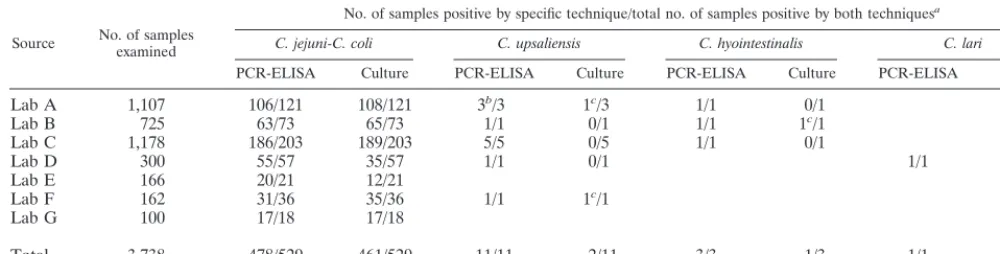
TABLE 1. Detection ofCampylobacterspecies
Source No. of samplesexamined
No. of samples positive by specific technique/total no. of samples positive by both techniquesa
C. jejuni-C. coli C. upsaliensis C. hyointestinalis C. lari
PCR-ELISA Culture PCR-ELISA Culture PCR-ELISA Culture PCR-ELISA Culture
Lab A 1,107 106/121 108/121 3b/3 1c/3 1/1 0/1
Lab B 725 63/73 65/73 1/1 0/1 1/1 1c/1
Lab C 1,178 186/203 189/203 5/5 0/5 1/1 0/1
Lab D 300 55/57 35/57 1/1 0/1 1/1 0/1
Lab E 166 20/21 12/21
Lab F 162 31/36 35/36 1/1 1c/1
Lab G 100 17/18 17/18
Total 3,738 478/529 461/529 11/11 2/11 3/3 1/3 1/1 0/1
aFor example, in the case ofC. jejuni-C. coli, a total of 461 of 3,738 isolates were detected by culture, while 478 of 529 were identified by the PCR algorithm described
in Fig. 1 (PCR-ELISA). A total of 529 were found by a combination of methods; i.e., 68 were not detected by culture and 51 were not identified by PCR-ELISA.
bIncludes one sample with a mixed infection withC. upsaliensisandC. jejunidetected by PCR-ELISA.
cFor these samples, culture identified the strain only as a “Campylobactersp.” Subsequent species identifications were by PCR-ELISA.
on May 15, 2020 by guest
http://jcm.asm.org/
In the course of the study several key practical issues of importance to any large-scale PCR-based survey were identi-fied. These included the necessity of a dedicated PCR suite to reduce the risk of contamination, the robustness of the ther-mocyclers, and the regular monitoring of the performance and detection threshold for PCR primers (their titers and qualities could deteriorate over time or could vary between batches and manufacturers).
C. jejuni and C. coli, as expected, represented the largest proportion of PCR-positive samples. There was congruence between PCR and culture for 77.5% of the 529 positive sam-ples, while 12.9% were found to be positive only by PCR and 9.6% were found to be positive only by culture. In any com-parison of two detection methods with a sizeable sample num-ber, one would not expect a complete correlation. Nonetheless, we note that in this investigation there are further factors to consider. Culture positive-only samples may have been PCR negative due to degradation ofCampylobactercells and DNA in the period (up to 10 days) between culture and receipt of the fecal sample for DNA extraction. In some cases inhibitory substances present in feces may have reduced the sensitivities of the PCR assays. There is also a possibility that certain wild-type C. jejuni or C. coli strains might have 16S rDNA sequences which are sufficiently divergent that they are not detected by PCR-ELISA, despite its detection of all Penner serotype reference strains (18). Isolates from a proportion of the specimens (laboratory C) which were negative by PCR were all successfully identified asC. jejuniwith the PCR algo-rithm (Fig. 1). This suggests that culture-positive, PCR-nega-tive samples occurred due to sampling factors rather than variations in target DNA sequences.
Culture has been found to be more sensitive than PCR in seeding experiments with logarithmic-phase cultures of labo-ratory strains ofC. jejuni(14). That finding may be due to the amount of fecal material which is sampled when inoculating a selective agar plate, as opposed to the small volume of diluted material (2.5l) sampled by PCR.
Nonetheless, our study found more positive samples by PCR-ELISA than by culture alone. This probably reflects the detection of Campylobacter cells in metabolic states that are less amenable to culture on selective media (sublethally dam-aged cells, viable but nonculturable cells, or even dead cells). A key feature of the PCR algorithm was that it provided both detection and identification.
The 11C. upsaliensisisolates detected by PCR-ELISA rep-resent an incidence of 0.29%. Only two of these samples had been positive by culture, and for each sample the isolate was reported as a “Campylobactersp.” The sex/age (in years) dis-tributions for the patients who providedC. upsaliensis-positive samples (m/2, m/5, m/10, f/23, m/25, f/27, f/33, m7/8, m/u, m/u, and u/u, where m is male, f is female, and u is unspecified) showed no evidence of an association with pediatric gastroen-teritis, as has been previously reported in the literature (2, 10). The three cases ofC. hyointestinalisinfection represent an incidence of 0.08%. Only one of these samples was reported as “Campylobactersp.” positive by culture. The sex/age distri-butions were m/39, f/66, and f/u. This represents the highest incidence ofC. hyointestinalisyet reported as a cause of human gastroenteritis. The only other incidence figure available, based on culture, was 0.01% (2 of 15,185 cases) (11). Earlier pilot PCR studies by our group had detectedC. hyointestinalis in 1 of 25 (16) and 1 of 200 (14) patients with gastroenteritis. Altogether, this would give an incidence of 0.13% (5 of 3,963). AlthoughC. hyointestinalisis associated with proliferative en-teritis in pigs (6), it has previously been considered to be only a very rare cause of gastroenteritis in humans (3, 5, 19). Our
results suggest that further investigation ofC. hyointestinalisas a human enteropathogen would be appropriate.
We detected C. lari (by PCR-ELISA alone) in only one sample (an incidence of 0.03%), although this species is often cited (15) as the third most commonly isolated enteropatho-genic campylobacter from humans. The low incidence in our survey suggests thatC. lariis less important thanC. upsaliensis andC. hyointestinalisin human gastroenteritis.
In 19 samples there was evidence from supplementary PCR assays of a mixed infection withC. jejuniand C. colinot ap-parent from culture. Seven of these occurred in a laboratory (laboratory A) which had the capacity to identify some isolates to the species level and which reported that five wereC. jejuni and two wereC. coli. It is likely, therefore, that only the pre-dominant colony type had been selected for identification. It is interesting that there were slightly more mixed C. jejuniand C. coli infections thanC. coli infections alone. Mixtures of Campylobacter species (and serotypes) in human infections may be more common than was heretofore assumed. Another mixed Campylobacter infection noted in the survey was of C. upsaliensis and C. jejuni, which were codetected by PCR-ELISA; here, only theC. jejunicomponent was apparent from culture. There were also instances of coinfections ofC. jejuni-C. coliwith other enteropathogens detected by culture and/or microscopy at the contributing laboratories. There were mix-tures of C. jejuni with both Shigella sonnei and Blastocystis hominis cysts (one sample), with aSalmonellasp. (two sam-ples), and with Cryptosporidium parvum oocysts (three sam-ples). The mixtures ofCryptosporidiumandC. jejuni-C. coliare of interest since the presence of a coccidian, which is most frequently associated with waterborne infections (25), suggests that the route of transmission of theC. jejunimay have been water for these samples.
Although PCR is more expensive and labor-intensive than culture, it offers a nonselective way to monitor the incidence of enteropathogenic bacteria. We have shown that it is of value for epidemiological purposes, and it will ultimately be amena-ble to automation. The use of a broad-specificity screening PCR greatly reduces the number of assays required for a com-prehensive survey. We have also demonstrated that our previ-ously published PCR-ELISA (18) can be successfully applied in this context. In summary, the PCR algorithm presented here offers a different perspective onCampylobactergastroenteritis than that provided by culture, giving information on the iden-tity and occurrence of species that are not detected by culture. PCR-based analysis has a role in large-scale epidemiological surveys ofCampylobacter.
ACKNOWLEDGMENTS
We are most grateful to the staff of Ashford PHL, Bangor PHL, Central Middlesex PHL, Chelmsford PHL, Dorchester PHL, Exeter PHL, and Preston PHL for the clinical samples and culture data.
This work was supported by a grant (grant DH220B) from the Department of Health, London, United Kingdom.
REFERENCES
1.Advisory Committee on the Microbiological Safety of Food, Department of Health.1993. Interim report on campylobacter. Her Majesty’s Stationery Office, London, United Kingdom.
2.Bourke, B., V. L. Chan, and P. Sherman.1998.Campylobacter upsaliensis:
waiting in the wings. Clin. Microbiol. Rev.11:440–449.
3.Edmonds, P., C. M. Patton, P. M. Griffin, T. J. Barrett, G. P. Schmid, C. N. Baker, M. A. Lambert, and D. J. Brenner.1987.Campylobacter hyointestinalis
associated with human gastrointestinal disease in the United States. J. Clin.
Microbiol.25:685–691.
4.Eyers, M., S. Chapelle, G. Van Camp, H. Goossens, and R. De Wachter.
1993. Discrimination among thermophilicCampylobacterspecies by
poly-merase chain reaction amplification of 23S rRNA gene fragments. J. Clin.
on May 15, 2020 by guest
http://jcm.asm.org/
Microbiol.31:3340–3343. (Erratum,32:1623, 1994.)
5.Fennell, C. L., A. M. Rompalo, P. A. Totten, K. L. Bruch, B. M. Flores, and W. E. Stamm.1986. Isolation of “Campylobacter hyointestinalis” from a
human. J. Clin. Microbiol.24:146–148.
6.Gebhart, C. J., P. Edmonds, G. E. Ward, H. J. Kurtz, and D. J. Brenner.
1985. “Campylobacter hyointestinalis” sp. nov.: a new species of
campy-lobacter found in the intestines of pigs and other animals. J. Clin. Microbiol.
21:715–720.
7.Giesendorf, B. A., and W. G. Quint.1995. Detection and identification of
Campylobacterspp. using the polymerase chain reaction. Cell. Mol. Biol.
41:625–638.
8.Gonzalez, I., K. A. Grant, P. T. Richardson, S. F. Park, and M. D. Collins.
1997. Specific identification of the enteropathogensCampylobacter jejuniand
Campylobacter coliby using a PCR test based on theceuEgene encoding a
putative virulence determinant. J. Clin. Microbiol.35:759–763.
9.Goossens, H., and J. P. Butzler.1992. Isolation and identification of Campy-lobacterspp., p. 93–109.InI. Nachamkin, M. J. Blaser, and L. S. Tompkins
(ed.),Campylobacter jejuni: current status and future trends. American
So-ciety for Microbiology, Washington, D.C.
10. Goossens, H., B. Pot, L. Vlaes, C. Van den Borre, R. Van den Abbeele, C. Van Naelten, J. Levy, H. Cogniau, P. Marbehant, J. Verhoef, K. Kersters, J. P. Butzler, and P. Vandamme. 1990. Characterization and description of “Campylobacter upsaliensis” isolated from human feces. J. Clin. Microbiol.
28:1039–1046.
11. Goossens, H., L. Vlaes, M. De Boeck, B. Pot, K. Kersters, J. Levy, P. De Mol, J. P. Butzler, and P. Vandamme.1990. Is ’Campylobacter upsaliensis’ an
unrecognised cause of human diarrhoea? Lancet335:584–586.
12. Lawson, A. J., D. Linton, and J. Stanley.1998. 16S rRNA gene sequences of ‘CandidatusCampylobacter hominis’, a novel uncultivated species, are found
in the gastrointestinal tract of healthy humans. Microbiology144:2063–2071.
13. Lawson, A. J., D. Linton, J. Stanley, and R. J. Owen.1997. Polymerase chain
reaction detection and speciation ofCampylobacter upsaliensisandC.
hel-veticusin human faeces and comparison with culture techniques. J. Appl.
Microbiol.83:375–380.
14. Lawson, A. J., M. S. Shafi, K. Pathak, and J. Stanley.1998. Detection of
Campylobacterin gastroenteritis: comparison of direct PCR assay of faecal
samples with selective culture. Epidemiol. Infect.121:547–553.
15. Linton, D.1996. Old and newCampylobacters: a review. PHLS Microbiol.
Digest13:10–15.
16. Linton, D., A. J. Lawson, R. J. Owen, and J. Stanley.1997. PCR detection,
identification to species level, and fingerprinting ofCampylobacter jejuniand
Campylobacter colidirect from diarrheic samples. J. Clin. Microbiol.35:
2568–2572.
17. Linton, D., R. J. Owen, and J. Stanley.1996. Rapid identification by PCR of
the genusCampylobacterand of fiveCampylobacterspecies enteropathogenic
for man and animals. Res. Microbiol.147:707–718.
18. Metherell, L. A., J. M. Logan, and J. Stanley.1999. PCR–enzyme-linked
immunosorbent assay for detection and identification ofCampylobacter
spe-cies: application to isolates and stool samples. J. Clin. Microbiol.37:433–435.
19. Minet, J., B. Grosbois, and F. Megraud.1988.Campylobacter hyointestinalis:
an opportunistic enteropathogen? J. Clin. Microbiol.26:2659–2660.
20. Oyofo, B. A., S. A. Thornton, D. H. Burr, T. J. Trust, O. R. Pavlovskis, and P. Guerry. 1992. Specific detection ofCampylobacter jejuniand Campy-lobacter coliby using polymerase chain reaction. J. Clin. Microbiol.30:2613– 2619.
21. Skirrow, M. B.1994. Diseases due toCampylobacter,Helicobacterand
re-lated bacteria. J. Comp. Pathol.111:113–149.
22. Swinscow, S. D. V.1996. Statistics at square one, 9th ed. BMJ Publishing Group, London, United Kingdom.
23. Thompson, J. D., D. G. Higgins, and T. J. Gibson.1994. CLUSTAL W: improving the sensitivity of progressive multiple sequence alignment through sequence weighting, position-specific gap penalties and weight matrix choice.
Nucleic Acids Res.22:4673–4680.
24. Thompson, L. M. I., R. M. Smibert, J. L. Johnson, and N. R. Krieg.1988.
Phylogenetic study of the genusCampylobacter. Int. J. Syst. Bacteriol.38:
190–200.
25. Tzipori, S., and J. K. Griffiths.1998. Natural history and biology of Crypto-sporidium parvum. Adv. Parasitol.40:5–36.
26. van Camp, G., Y. van de Peer, S. Nicolai, J. M. Neefs, P. Vandamme, and R. de Wachter.1993. Structure of 16S and 23S ribosomal RNA genes in Campy-lobacterspecies: phylogenetic analysis of the genusCampylobacterand
pres-ence of internal transcribed spacers. Syst. Appl. Microbiol.16:361–368.
27. van Doorn, L. J., B. A. Giesendorf, R. Bax, B. A. van der Zeijst, P. Van-damme, and W. G. Quint.1997. Molecular discrimination between Campy-lobacter jejuni,Campylobacter coli,Campylobacter lariandCampylobacter upsaliensisby polymerase chain reaction based on a novel putative GTPase
gene. Mol. Cell. Probes11:177–185.
28. Vanniasinkam, T., J. A. Lanser, and M. D. Barton.1999. PCR for the
detection ofCampylobacterspp. in clinical specimens. Lett. Appl. Microbiol.
28:52–56.
29. Waegel, A., and I. Nachamkin.1996. Detection and molecular typing of
Campylobacter jejuniin fecal samples by polymerase chain reaction. Mol.
Cell. Probes10:75–80.
30. Wegmuller, B., J. Luthy, and U. Candrian.1993. Direct polymerase chain
reaction detection ofCampylobacter jejuniandCampylobacter coliin raw
milk and dairy products. Appl. Environ. Microbiol.59:2161–2165.