0095-1137/06/$08.00
⫹
0
doi:10.1128/JCM.44.4.1447–1452.2006
Copyright © 2006, American Society for Microbiology. All Rights Reserved.
Use of a Serotype-Specific DNA Microarray for Identification of
Group B
Streptococcus
(
Streptococcus agalactiae
)
Linyan Wen,
1,2Quan Wang,
1,2Yayue Li,
1,2Fanrong Kong,
3Gwendolyn L. Gilbert,
3Boyang Cao,
1,2Lei Wang,
1,2and Lu Feng
1,2*
TEDA School of Biological Sciences and Biotechnology, Nankai University, 23# HongDa Street, TEDA, Tianjin 300457,
People’s Republic of China
1; Tianjin Key Laboratory for Microbial Functional Genomics, TEDA College,
Nankai University, 23# HongDa Street, TEDA, Tianjin 300457, People’s Republic of China
2; and
Centre for Infectious Diseases and Microbiology, Institute of Clinical Pathology and Medical Research,
Westmead Hospital, Westmead, 2145 New South Wales, Australia
3Received 20 October 2005/Returned for modification 3 January 2006/Accepted 25 January 2006
Group B
Streptococcus
(GBS;
Streptococcus agalactiae
) is an important cause of sepsis and meningitis. Nine
GBS serotypes, based on capsular polysaccharide (CPS) antigens, have been described. Their distribution
varies worldwide and needs to be monitored to understand the epidemiology of GBS disease and inform the
development of vaccines. In this study, we sequenced
cpsH
of GBS serotype II (
cpsHII
) and compared it with
that of the other eight serotypes to identify serotype-specific regions. We then developed a DNA microarray
based on the
cpsH
gene and used it to test 88 GBS isolates—9 serotype reference strains and 79 clinical
isolates—and 7 other bacterial and fungal species which are commonly present in the vagina flora. The
microarray was shown to be specific and reproducible. This is the first report of a microarray which can
identify the nine GBS serotypes. The use of a microarray has advantages over traditional serotyping methods
and will be of practical value in both reference and diagnostic laboratories.
Group B
Streptococcus
(GBS;
Streptococcus agalactiae
) is an
important cause of sepsis and meningitis in neonates and
in-fants and of invasive disease in pregnant women, nonpregnant,
presumably immunocompromised adults, and the elderly (8,
22). Capsular polysaccharide (CPS) is external to the
gram-positive bacterial cell wall and enables the bacterium to evade
host immune defenses. CPS is essential to the virulence of
GBS, and serotype-specific antibody is protective. Therefore, it
is the target for most experimental GBS vaccines. Nine distinct
CPS serotypes (Ia, Ib, and II to VIII) have been described (12,
13), and their distribution varies worldwide. For example,
se-rotypes Ia and III have been reported to be the predominant
ones in the maternal genital tract in the United States (2, 7),
China (23), Germany (9), and Australia and New Zealand
(16). In Korea, serotype Ib is most common (25), and in Japan
serotypes VI and VIII are predominant among GBS isolated
from pregnant women (18). Epidemiological studies of GBS
are important for the development of GBS vaccines that are
suitable for all geographic areas. Therefore, a convenient and
rapid method to identify all nine GBS serotypes would be
useful.
Many GBS serotyping methods have been reported
previ-ously, including immunoprecipitation (26), enzyme
immunoas-say (14), coagglutination (11), counterimmunoelectrophoresis
and capillary precipitation (24), latex agglutination (27),
fluo-rescence microscopy (6), and inhibition enzyme-linked
immu-nosorbent assay (1), and antisera are necessary for most of
these methods. Commercial antisera are expensive and
avail-able for only six serotypes (Ia to V), which does not satisfy the
needs of traditional serotyping methods (1). Moreover,
sero-typing is often subjective, and a significant proportion of
iso-lates are nontypeable. Molecular typing methods, such as
pulsed-field gel electrophoresis (20) and restriction
endonucle-ase analysis (19), have also been used for epidemiological
studies, but they do not directly identify serotypes and are
generally time-consuming and expensive.
A previous report, based on analysis of
cpsH
in GBS
sero-types Ia and III (3), suggested that
cpsH
, which encodes CPS
polymerases, was serotype specific in bacteria which produce
complex polysaccharides. We have developed a molecular
se-rotyping method based on PCR and sequencing of various
genes in the
cps
cluster, including
cpsH
, which has been
cently adapted to a more practicable multiplex PCR and
re-verse line blot assay format, targeting serotype-specific
se-quences (16a). Sese-quences of
cpsH
for all GBS serotypes
have been deposited in GenBank (see Table 2). Sequence
alignment shows that the
cpsHII
(GenBank accession number
AY375362) and
cpsHIII
(AF363056) sequences are identical,
which is inconsistent with the different CPS structures of these
two serotypes (4, 17). Therefore, we sequenced another
cpsHII
strain in preparation for this study, in which we developed a
DNA microarray, based on serotype-specific sequences of
cpsH
, for identification of all GBS serotypes.
DNA microarray is a newly developed technology used for
the detection of pathogens and is rapid and sensitive. It
con-sists of four steps: extraction of genomic DNA, amplification of
targeted DNA, hybridization of labeled DNA with
oligonucle-otide probes immobilized on a microarray, and results analysis.
We believe it has significant advantages over other methods for
routine serotyping of GBS.
* Corresponding author. Mailing address: TEDA School of
Bio-logical Sciences and Biotechnology, Nankai University, 23 HongDa
Street, TEDA, Tianjin 300457, People’s Republic of China. Phone:
86-22-66229592. Fax: 86-22-66229596. E-mail: [email protected].
1447
on May 16, 2020 by guest
http://jcm.asm.org/
MATERIALS AND METHODS
Bacterial strains.Nine GBS reference strains, 79 GBS clinical isolates, which were isolated in different countries between 1994 and 2001, and isolates of 7 other bacterial and fungal species, namely,Staphylococcus aureus,Staphylococcus epidermidis,Streptococcus pneumoniae,Escherichia coli,Lactobacillus acidophi-lus,Lactobacillus delbrueckii, andCandida albicans, were used in this study (Table 1). All of the GBS isolates had been previously characterized by conven-tional and molecular serotyping (MS) as previously described (15).
The GBS isolates were inoculated into Todd-Hewitt broth (10.0 g/liter beef heart infusion, 20.0 g/liter tryptone, 2.0 g/liter glucose, 2.0 g/liter sodium bicar-bonate, 2.0 g/liter sodium chloride, 0.4 g/liter disodium phosphate [pH 7.8⫾ 0.2]) and incubated overnight at 37°C.
Sequence analysis.Sequencing ofcpsHIIwas carried out using an ABI 3730 automated DNA sequencer. Two primers, wl-3401 (5⬘-GATTGTTATCACACA TGGC-3⬘) and wl-3402 (3⬘-ATATATTTTTTTCA/GTAA/TTAACC-5⬘), were used to amplify the gene, and the amplicon is 1,881 bp. The sequence data used in this study forcpsHof other serotypes were obtained from GenBank with the following accession numbers: AF332905, AF332906, AF363037, AF363044, AF363045, AF363053, AF363054, and AY375362. Sequence alignment and com-parisons ofcpsHsequences of all nine GBS serotypes were performed using the ClustalW program (http://www.ebi.ac.uk/clustalw). The phylogenetic tree was generated using Mega 3.
Serotype II-specific PCR.Because of differences between ourcpsHIIsequence and the one available in GenBank (accession number AY375362), we designed three serotype II-specific primer pairs based on our sequence and two pairs based on the original sequence. We then tested the 10 serotype II strains in our collection by PCR and identified the products by gel electrophoresis. Primer sequences are shown in Table 2.
Genomic DNA extraction.Genomic DNA extraction was performed as follows. Overnight broth cultures (1.5 ml) were centrifuged for 10 min at 3,000⫻g. The deposit was resuspended in 500l of SET (75 mM NaCl, 25 mM EDTA, 20 mM Tris [pH 7.5]). Lysozyme (50 mg/ml) was added to the tube, and after incubation at 37°C for 2 h, 1/3 volumes of 5 M NaCl, 1/10 volumes of 10% sodium dodecyl sulfate (SDS), and 0.5 mg/ml proteinase K were added. Tubes were incubated at 55°C with occasional inversion for 2 h, then 1 volume of chloroform was added, and the tubes were centrifuged at 4,500⫻gfor 15 min twice to purify the DNA. The resulting supernatant was transferred to a new tube, and 1 volume of isopropanol was added. Tubes were held at⫺20°C for 20 min and then centri-fuged at 4,500⫻g for 10 min. The aqueous phase was discarded, and the remaining contents were rinsed with 70% ethanol and dried at 37°C. Purified DNA was dissolved in 30l Tris-EDTA and stored at⫺20°C.
Multiplex PCR.Based on analysis ofcpsHsequences of all nine serotypes, primer pairs suitable for multiplex PCR were designed. From 69 primers initially tested, 13 were chosen (Table 2) based on testing of 60 GBS isolates (9 serotype reference strains and 51 clinical isolates [Table 1]). These 13 primers include a universal forward primer for serotypes Ia to VII targetingcpsG, which apparently encodesN-acetylglucosaminyl transferase (21) and is conserved in all serotypes except VIII (4). The forward primer for serotype VIII targetscpsHVIII. Sero-type-specific reverse primers were designed for each of the nine serotypes as well as a pair of primers targeting the GBS species-specific genecfb. The PCR products range from 240 bp to 1,310 bp in length.
For multiplex PCR, DNA was prepared as above, and 50 to 100 ng was used as a template in a final volume of 50l of PCR mixture containing the following: 1⫻PCR buffer (50 mM KCl, 10 mM Tris-HCl [pH 8.3]); 0.5 mM MgCl2; 200M concentrations of dATP, dCTP, dGTP, and dTTP; 42 nM each of two primers based on thecfbgene (housekeeping gene of GBS); 140 nM each of 11 other primers; and 0.03 UTaqDNA polymerase. The PCR cycle was performed with the initial denaturation at 95°C for 10 min, followed by 30 cycles of 95°C for 30 s, 55°C for 45 s, and 72°C for 1 min, and concluding with a cycle of 72°C for 10 min.
Labeling of the targeted DNA.For labeling of PCR products, multiplex PCR mixtures contained 1⫻PCR buffer (50 mM KCl, 10 mM Tris-HCl [pH 8.3]); 0.8 mM MgCl2; 334M concentrations of dATP, dCTP, dGTP, and dTTP; 187 nM each of 10 reverse primers; 0.25 nM cyanine dye Cy3-dUTP; and 0.05 UTaq
DNA polymerase complemented to a final volume of 30l with amplified multiplex PCR products from the above amplification step. The PCR cycle was performed with the initial denaturation at 95°C for 10 min followed by 35 cycles of denaturation for 30 s at 95°C, annealing for 45 s at 50°C, and extension for 1 min at 72°C, and cycling was concluded with a final elongation for 10 min at 72°C. All labeled DNA was purified in refining tubes (Millipore Co.) and then stored at⫺20°C in the dark.
Oligonucleotide probe set and microarray construction.Probes used in this study were designed using Oligoarray 2.0 and synthesized with a 5⬘amidocyanogen
[image:2.585.302.541.79.498.2]mod-ifier, and 15 poly(T) oligonucleotides were added. From 178 probes tested, 35 were selected based on preliminary testing of 60 GBS strains representing all serotypes (Table 3). Glass slides, modified by an aldehyde group, were purchased from CEL Corporation. There are four arrays on each glass slide. The construction of a single array and the meaning of each spot are shown in Fig. 1. Oligonucleotide
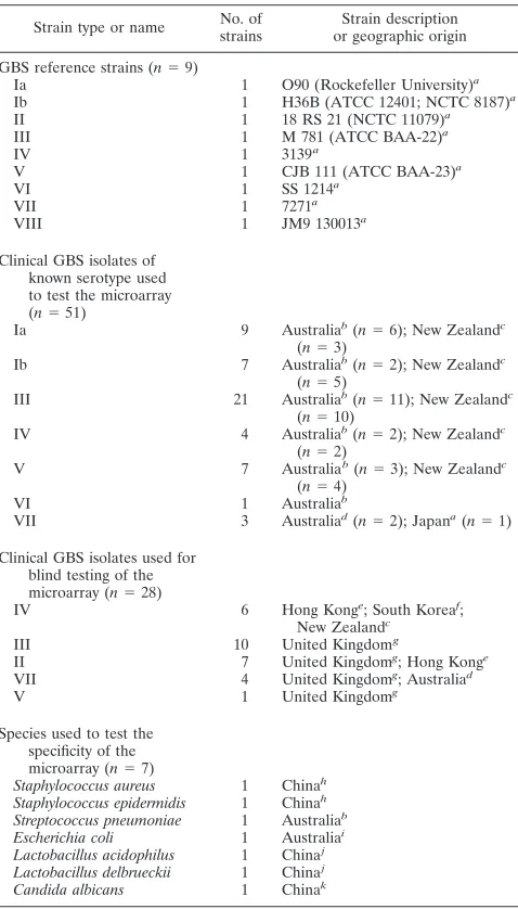
TABLE 1. Isolates used in this study
Strain type or name No. of strains
Strain description or geographic origin
GBS reference strains (n⫽9)
Ia 1 O90 (Rockefeller University)a
Ib 1 H36B (ATCC 12401; NCTC 8187)a
II 1 18 RS 21 (NCTC 11079)a
III 1 M 781 (ATCC BAA-22)a
IV 1 3139a
V 1 CJB 111 (ATCC BAA-23)a
VI 1 SS 1214a
VII 1 7271a
VIII 1 JM9 130013a
Clinical GBS isolates of known serotype used to test the microarray (n⫽51)
Ia 9 Australiab(n⫽6); New Zealandc (n⫽3)
Ib 7 Australiab(n⫽2); New Zealandc (n⫽5)
III 21 Australiab(n⫽11); New Zealandc (n⫽10)
IV 4 Australiab(n⫽2); New Zealandc (n⫽2)
V 7 Australiab(n⫽3); New Zealandc (n⫽4)
VI 1 Australiab
VII 3 Australiad(n⫽2); Japana(n⫽1) Clinical GBS isolates used for
blind testing of the microarray (n⫽28)
IV 6 Hong Konge; South Koreaf; New Zealandc
III 10 United Kingdomg
II 7 United Kingdomg; Hong Konge
VII 4 United Kingdomg; Australiad
V 1 United Kingdomg
Species used to test the specificity of the microarray (n⫽7)
Staphylococcus aureus 1 Chinah
Staphylococcus epidermidis 1 Chinah
Streptococcus pneumoniae 1 Australiab
Escherichia coli 1 Australiai
Lactobacillus acidophilus 1 Chinaj
Lactobacillus delbrueckii 1 Chinaj
Candida albicans 1 Chinak
aObtained from Channing Laboratory, Boston, Mass.
bObtained from the Centre for Infectious Diseases and Microbiology labora-tory services (CIDM), Sydney, Australia.
cObtained from the Streptococcus Reference Laboratory, ESR, Wellington, New Zealand.
dObtained from the Department of Microbiology and Immunology, Univer-sity of Melbourne, Australia.
eObtained from the Department of Microbiology, The Chinese University of Hong Kong, Prince of Wales Hospital, Hong Kong.
fObtained from the Research Institute of Bacterial Resistance, Yonsei Uni-versity College of Medicine, Seoul, South Korea.
gObtained from the Nuffield Department of Clinical Laboratory Sciences, Insti-tute for Molecular Medicine, John Radcliffe Hospital, Oxford, United Kingdom.
hObtained from the National Institute for The Control of Pharmaceutical and Biological Products, CMCC, Beijing, People’s Republic of China.
iObtained from the University of Sydney, Sydney, Australia.
jObtained from the Tianjin University of Science and Technology, Tianjin, People’s Republic of China.
kObtained from the Institute of Dermatology, Peking Union Medical College & Chinese Academy of Medical Science, Nanjing, People’s Republic of China.
on May 16, 2020 by guest
http://jcm.asm.org/
probes were diluted in 50% dimethyl sulfoxide to a final concentration of 1g/l and spotted onto the slides using SpotArray 7.2 (Perkin Elmer Corporation). Each probe was duplicated three times. Printed slides were dried for 24 h at room temperature. In order to immobilize probes, slides were treated with a UV Cross-linker (UVP Corporation) and then stored at room temperature.
[image:3.585.40.549.81.228.2]DNA microarray hybridization.The purified product was baked for 90 min at 65°C in a dry oven, diluted in 12l hybridization buffer (25% formamide, 0.1% SDS, 6⫻SSPE [1⫻SSPE is 0.18 M NaCl, 10 mM NaH2PO4, and 1 mM EDTA {pH 7.7}]) and hybridized for 16 h at 40°C. After hybridization, the chip was rinsed in solution A (1⫻SSC [1⫻SSC is 0.15 M NaCl plus 0.015 M sodium
TABLE 2. Oligonucleotide primers used in this study
Primer name Gene Tm(°C)
GenBank
accession no. Sequence
wl-4432
cfb
a42.5
X72754
328-GATGTATCTATCTGGAACTCTAGTG-352
wl-4433
cfb
48.4
X72754
568-TTTTTCCACGCTAGTAATAGCCTC-545
wl-4347
cpsGIa-VII
b49.5
AF363060
2126-GAACCTCAGAATTGTCAGTGGTCA-2149
wl-4596
cpsHIa
49.5
AF363060
3381- ATTATTTATAACGATGTTTACTGTA-3357
wl-4597
cpsHIb
63.1
AB050723
3718-AGGTCGAATGCGATCCTAGTGG-3697
wl-4598
cpsHII
52.7
DQ234264
896-CTTAGTGCAAGTGTTGAAGAATA-874
wl-4599
cpsHIII
60.5
AF363056
3015-TTGGGGTTTCAAAAAAAGTATGG-2993
wl-4600
cpsHIV
49
AF355776
7831-CAACTACATTGACTTTTTTACTAT-7808
wl-4601
cpsHV
51.4
AF349539
7459-ATAATACCCTGTCCTTGAAAA-7439
wl-4602
cpsHVI
53.9
AF337958
7840-TACTAATGTATGATGAATGTGAACC-7816
wl-4603
cpsHVII
48.1
AY376403
4820-GTTCAACTATTCTTATTTCTTTAC-4797
wl-4604
cpsHVIII
c51.6
AY375363
4615-GGTGCCACTTCTTTAGGTT-4633
wl-4605
cpsHVIII
50.9
AY375363
5840-TCACAAATCGCTTCTTCAT-5822
a
Thecfbgene is a housekeeping gene which encodes cyclic AMP, which is used as the positive control of GBS. b
The universal upload primer of types Ia to VII was designed in thecpsGgene. The N-terminal regions of this gene in all nine serotypes expect serotype VIII share a high level of identity.
c
The upload primer of GBS type VIII was designed in thecpsHVIIIgene.
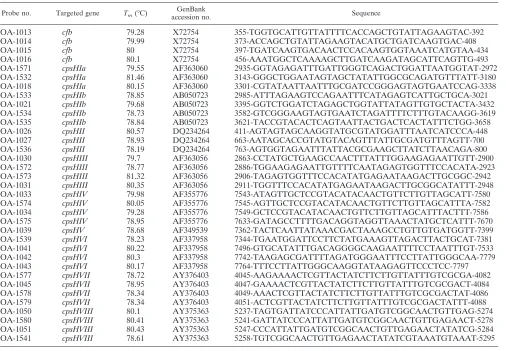
TABLE 3. Oligonucleotide probes used in this study
Probe no. Targeted gene Tm(°C)
GenBank
accession no. Sequence
OA-1013
cfb
79.28
X72754
355-TGGTGCATTGTTATTTTCACCAGCTGTATTAGAAGTAC-392
OA-1014
cfb
79.99
X72754
373-ACCAGCTGTATTAGAAGTACATGCTGATCAAGTGAC-408
OA-1015
cfb
80
X72754
397-TGATCAAGTGACAACTCCACAAGTGGTAAATCATGTAA-434
OA-1016
cfb
80.1
X72754
456-AAATGGCTCAAAAGCTTGATCAAGATAGCATTCAGTTG-493
OA-1571
cpsHIa
79.55
AF363060
2935-GGTAGAGATTTGATTGGGTCAGACTGGATTAATGGTAT-2972
OA-1532
cpsHIa
81.46
AF363060
3143-GGGCTGGAATAGTAGCTATATTGGCGCAGATGTTTATT-3180
OA-1018
cpsHIa
80.15
AF363060
3301-CGTATAATTAATTTGCGATCCGGGAGTAGTGAATCCAG-3338
OA-1533
cpsHIb
78.85
AB050723
2985-ATTTAGAAGTCCAGAATTTCATAGAGTCATTGCTGCA-3021
OA-1021
cpsHIb
79.68
AB050723
3395-GGTCTGGATCTAGAGCTGGTATTATAGTTGTGCTACTA-3432
OA-1534
cpsHIb
78.73
AB050723
3582-GTCGGGAAGTAGTGAATCTAGATTTTCTTTGTACAAGG-3619
OA-1535
cpsHIb
78.84
AB050723
3621-TACCGTACACTCAGTAATTACTGACTCACTATTTCTGG-3658
OA-1026
cpsHII
80.57
DQ234264
411-AGTAGTAGCAAGGTATGCGTATGGATTTAATCATCCCA-448
OA-1027
cpsHII
78.93
DQ234264
663-AATAGCACCGTATGTACAGTTTATTGCGATGTTTAGTT-700
OA-1536
cpsHII
78.19
DQ234264
763-AGTGGTAGAATTTATTACGCGAAGCTTATCTTAACAGA-800
OA-1030
cpsHIII
79.7
AF363056
2863-CCTATGCTGAAGCCAACTTTATTTGGAAGAGAATTGTT-2900
OA-1572
cpsHIII
78.77
AF363056
2886-TGGAAGAGAATTGTTTTCAATAGAGTGGTTTCCACATA-2923
OA-1573
cpsHIII
81.32
AF363056
2906-TAGAGTGGTTTCCACATATGAGAATAAGACTTGCGGC-2942
OA-1031
cpsHIII
80.35
AF363056
2911-TGGTTTCCACATATGAGAATAAGACTTGCGGCATATTT-2948
OA-1033
cpsHIV
79.98
AF355776
7543-ATAGTTGCTCCGTACATACAACTGTTCTTGTTAGCATT-7580
OA-1574
cpsHIV
80.05
AF355776
7545-AGTTGCTCCGTACATACAACTGTTCTTGTTAGCATTTA-7582
OA-1034
cpsHIV
79.28
AF355776
7549-GCTCCGTACATACAACTGTTCTTGTTAGCATTTACTTT-7586
OA-1575
cpsHIV
78.95
AF355776
7633-GATAGCCTTTTGACAGGTAGGTTAAACTATGCTCATTT-7670
OA-1039
cpsHV
78.68
AF349539
7362-TACTCAATTATAAACGACTAAAGCCTGTTGTGATGGTT-7399
OA-1539
cpsHVI
78.23
AF337958
7344-TGAATGGATTCCTTCTATGAAAGTTAGACTTACTGCAT-7381
OA-1041
cpsHVI
80.22
AF337958
7496-GTGCATATTTGACAGGGGCAAGAATTTTCCTAATTTGT-7533
OA-1042
cpsHVI
80.3
AF337958
7742-TAAGAGCGATTTTAGATGGGAATTTCCTTATTGGGCAA-7779
OA-1043
cpsHVI
80.17
AF337958
7764-TTTCCTTATTGGGCAAGGTATAAGAGTTCCCTCC-7797
OA-1577
cpsHVII
78.72
AY376403
4045-AAGAAAACTCGTTACTATCTTCTTGTTATTTGTCGCGA-4082
OA-1045
cpsHVII
78.95
AY376403
4047-GAAAACTCGTTACTATCTTCTTGTTATTTGTCGCGACT-4084
OA-1578
cpsHVII
78.34
AY376403
4049-AAACTCGTTACTATCTTCTTGTTATTTGTCGCGACTAT-4086
OA-1579
cpsHVII
78.34
AY376403
4051-ACTCGTTACTATCTTCTTGTTATTTGTCGCGACTATTT-4088
OA-1050
cpsHVIII
80.1
AY375363
5237-TAGTGATTATCCCATTATTGATGTCGGCAACTGTTGAG-5274
OA-1580
cpsHVIII
80.41
AY375363
5241-GATTATCCCATTATTGATGTCGGCAACTGTTGAGAACT-5278
OA-1051
cpsHVIII
80.43
AY375363
5247-CCCATTATTGATGTCGGCAACTGTTGAGAACTATATCG-5284
OA-1541
cpsHVIII
78.61
AY375363
5258-TGTCGGCAACTGTTGAGAACTATATCGTAAATGTAAAT-5295
on May 16, 2020 by guest
http://jcm.asm.org/
[image:3.585.43.549.378.729.2]citrate], 0.1% SDS) for 3 min, solution B (0.05⫻SSC) for 3 min, and solution C (95% ethanol) for 1.5 min. The chip was then dried, in the dark, at room temperature.
Signal detection and data analysis.The hybridized microarray was scanned with a 532-nm laser beam using the biochip scanner LuxScan-10K/A (Capitalbio Corporation) using the following parameters: laser intensity, 80%; photomulti-plier tube gain, 70%; scan resolution, 5 nm.
Nucleotide sequence accession number.The DNA sequence ofcpsHIIhas been deposited in GenBank under the accession number DQ234264.
RESULTS
Sequencing and analysis of
cpsHII
.
The sequence we
ob-tained for
cpsHII
from the GBS serotype II reference strain
(Table 1) was significantly different from that previously
de-posited in GenBank (accession number AY375362). All 10
GBS serotype II strains tested by PCR produced amplicons of
the expected length with primers (wl-4184, 5
⬘
-ATACAGGTG
TTTACAGGGAC-3
⬘
; wl-4185, 3
⬘
-GATAAATAGATGGCA
AAGAA-5
⬘
; wl-4186, 5
⬘
-TAGGGAGTAGGAAGATAGC-3
⬘
;
wl-4187, 3
⬘
-TAACGCTACAAATCAAACA-5
⬘
; wl-4184, 5
⬘
-A
TACAGGTGTTTACAGGGAC-3
⬘
; and wl-4188, 3
⬘
-GCTTT
CAATCACGTCCTAGT-5
⬘
) based on the new sequence but
not with those based on the original sequence. These results
confirm that the original sequence of
cpsHII
was incorrect.
Analysis of
cpsH
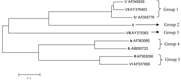
sequences of all nine GBS serotypes.
Se-quence alignment of
cpsH
sequences of all nine GBS serotypes
showed that they fell into five groups (Fig. 2), each consisting
of one to three sequences, which is consistent with CPS
struc-tural data (4). The three
cpsH
sequences in group 1 shared 85.5
to 91% identity, and the two sequences in groups 4 and 5
shared 75% and 76.7% identity, respectively. Despite the
sim-ilarities between
cpsH
sequences within groups, we were able
to identify serotype-specific regions, which could be probed to
differentiate these closely related serotypes. Therefore,
cpsH
appears to be an appropriately variable, serotype-specific
re-gion within the
cps
gene cluster for use as the targeted gene for
differentiation of GBS serotypes.
Optimization of multiplex PCR.
Initially, we used the same
concentration (140 nM) for all 13 primers, but we could not
consistently achieve expected signals after hybridization for
some serotypes. The multiplex PCR products were tested by
gel electrophoresis, which showed that only some of the
tar-geted serotype sequences which produce long fragments were
amplified. Therefore, we tested various proportions of the
primers which produce amplicons of short and long fragments
in ratios of 1:2, 1:3, and 1:4. By repeating hybridization and gel
electrophoresis, we found that expected signals were achieved
consistently when the ratio of primers producing short fragments
to primers producing long fragments was approximately 1:3.
Specificity.
The DNA microarray was tested initially using
60 GBS isolates, including 9 serotype reference strains and 51
clinical isolates of known serotype, mainly from Australia and
New Zealand (Table 1). Through 242 hybridization reactions,
35 specific probes and 13 primers were selected for use in the
microarray. The microarray identified all 60 GBS isolates
cor-rectly when results were compared with those of our MS
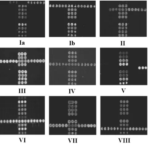
method (15) (Fig. 3), and it gave negative results for all 7
[image:4.585.133.450.572.713.2]FIG. 1. Schematic diagram of the microarray, showing positions of immobilized probes spotted within a single well of the four-well glass slides.
Each array contains 150 spots arranged in 15 columns. Cy3 is a fluorescent dye. Sequences of probes immobilized at each location are shown in
Table 3.
FIG. 2. A phylogenetic tree generated based on
cpsH
of all nine GBS serotypes by the neighbor-joining method.
on May 16, 2020 by guest
http://jcm.asm.org/
isolates of other bacterial and fungal species likely to be
present in the vagina or to cause bacteremia (Table 3).
Double-blind test.
Twenty-eight clinical isolates (shown in
Table 1) were randomly selected from all strains which were
stored in the Centre for Infectious Diseases and
Microbiol-ogy for double-blind testing in order to prove the stability
and veracity of the GBS DNA microarray. After
hybridiza-tion, we found that 7 isolates are serotype II, 10 isolates are
serotype III, 6 isolates are serotype IV, 1 isolate is serotype
V, and 4 isolates are serotype VII, which is consistent with
MS results.
DISCUSSION
Optimization of multiplex PCR.
We found that not only
melting temperatures (
T
m) and cross-dimer formation between
primer pairs but also the concentrations of individual primers
in the reaction mixture affected the success of the multiplex
PCR. A ratio of 1:3 of primers producing short fragments to
primers producing long fragments gave optimal results,
prob-ably because primers producing short fragments consume
re-agents, such as deoxynucleoside triphosphate and
Taq
DNA
polymerase, preferentially unless present in a lower
concen-tration.
Two-step multiplex PCR was used in this study. The first
step ran without labeling, and the second one ran with labeling.
Although this test format prolongs the time of detection, it has
many advantages. First, two-step PCR can amplify more
am-plicons, which enhance the hybridization signal, and in the
meantime make the microarray method more sensitive. In
addition, if the amplification and labeling of target DNA are
finished at the same time, the labeled target DNA must be
denatured before hybridization, and the denatured DNA must
always be annealed, which would reduce the amount of
single-stranded DNA. To avoid such situations, two-step multiplex
PCR was selected. In the second step, we used only
down-stream primers, which produce single-stranded labeled target
DNA directly.
Hybridization.
The final sets of probes and primers were
selected according to the following criteria: the probes were in
the amplified region and specific to their corresponding
sero-type, and there was no significant cross-dimer formation
be-tween members of primer pairs. Although the probes and
primers were designed to be specific, repeated hybridization
was needed to select the best primers and probes for the DNA
microarray.
According to a previous report, 15 poly(T) was the best
spacer length to ensure high-quality hybridization results (10).
All probes on this DNA microarray have approximately the
same
T
mvalues and GC contents. There is no obvious
rela-tionship between probe length and consistency of
hybridiza-tion, and probes of the same length do not necessarily
hybrid-ize under the same conditions. On the other hand, we found
that probes with relatively similar GC contents and
T
mhybrid-ize consistently under similar conditions. Furthermore,
some-times short labeled single-stranded amplicons are more
diffi-cult to hybridize with corresponding probes than the larger
ones. This phenomenon is infrequent and may be due to the
secondary structure of DNA.
Practicability.
We tested the specificity and reproducibility
of the microarray with GBS isolates of unknown (at the time of
testing) serotype and other bacterial and fungal species likely
to be encountered in similar clinical specimens. The results
confirmed that
cpsH
is a suitable gene to identify GBS
sero-types and that our GBS DNA microarray, the first of its kind to
be described, is a practicable and reliable tool for routine
identification and serotyping of GBS.
According to the 2002 Centers for Disease Control and
Prevention guidelines, all pregnant women should be screened
for GBS at 35 to 37 weeks of pregnancy (http://www.cdc.gov
/mmwr/preview/mmwrhtml/rr5111a1.htm). In this study, we
did not compare the sensitivity of our method with
conven-tional culture methods for direct detection of GBS. However,
we believe that its use for routine screening of vaginal
secre-tions of pregnant women, either directly or after preliminary
enrichment, would be feasible. This requires further
evalua-tion. This DNA microarray will certainly be useful for testing
GBS isolates in studies of the epidemiology and pathogenesis
of GBS infection and vaccine research.
It would be useful to add probes for surface protein antigens
to the microarray. A multiplex PCR assay has been reported to
identify GBS proteins (5). In the future it should be practicable
to combine multiplex PCR with the DNA microarray to
iden-tify both capsular polysaccharide and protein antigens
simul-taneously.
ACKNOWLEDGMENTS
[image:5.585.44.284.69.304.2]This work was supported by the NSFC Key Program (30530010) and
funds from the Science and Technology Committee of Tianjin City
(05YFGZGX04700) to L.W. and L.F.
FIG. 3. Representative hybridization results of GBS strains of nine
serotypes. The number under each array shows the serotype of GBS
tested. Three lines of dots located in the middle of the array are
positive controls based on
cfb
of GBS.
on May 16, 2020 by guest
http://jcm.asm.org/
REFERENCES
1.Arakere, G., A. E. Flores, P. Ferrieri, and C. E. Frasch.1999. Inhibition enzyme-linked immunosorbent assay for serotyping of group B streptococcal isolates. J. Clin. Microbiol.37:2564–2567.
2.Blumberg, H. M., D. S. Stephens, C. Licitra, N. Pigott, R. Facklam, B. Swami-nathan, and I. K. Wachsmuth.1992. Molecular epidemiology of group B strep-tococcal infections: use of restriction endonuclease analysis of chromosomal DNA and DNA restriction fragment length polymorphisms of ribosomal RNA genes (ribotyping). J. Infect. Dis.166:574–579.
3.Chaffin, D. O., S. B. Beres, H. H. Yim, and C. E. Rubens.2000. The serotype of type Ia and III group B streptococci is determined by the polymerase gene within the polycistronic capsule operon. J. Bacteriol.182:4466–4477. 4.Cieslewicz, M. J., D. Chaffin, G. Glusman, D. Kasper, A. Madan, S.
Ro-drigues, J. Fahey, M. R. Wessels, and C. E. Rubens.2005. Structural and genetic diversity of group BStreptococcuscapsular polysaccharides. Infect. Immun.73:3096–3103.
5.Creti, R., F. Fabretti, G. Orefici, and C. von Hunolstein.2004. Multiplex PCR assay for direct identification of group B streptococcal alpha-protein-like protein genes. J. Clin. Microbiol.42:1326–1329.
6.Cropp, C. B., R. A. Zimmerman, J. Jelinkova, A. H. Auernheimer, R. A. Bolin, and B. C. Wyrick.1974. Serotyping of group Bstreptococciby slide agglutination fluorescence microscopy, and microimmunodiffusion. J. Lab. Clin. Med.84:594–603.
7.Edwards, M. S.1990. Group B streptococcal infections. Pediatr. Infect. Dis. J.9:778–781.
8.Farley, M. M.2001. Group B streptococcal disease in nonpregnant adults. Clin. Infect. Dis.33:556–561.
9.Fluegge, K., S. Supper, A. Siedler, and R. Berner.2005. Serotype distribution of invasive group B streptococcal isolates in infants: results from a nation-wide active laboratory surveillance study over 2 years in Germany. Clin. Infect. Dis.40:760–763.
10.Guo, Z., R. A. Guilfoyle, A. J. Thiel, R. Wang, and L. M. Smith.1994. Direct fluorescence analysis of genetic polymorphisms by hybridization with oligo-nucleotide arrays on glass supports. Nucleic Acids Res.22:5456–5465. 11.Hakansson, S., L. G. Burman, J. Henrichsen, and S. E. Holm.1992. Novel
coagglutination method for serotyping group B streptococci. J. Clin. Micro-biol.30:3268–3269.
12.Harrison, L. H., J. A. Elliott, D. M. Dwyer, J. P. Libonati, P. Ferrieri, L. Billmann, A. Schuchat, and Maryland Emerging Infections Program.1998. Serotype distribution of invasive group B streptococcal isolates in Maryland: implications for vaccine formulation. J. Infect. Dis.177:998–1002. 13.Hickman, M. E., M. A. Rench, P. Ferrieri, and C. J. Baker.1999. Changing
epidemiology of group B streptococcal colonization. Pediatrics104:203–209. 14.Holm, S. E., and S. Hakansson.1988. A simple and sensitive enzyme
im-munoassay for determination of soluble type-specific polysaccharide from group Bstreptococci. J. Immunol. Methods106:89–94.
15.Kong, F., S. Gowan, D. Martin, G. James, and G. L. Gilbert.2002. Serotype identification of group B streptococci by PCR and sequencing. J. Clin. Microbiol.40:216–226.
16.Kong, F., D. Martin, G. James, and G. L. Gilbert.2003. Towards a geno-typing system forStreptococcus agalactiae(group Bstreptococcus): use of mobile genetic elements in Australasian invasive isolates. J. Med. Microbiol.
52:337–344.
16a.Kong, F., L. Ma, and G. L. Gilbert.2005. Simultaneous detection and serotype identification ofStreptococcus agalactiaeusing multiplex PCR and reverse line blot hybridization. J. Med. Microbiol.54:1133–1138. 17.Kuypers, J. M., L. M. Heggen, and C. E. Rubens.1989. Molecular analysis of
a region of the group B streptococcus chromosome involved in type III capsule expression. Infect. Immun.57:3058–3065.
18.Lachenauer, C. S., D. L. Kasper, J. Shimada, Y. Ichiman, H. Ohtsuka, M. Kaku, L. C. Paoletti, P. Ferrieri, and L. C. Madoff.1999. Serotypes VI and VIII predominate among group Bstreptococciisolated from pregnant Jap-anese women. J. Infect. Dis.179:1030–1033.
19.Nagano, Y., N. Nagano, S. Takahashi, K. Murono, K. Fujita, F. Taguchi, and Y. Okuwaki.1991. Restriction endonuclease digest patterns of chromosomal DNA from group B beta-haemolyticstreptococci. J. Med. Microbiol.35:297– 303.
20.Rolland, K., C. Marois, V. Siquier, B. Cattier, and R. Quentin.1999. Genetic features ofStreptococcus agalactiaestrains causing severe neonatal infec-tions, as revealed by pulsed-field gel electrophoresis andhylBgene analysis. J. Clin. Microbiol.37:1892–1898.
21.Rubens, C. E., R. F. Haft, and M. R. Wessels.1995. Characterization of the capsular polysaccharide genes of group Bstreptococci. Dev. Biol. Stand.
85:237–244.
22.Schuchat, A.1999. Group Bstreptococcus. Lancet353:51–56.
23.Shen, A., Y. Zhu, G. Zhang, Y. Yang, and Z. Jiang.1998. Experimental study on distribution of serotypes and antimicrobial patterns of group B strepto-coccusstrains. Chin. Med. J. (Engl. Ed.)111:615–618.
24.Triscott, M. X., and G. H. Davis.1979. A comparison of four methods for the serotyping of group Bstreptococci. Aust. J. Exp. Biol. Med. Sci.57:521–527. 25.Uh, Y., I. H. Jang, K. J. Yoon, C. H. Lee, J. Y. Kwon, and M. C. Kim.1997. Colonization rates and serotypes of group Bstreptococciisolated from preg-nant women in a Korean tertiary hospital. Eur. J. Clin. Microbiol. Infect. Dis.
16:753–756.
26.Wilkinson, H. W., and M. D. Moody.1969. Serological relationships of type I antigens of group B streptococci. J. Bacteriol.97:629–634.
27.Zuerlein, T. J., B. Christensen, and R. T. Hall.1991. Latex agglutination detection of group-B streptococcal inoculum in urine. Diagn. Microbiol. Infect. Dis.14:191–194.