Copyright © 2001, American Society for Microbiology. All Rights Reserved.
Determination of
Enterococcus faecalis groESL
Full-Length
Sequence and Application for Species Identification
LEE-JENE TENG,1,2* PO-REN HSUEH,2YI-HUI WANG,1HSIAO-MANN LIN,1
KWEN-TAY LUH,2ANDSHEN-WU HO1,2
School of Medical Technology, National Taiwan University College of Medicine,1and Department of Laboratory Medicine, National Taiwan University Hospital,2Taipei, Taiwan
Received 20 March 2001/Returned for modification 14 May 2001/Accepted 3 July 2001
Amplification of the partial Cpn60 (or GroEL) gene segment has been used for identification of many bacteria, includingEnterococcusspecies. To obtain more sequence data from groESLgenes of Enterococcus
faecalis, the full-length sequence of theE. faecalis groESLgenes containinggroES(285 bp), spacer (57 bp), and
groEL(1,626 bp) was determined. A database search of GenBank revealed that the deducedE. faecalisGroES
and GroEL proteins show significant homology to the GroES and GroEL proteins of other bacteria. The GroEL
(groEL) of E. faecalis had the highest identity with Streptococcus pneumoniae (81.8% amino acid sequence
identity and 73.0% nucleotide sequence identity), followed byLactococcus zeae, while GroES (groES) had 60.2% (64.6%) identity withLactobacillus zeae and 58.5% (66.2%) identity withLactococcus lactis, followed by 57.0% (65.5%) identity with Bacillus subtilis.Based on the groES sequence, an E. faecalis-specific PCR assay was developed, and this PCR assay was positive for all theE. faecalisstrains tested. Dot blot hybridization using eithergroESorgroELas the probe distinguishedE. faecalisclearly from other species, indicating that both genes can be used as suitable targets forE. faecalisidentification. Moreover, broad-range PCR-restriction fragment length polymorphism ofgroESLwas designed to differentiate eight commonly encountered
Entero-coccus species. TheEnterococcus species of reference strains could be easily differentiated on the basis of
restriction patterns produced byHaeIII andRsaI. The DNA-based assays developed in this study provide an alternative to currently used methods of identification for clinically important enterococcal species.
Once considered harmless commensals of the intestinal tract, enterococci now rank among the leading causes of nosocomial infections (19). There are two major pathogenic species in humans,Enterococcus faecalisandE. faecium,with occasional infections being caused by other species (19). The increasing occurrence of high-level gentamicin-resistant (HLGR) and vancomycin-resistant enterococci (VRE) has become a major concern worldwide (13, 27).
Rapid identification of bacteria is important for effective patient management and reducing the spread of antibiotic resistance (2). Conventional identification methods, which are based on phenotypic and culture characteristics, require 2 to 3 days to provide results (7, 32). Currently, species identification of enterococci in many laboratories relies on automation or rapid kits. However, errors in automated identification systems are not easily detected, as species identification is based on an inbuilt database (8, 29). In addition, atypical phenotypic char-acteristics can lead to misidentification. Another approach to species identification may be the use of molecular methods. Several DNA-based techniques for identifying clinical isolates have been developed (5, 6, 15, 18). A variety of conserved genes, including 16S rRNA genes, the tRNA intergenic spacer, theD-alanine:D-alanine ligase gene (ddlgene), thesodAgene
encoding superoxide dismutase, penicillin-binding protein 5, and the elongation factortufgene have been used for identi-fication of enterococci (15, 20, 21, 23, 30).
The groESL genes (also known as cpn10/60 or hsp10/60), which encode 10-kDa (GroES) and 60-kDa (GroEL) heat shock proteins, are ubiquitous and evolutionarily highly con-served among bacteria (12). Recently, Goh et al. developed reverse checkerboard hybridization to identifyStaphylococcus andEnterococcusspecies on the basis of amplification of par-tial chaperonin 60 gene sequences (10, 11). The goals of this study were to obtain the full-length sequences ofgroESLgenes ofE. faecalisand provide another approach for species iden-tification.
MATERIALS AND METHODS
Bacterial strains.E. faecalisATCC 29212,E. faeciumATCC 19434,E. avium
ATCC 14025,E. casseliflavus ATCC 25788, E. gallinarumATCC 49573, E. raffinosusATCC 49427,E. hiraeATCC 8043, andE. duransATCC 19432 were obtained from the American Type Culture Collection (ATCC), Rockville, Md. Clinical isolates, including eightE. faecalisisolates, fourE. faeciumisolates, two
E. aviumisolates, fourE. casseliflavusisolates,fourE. gallinarumisolates, and two E. raffinosus isolates, were obtained from the Bacteriology Laboratory, National Taiwan University Hospital, a 2,000-bed teaching hospital in northern Taiwan.
DNA amplification and sequencing of a partial fragment by PCR.Initially,
degenerate PCR primers, 590F and 590R (Table 1), complementary to highly conserved regions of thegroELgene among eubacteria, were designed and used to amplify a 590-bp internal fragment of thegroELgene from enterococcal species. Genomic DNA was isolated and purified fromEnterococcusspecies with a DNA isolation kit, Puregene (Gentra Systems, Inc., Minneapolis, Minn.), according to the manufacturer’s instructions. The thermal cycling conditions were 1 min at 94°C, 1 min at 50°C, and 1 min at 72°C, followed by a final extension of 7 min at 72°C. An amplified product of the expected size was subsequently sequenced.
Southern blot hybridization.A 590-bp DNA product internal to thegroEL
gene ofE. faecaliswas used as the probe. Probes were produced by the PCR method described above and simultaneously labeled by incorporation of digoxi-genin-11-dUTP (Boehringer Mannheim, Mannheim, Germany).E. faecalisDNA
* Corresponding author. Mailing address: School of Medical Tech-nology, National Taiwan University College of Medicine, No 1, Chang-Te St., Taipei, Taiwan. Phone: 886-2-23123456, ext. 6918. Fax: 886-2-23711574. E-mail: [email protected].
3326
on May 15, 2020 by guest
http://jcm.asm.org/
was digested withEcoRI orBamHI and then separated by agarose gel electro-phoresis and transferred to nylon membranes (Hybond-N; Amersham Phamacia Biotech Inc., Piscataway, N.J.) using standard techniques. After prehybridiza-tion, membranes were hybridized with digoxigenin-labeled DNA fragments in 6⫻SSC (1⫻SSC is 0.15 M NaCl plus 0.015 M sodium citrate)–0.5% sodium dodecyl sulfate (SDS)–50% formamide at 42°C for 16 h. After high-stringency washing (68oC with 0.1⫻SSC–0.1% SDS), the detection of hybridization was performed by using an antidigoxigenin antibody conjugated to alkaline phospha-tase as a substrate (Boehringer Mannheim) according to the manufacturer’s instructions.
Cloning and sequencing ofgroESLgenes ofE. faecalisby LA-PCR.In order to
obtain the entiregroESL gene sequences, an LA-PCR in vitro cloning kit (Takara Shuzo Co., Tokyo, Japan) was used. The amplification was performed with one cassette primer (C1 or C2) supplied by the manufacturer and a target gene-specific primer (Table 1). Amplification fragments were subsequently se-quenced on an Applied Biosystem model 377 sequencing system (Applied Bio-systems, Foster City, Calif.) using theTaqBigDye-Deoxy Terminator cycle se-quencing kit (Applied Biosystems) according to the manufacturer’s instructions. The completeE. faecalis groESLsequence was collected by aligning and com-bining amplification fragments obtained by LA-PCR.
E. faecalis-specific PCR.The sequence ofE. faecalis groESLgenes was com-pared with the published sequences of other bacteria. A pair of PCR primers derived fromgroESsequence was used to amplify a target region of 185 bp from
E. faecalis. These primers were named EfGroES-F and EfGroES-R (Table 1). The PCR was carried out in a DNA thermal cycler (MJ Research, Inc., Water-town, Mass.) with 35 cycles of denaturation (94°C, 30 s), annealing (52°C, 1 min), and extension (72°C, 1 min), followed by a final extension step (72°C, 7 min). The PCR amplification products were analyzed by agarose gel electrophoresis in 1.5% agarose (FMC BioProducts, Rockland, Maine) and stained with ethidium bromide. A visible band of the appropriate size (185 bp) was considered to indicate a positive reaction.
Dot blot hybridization.Probes were produced with the PCR method and
simultaneously labeled by incorporation of digoxigenin-11-dUTP (Boehringer Mannheim). For each strain tested, 300 ng of chromosomal DNA was denatured by heating at 96°C for 10 min and spotted onto Hybond-N nylon membranes (Amersham Pharmacia Biotech Inc.). DNA was then fixed onto the filter by UV treatment at an intensity of 120 mJ/cm2for 3 min on a UV cross-linker. The prehybridization and hybridization temperatures were both 42°C. All filters were prehybridized for 1 h in 5⫻SSC. Hybridization was carried out overnight with the heat-denatured probe. Detection was performed by using an antidigoxigenin antibody conjugated to alkaline phosphatase as a substrate (Boehringer Mann-heim) according to the manufacturer’s instructions.
Intraspecies polymorphism.The intraspecies polymorphism ofgroESorgroEL
was investigated by sequencinggroESandgroELpartial fragments amongE. faecalis clinical isolates including two vancomycin-susceptible Enterococcus
(VSE) and two VRE isolates.
Broad-range PCR-RFLP.Based on the sequence obtained, primers EntGroES-F
and EntGroEL-R were derived to amplify the entiregroESLregion of DNA fromEnterococcusspecies by PCR-restriction fragment length polymorphism (RFLP). The amplification product was subsequently digested with the restric-tion enzymesHaeIII andRsaI (Gibco-BRL, Gaithersburg, Md.). After incuba-tion, the DNA fragments were subjected to gel electrophoresis (FMC BioProd-ucts), stained with ethidium bromide, and photographed under UV light.
Nucleotide sequence accession number.The complete sequence of theE.
faecalis groESLhas been submitted to the GenBank database under accession number AF335185.
RESULTS
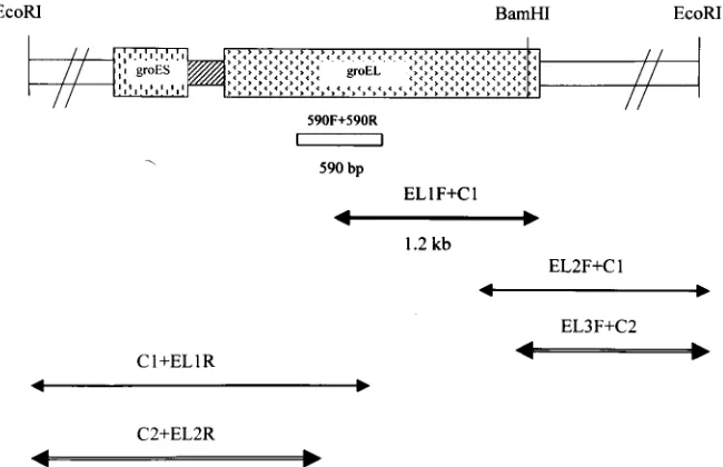
Nucleotide sequence of the groESL genes. Initially, PCR primers complementary to highly conserved regions of the groELgene among eubacteria were derived and used to am-plify a 590-bp portion of the groEL gene from E. faecalis. Genomic E. faecalis DNA was used as a template in PCR. After sequencing and homology searches with databases from gene banks, this fragment yielded the highest mating scores for bacterial groEL genes. Southern hybridization of the 590-bp groEL partial fragment toE. faecalisgenomic DNA digested withBamHI andEcoRI showed the specific hybridization of a 3-kb BamHI fragment and a 5-kbEcoRI fragment (Fig. 1). Then, the LA-PCR was performed. A 1.2-kb DNA fragment was amplified from BamHI-digested genomic DNA by the primers EL-1F and C1 (Fig. 2). Subsequently, two other PCR fragments were obtained by nested LA-PCR from Eco RI-di-gested genomic DNA, and the sequences of these fragments (EL3F⫹C2 and C2⫹EL2R) were determined (Fig. 2). There-fore, the full-length sequence ofE. faecalis groESLoperon was obtained.
[image:2.612.56.552.87.283.2]The sequence obtained revealed the presence of two open reading frames (ORF) of 285 nucleotides and 1,626 nucleo-tides separated by 57 nucleonucleo-tides. Comparative analysis of these nucleotide sequences with those in the genetic databases showed that the deducedE. faecalisORF 1 and ORF 2 pro-teins showed significant homology with the GroES and GroEL proteins of other bacteria (Table 2). The GroES (groES) ho-molog ofE. faecalishad 60.2% amino acid sequence identity TABLE 1. PCR primers used in this study
Primer name Sequencea(5⬘to 3⬘) Gene and nucleotide positions
Universal amplification
590F GGNGACGGNACNACNACNGCAACNGT groEL, 255–280
590R TCNCCRAANCCNGGYGCNTTNACNGC groEL, 844–819
LA-PCR
C1 GTACATATTGTCGTTAGAACGCGTAATACGACTCA
C2 CGTTAGAACGCGTAATACGACTCACTATAGGGAGA
EL-1F CAAGTCGCTGCTGTTTCATC groEL, 435–454
EL-2F AACCAAATCGGCGAAACAAC groEL, 1047–1066
EL-3F GGTGAATGGGTAAACATGGTTGAA groEL, 1440–1463
EL-1R CATCCGCAATAATCAATAGT groEL, 753–734
EL-2R CCTGATGAAACAGCAGCGAC groEL, 457–438
E. faecalisspecific
EfGroES-F GGAATTGTTCTTGCATCCGT groES, 67–86
EfGroES-R ACAATTAAGTATTCTACGCC groES, 251–232
Broad-range PCR-RFLP
EntGroES-F TTAAAACCATTAGGCGATCG groES, 4–23
EntGroEL-R CCCATNCCCATNGANGGRTCCAT groEL, 1613–1591
aN, any nucleotide; R, purine; Y, pyrimidine.
on May 15, 2020 by guest
http://jcm.asm.org/
(64.6% nucleotide sequence identity) withLactobacillus zeae and 58.5% (66.2%) identity withLactococcus lactis, followed by 57.0% (65.5%) identity with Bacillus subtilis, while the GroEL (groEL) homolog ofE. faecalishad highest homology (81.8% amino acid sequence identity and 73.0% nucleotide sequence identity) withStreptococcus pneumoniae, followed by 79.7% (71.7%) identity withLactococcus zeae.ThegroES ho-molog (ORF 1) is 285 bp long, and 94 amino acids were deduced. A putative ribosome-binding site (GGAGG) was lo-cated 8 nucleotides upstream of the start codon (GTG) of ORF 1. A potential translation initiation codon (AUG) of the second ORF (ORF 2) was located 57 bp downstream of the stop codon (TAA) of ORF 1. ORF 2 (groELhomolog) is 1,626
bp long, and 541 amino acids were deduced. A repeat sequence (positions 766 to 792 in GenBank accession number AF335185) was observed upstream of ORF 1 and was identical with CIR-CE-like element, a well-conserved inverted repeat involved in groE regulation in gram-positive bacteria. A large inverted repeat which may function as a rho-independent transcrip-tional terminator was located downstream of OFR 2.
[image:3.612.74.270.76.241.2]Development of anE. faecalis-specific PCR.Since thegroES sequences vary more thangroELamong organisms,E. faecalis -specific identification of DNA was tested with the primers based onE. faecalis groES sequences. The specificity of the primers was tested with eight ATCC strains and 24 clinical isolates of variousEnterococcusspecies. PCRs with the primer pair EfGroES-F/EfGroES-R identified allE. faecalisisolates. AllE. faecalis isolates tested produced the expected 185-bp amplicon (Fig. 3). Moreover, the sequences of the 185-bp amplicon generated from four clinical isolates perfectly matched the reference sequence from the ATCC strain. There FIG. 1. Southern hybridization of the 590-bpgroELinternal
frag-ment toE. faecalisgenomic DNA digested with restriction enzymes shows the probe hybridized to a 5-kbEcoRI fragment and 3-kbBamHI fragment. Lane M, markers; lanes 1 to 5, digested withXbaI, PstI, HindIII, EcoRI, andBamHI, respectively.
FIG. 2. Schematic illustration of amplification fragments and restriction sites ofE. faecalis groESLgenes. The box betweengroESandgroEL
[image:3.612.310.551.100.205.2]is spacer. The 590-bp fragment was used as the probe for Southern blot. The arrows below the restriction map indicate the amplified and sequenced fragments. The primers used in LA-PCR are indicated above the arrows.
TABLE 2. Sequence identity among inferred GroES and GroEL genes fromE. faecalisand related bacteria
Speciesa
Amino acid (nucleotide) sequence identity withE. faecalisgene
product (gene)
GroES (groES) GroEL (groEL)
Lactobacillus zeae 60.2 (64.6) 79.7 (71.7)
Lactococcus lactis 58.5 (66.2) 79.5 (74.6)
Bacillus subtilis 57.0 (65.5) 77.5 (70.2)
Bacillus stearothermophilus 57.0 (64.6) 77.7 (73.4)
Streptococcus pneumoniae 46.8 (58.5) 81.8 (73.0)
Staphylococcus aureus 47.8 (58.6) 71.2 (71.3)
aGenBank accession numbers are as follows: forL. zeae, AF010281; forL.
lactis, X71132; forB. subtilis, D10972; forB. stearothermophilus, L10132; forS. pneumoniae, AF117741; and forS. aureus, D14711.
on May 15, 2020 by guest
http://jcm.asm.org/
[image:3.612.142.467.491.701.2]was no amplification product of 185 bp from 16 clinical isolates of other species.
Dot blot hybridization. Dot blot hybridization was per-formed on eight reference strains and 24 clinical isolates. The amplification products ofgroESorgroELfromE. faecaliswere used as probes. Examples of results usingE. faecalis groESas probe are shown in Fig. 4. Only DNAs fromE. faecalisshowed a strong hybridization signal. No hybridization signal was de-tected by dot blot analysis of DNAs from non-E. faecalis strains.
Differentiation of Enterococcus species by groESL broad-range PCR-RFLP.A PCR amplification was performed with a pair of primers, EntGroES-F and EntGroEL-R, based on the E. faecalis groESLgenes sequence obtained in this study. PCR-RFLP analysis was carried out with eightEnterococcusATCC reference strains. A major PCR-amplified product of approx-imately 1.9 kb in length was detected. These fragments corre-spond to nearly the entire length ofgroESLgenes. The ampli-fied PCR products were subsequently subjected to two sets of restriction enzyme digestion, withRsaI andHaeIII being used individually. The analysis showed that the RFLP profiles of PCR products from each species ofEnterococcuswere quite distinguishable, except for HaeIII digests between E. avium andE. hirae(Fig. 5).
A total of eightE. faecalisclinical isolates were tested by this analysis. All isolates tested, including vancomycin-susceptible
and vancomycin-resistant E. faecalis isolates, showed RFLP patterns identical to that of the reference strain, indicating intraspecies uniformity.
DISCUSSION
Accurate species identification of enterococci has become important with the wide prevalence of acquired vancomycin resistance. Conventional methods of identification of Entero-coccusspecies are time-consuming (7, 32). Errors can happen and are not easily detected in automated identification sys-tems, and supplemental testing is sometimes required for iden-tification (14). Tsakris et al. found that 14E. faecalisisolates with HLGR were misidentified as E. durans when using a semiautomated system (29). The development of rapid and sensitive DNA-based assays may improve the speed and accu-racy of diagnosis of enterococcal infections. In this study, we describe the full-length sequencing ofgroESLgenes fromE. faecalisand the application of species identification.
[image:4.612.53.295.77.182.2]Using the LA-PCR method, the complete sequence of theE. faecalis groESLgenes containing the putative promoter region, ORF 1 (groEShomolog, 285 bp), spacer (57 bp), and ORF 2 (groEL homolog, 1,626 bp) was determined. The sequence data ofgroESLgenes from E. faecalis showed that the gene structure was similar to those of most bacterial species studied (3, 24, 26, 28). The deduced amino acid sequences of ORF 1 (94 amino acids) and ORF 2 (541 amino acids) proteins ex-hibited a high degree of overall identity with homologous bac-terial GroESs and GroELs, respectively. The homology search with published gene sequences in the database revealed that the GroES (groES) sequence ofE. faecalishad highest identity withLactobacillus zeae(60.2% amino acid identity and 64.6% nucleotide identity) andLactococcus lactis(58.5% amino acid identity and 66.2% nucleotide identity), followed byBacillus subtilis, while the GroEL (groEL) sequence ofE. faecalishad highest identity withS. pneumoniae(81.8% amino acid identity and 73.0% nucleotide identity), followed byLactococcus zeae (79.7% amino acid identity and 71.7% nucleotide identity). The data presented here suggest a close relationship between these organisms. This result is generally in agreement with the data obtained from 5S rRNA, 16s RNA andgrpEreported by Ahmad et al. (1). Studies of the phylogenetic relationship by Ahmad et al. also found thatStreptococcusspecies, L. lactis, FIG. 3.E. faecalis-specific PCR amplification. Specific
amplifica-tion of the 185-bp DNA fragment was detected only inE. faecalis
isolates. M, 100-bp DNA ladder (Gibco-BRL). Lanes 1 to 4,E. faecalis
clinical isolates; lane 5,E. faecium; lane 6,E. durans; lane 7,E. hirae; lane 8,E. avium; lane 9,E. gallinarum; lane 10,E. casseliflavus; lane 11, negative control.
FIG. 4. Dot blot hybridization usingE. faecalis groESas the probe, showing the species specificity. All positive hybridization (1A, 1B, and 1C) was obtained fromE. faecalisisolates. 1A,E. faecalisATCC 29212; 1B and 1C,E. faecalisclinical isolates. All negative reactions were from
E. faecium(1D),E. cecorum(1E),E. durans(1F),E. casseliflavus(2A),
[image:4.612.335.523.79.181.2]E. gallinarum(2B and 2C),E. hirae(2D), andE. avium(2E and 2F).
FIG. 5. Broad-range PCR-RFLP ofgroESLgenes among eight en-terococcal species. Lane M, DNA size markers. Lanes 1 to 8,RsaI digestion; lanes 10 to 16,HaeIII. Lanes 1 and 9,E. faecalis; lanes 2 and 10, E. faecium; lanes 3 and 11, E. casseliflavus; lanes 4 and 12,E. gallinarum; lanes 5 and 13,E. avium; lanes 6 and 14,E. raffinosus; lanes 7 and 15,E. durans; and lanes 8 and 16,E. hirae.
on May 15, 2020 by guest
http://jcm.asm.org/
[image:4.612.62.288.579.675.2]andE. faecalisformed groups within the low-G⫹C gram-positive bacteria.
E. faecalis groESandgroEL each have a ribosome-binding site. The putative ribosome-binding site sequence (GGAGG) of E. faecalis GroES was identical to that ofS. pneumoniae. The putative ribosome-binding site sequence of E. faecalis GroEL was GGTGA. The sequence data obtained in this study suggest that theE. faecalis groESgene may utilize an uncom-mon start codon, GTG. To confirm that this uncomuncom-mon start codon sequence is correct, we performed another PCR, am-plifying a fragment covering this region fromE. faecalisATCC reference strain and two clinical isolates. All revealed the same results. The importance of the GTG start codon in Enterococ-cus is unknown. Non-AUG initiation codons usually act to limit the expression of a gene product at the translational level (24). Others have reported thatL. helveticusutilizes UUG as the start codon of GroES genes (3). Upstream of thegroES, a putative CIRCE sequence was also identified. The sequence of CIRCE was conserved in most gram-positive bacteria and some gram-negative bacteria (28, 31, 33). In thegroESLand dnaKoperons ofBacillus subtilisand many other gram-positive bacteria, CIRCE is located between the transcription start point and the start codon of the first ORF (31, 33).
The C terminus ofE. faecalisGroEL was PSMGMGGMM. This sequence is also conserved in many gram-positive bacte-ria, such as PSMGMGGMI inL. lactisand PSMMGGMM in S. pneumoniae. Comparison of these sequences shows that the GroEL of gram-positive bacteria usually consists of only one GGM at the C terminus. In contrast, the C terminus of most gram-negative bacteria consists of three tandem repeats of the GGM. A large inverted repeat which may function as a rho-independent transcriptional terminator was located down-stream of the groEL gene. The termination sequence of the inverted repeat was similar to others (24).
The spacer length between the GroES translation termina-tion codon and the putative translatermina-tion start codon for GroEL was 57 nucleotides. The spacer length usually varies among different species. The spacer length is 15 bp inS. pneumoniae, 75 bp in Staphylococcus aureus, 87 bp inL. lactis, 36 bp inL. zeae, 46 bp inB. subtilis, and 45 bp inE. coli. Whether the spacer length or sequence is specific for each enterococcal species needs to be determined.
HSP60 genes are ubiquitous in both prokaryotes and eu-karyotes and encode highly conserved housekeeping proteins which are essential for the survival of cells. They are more variable than the 16S rRNA gene sequence and are therefore potentially useful for the identification of genetically related species. HSP60 gene has been used as a target for species identification of enterococci and many other bacteria. For ex-ample, analysis of thehsp60gene based on PCR, PCR-RFLP, or direct sequencing has previously been used for the identi-fication of Mycobacterium species, Staphylococcus species, Streptococcus iniae,Ehrlichiaspecies, and other species (4, 9, 11, 22, 25, 26). Since groESseems to be more variable than groEL among different organisms and the specific primers based ongroESmay be more easily found to differentiateE. faecalis from other organisms, we tried to use groES as an alternative target for species identification. Based on the se-quence determined, anE. faecalis-specific PCR was developed. No false positives or false negatives were observed. This assay
may facilitate the accurate identification ofE. faecalis. Other approaches using HSP10 as a target for identification have been reported. For example, LaVerda and Byrne used mono-clonal antibodies against HSP10 to identifyChlamydia tracho-matis(16).
The dot blot hybridization results from testing eight ATCC strains and 24 clinical isolates in this study and the DNA sequencing of the PCR fragments showing complete identity from four clinical isolates are highly suggestive that not only thegroELbut alsogroESgene can be a useful target for species identification.
The sequence obtained in this study was from E. faecalis ATCC 29212. It is different from the strain (ATCC 19434) used by Goh et al. for (10) Cpn60 partial sequencing. The deduced amino acid residues of groEL agreed with the pub-lished Cpn60 partial fragment (184 amino acids, 552 tides) but differed in one amino acid residue and two nucleo-tides. The observed difference in this region may represent strain-to-strain variations. Intraspecies variation in this region was further tested for four unrelated clinical isolates, two VRE and two VSE. The results showed that the similarity of nucle-otide sequences were very high (greater than 99% identity) in this 590-bp region, and the deduced amino acid sequences from three isolates were identical to that of ATCC 29212 and one is identical to that of ATCC 19434. Intraspecies variation of groEL among Bartonella and Ehrlichia species has been studied by Marston et al. and Sumner et al., respectively, and their reports revealed that some sequence divergence may be evident between strains from different countries (17, 26).
Besides the identification of E. faecalis by species-specific PCR assay, identification of other species was also be per-formed by broad-range PCR-RFLP. The PCR-RFLP of groESLwithRsaI distinguished clearly between the type strains of eight commonly encountered Enterococcus species. The HaeIII digestions distinguished most species but notE. avium and E. durans. Preliminary data from 24 clinical isolates of Enterococcus species showed that species-specific patterns were observed. However, whether more than one pattern ex-isted in clinical isolates of same species is unknown, and more isolates are needed to test.
In conclusion, our results indicated that theE. faecalis spe-cies-specific PCR, dot blot hybridization, and broad-range PCR-RFLP ofgroESLgenes used in this study are relatively simple and accurate in the identification of at least eight spe-cies of commonly encountered Enterococcus, especially for those with atypical phenotypes. The procedure described in this paper offers a rapid, convenient, and effective tool to identify E. faecalisand other species. The PCR-based assays developed in this study provide an alternative to currently used methods for identification of clinically important enterococcal species. Determining the sequences of groESL among other Enterococcus species has been undertaken. After obtaining these sequences, specific PCR of other species and phylogeny will be developed.
ACKNOWLEDGMENT
This work was supported by grant NSC 89–2314-B-002–269 from the National Science Council of Taiwan.
on May 15, 2020 by guest
http://jcm.asm.org/
REFERENCES
1.Ahmad, S., A. Selvapandiyan, and R. K. Bhatnagar.2000. Phylogenetic
analysis of Gram-positive bacteria based ongrpE, encoded by thednaK
operon. Int. J. Syst. Evol. Microbiol.50:1761–1766.
2.Bergeron, M. G., and M. Ouellette.1998. Preventing antibiotic resistance
through rapid genotypic identification of bacteria and of their antibiotic resistance genes in the clinical microbiology laboratory. J. Clin. Microbiol.
36:2169–2172.
3.Broadbent, J. R., C. J. Oberg, and L. Wei.1998. Characterization of the
Lactobacillus helveticus groESLoperon. Res. Microbiol.149:247–253.
4.Chae, J., J. E. Foley, J. Stephen, and J. E. Madigan.2000. Comparison of the
nucleotide sequences of 16S rRNA, 444Ep-ank, andgroESLheat shock operon genes in naturally occurringEhrlichia equiand human granulocytic ehrlichiosis agent isolates from northern California. J. Clin. Microbiol.38:
1364–1369.
5.Descheemaeker, P., C. Lammens, B. Pot, P. Vandamme, and H. Goossens.
1997. Evaluation of arbitrarily primed PCR analysis and pulsed-field gel electrophoresis of large genomic DNA fragments for identification of en-terocooi important in human medicine. Int. J. Syst. Bacteriol.47:555–561.
6.Dutka-Malen, S., S. Evers, and P. Courvalin.2000. Detection of
glycopep-tide resistance genotypes and identification to the species level of clinically relevant enterococci by PCR. J. Clin. Microbiol.33:24–27.
7.Facklam, R. R., and M. D. Collins.1989. Identification ofEnterococcus
species isolated from human infections by a conventional test scheme. J. Clin. Microbiol.27:731–734.
8.Garcia-Garrote, F., E. Cercenado, and E. Bouza.2000. Evaluation of a new
system, VITEK2:for identification and antimicrobial susceptibility testing of enterococci. J. Clin. Microbiol.38:2108–2111.
9.Goh, S. H., D. Driedger, S. Gillett, D. E. Low, S. M. Hemmingsen, M. Amos,
D. Chan, M. Lovgren, B. M. Willey, C. Shaw, and J. A. Smith.1998.
Strep-tococcus iniae, a human and animal pathogen: specific identification by the chaperonin 60 gene identification method. J. Clin. Microbiol.36:2164–2166.
10. Goh, S. H., R. R. Facklam, M. Chang, J. E. Hill, G. J. Tyrrell, E. C. M.
Burns, D. Chan, C. He, T. Rahim, C. Shaw, and S. M. Hemmingsen.2000.
Identification ofEnterococcusspecies and phenotypically similar Lactococ-cusandVagococcusspecies by reverse checkerboard hybridization to chap-eronin 60 gene sequences. J. Clin. Microbiol.38:3953–3959.
11. Goh, S. H., S. Potter, J. O. Wood, S. M. Hemmingsen, R. P. Reynolds, and
A. W. Chow.1996. HSP60 gene sequences as universal targets for microbial
species identification: studies with coagulase-negative staphylococci. J. Clin. Microbiol.34:818–823.
12. Hemmingsen, S. M., C. S. Woolford. M. van der Vies, K. Tilly, D. T. Dennis,
C. P. Georgopoulos, R. W. Hendrix and R. J. Ellis.1988. Homologous plant
and bacterial proteins chaperone oligomeric protein assembly. Nature (Lon-don)333:330–334.
13. Hsueh, P. R., L. J. Teng, H. J. Pan, Y. C. Chen, L. H. Wang, S. C. Chang,
S. W. Ho, and K. T. Luh.1999. Emergence of vancomycin-resistant
entero-cocci at a university hospital in Taiwan: persistence of multiple species and multiple clones. Infect. Control Hosp. Epidemiol.20:828–833.
14. Iwen, P. C., M. E. Rupp, P. C. Schreckenberger, and S. H. Hinrichs.1999.
Evaluation of the revised MicroScan dried overnight gram-positive identifi-cation panel to identifyEnterococcusspecies. J. Clin. Microbiol.37:3756– 3758.
15. Ke, D., F. J. Picard, F. Martineau, C. Me´nard, P. H. Roy, M. Ouellette, and
M. G. Bergeron.1999. Development of PCR assay for rapid detection of
enterococci. J. Clin. Microbiol.37:3497–3503.
16. LaVerda, D., and G. I. Byrne.1997. Use of monoclonal antibodies to
facil-itate identification, cloning, and purification ofChlamydia trachomatishsp10. J. Clin. Microbiol.35:1209–1215.
17. Marston, E. L., J. W. Sumner, and R. L. Regnery. 1999. Evaluation of
intraspecies genetic variation within the 60 kDa heat-shock protein gene (groEL) ofBartonellaspecies. Int. J. Syst. Bacteriol.49:1015–1023.
18. Monstein, H. J., M. Quednau, A. Samuelsson, S. Ahrne´, B. Lsaksson, and J.
Jonasson.1998. Division of the genusEnterococcusinto species groups using
PCR-based molecular typing methods. Microbiology144:1171–1179.
19. Mundy, L. M., D. F. Sahm, and M. Gilmore.2000. Relationships between
enterococcal virulence and antimicrobial resistance. Clin. Microbiol. Rev.
13:513–522.
20. Patel, R., K. E. Piper, M. S. Rouse, J. M. Steckelberg, J. R. Uhl, P. Kohner,
M. K. Hopkins, F. R. Cockerill III, and R. C. Kline.1998. Determination of
16S rRNA sequences of Enterococci and application to species identification of nonmotileEnterococcus gallinarumisolates. J. Clin. Microbiol.36:3399– 3407.
21. Poyart, C., G. Quesnes, and P. Trieu-Cuot.2000. Sequencing the gene
encoding manganese-dependent superoxide dismutase for rapid species identification of enterococci. J. Clin. Microbiol.38:415–418.
22. Rastogi, N., K. S. Goh, and M. Berchel.1999. Species-specific identification
ofMycobacterium lepraeby PCR-restriction fragment length polymorphism analysis of thehsp65gene. J. Clin. Microbiol.37:2016–2019.
23. Robbi, C., C. Signoretto, M. Boaretti, and P. Canepari.1996. The gene
coding for penicillin-binding protein 5 ofEnterococcus faecalisis useful for the development of a species-specific DNA probe. Microb. Drug Resist.
2:215–218.
24. Schmidt, A., M. Schiesswohl, U. Volker, M. Hecker, and W. Schumann.
1992. Cloning, sequencing, mapping, and transcriptional analysis of the
groESLoperon fromBacillus subtilis. J. Bacteriol.174:3993–3999.
25. Steingrube, V. A., J. L. Gibson, B. A. Brown, Y. Zhang, R. W. Wilson, M.
Rajagopalan, and R. J. Wallace, Jr.1995. PCR amplification and restriction
endonuclease analysis of a 65-kilodalton heat shock protein gene sequence for taxonomic separation of rapidly growing mycobacteria. J. Clin. Microbiol.
33:149–153.
26. Sumner, J. W., G. A. Storch, R. S. Buller, A. M. Liddell, S. L. Stockham, Y.
Rikihisa, S. Messenger, and C. D. Paddock.2000. PCR amplification and
phylogenetic analysis ofgroESLoperon sequences fromEhrlichia ewingiiand
Ehrlichia muris. J. Clin. Microbiol.38:2746–2749.
27. Teng, L. J., S. J. Liaw, P. R. Hsueh, S. W. Ho, and K. T. Luh.1998.
Heterogeneity of resistance elements in clinical isolates of enterococci with high-level gentamicin resistance. J. Formos. Med. Assoc.97:855–859.
28. Thies, F. L., A. Weishaupt, H. Karch, H. P. Hartung, and G. Giegerich.1999.
Cloning, sequencing and molecular analysis of the Campylobacter jejuni groESLbicistronic operon. Microbiology145:89–98.
29. Tsakris, A., N. S. Woodford, Pournaras, M. Kaufmann, and J. Douboyas.
1998. Apparent increased prevalence of high-level aminoglycoside-resistant
Enterococcus duransresulting from false identification by a semiautomated software system. J. Clin. Microbiol.36:1419–1421.
30. Tyrrell, G. J., R. N. Rethune, B. Willey, and D. E. Low.1997. Species
identification of enterococci via intergenic ribosomal PCR. J. Clin. Micro-biol.35:1054–1060. (Erratum,35:2434.)
31. Wetzstein, M., U. Volker, J. Dedio, S. Lobau, U. Zuber, M. Scheisswohl, M.
Hecker, and W. Schumann.1992. Cloning, sequencing, and molecular
anal-ysis ofdnaKlocus fromBacillus subtilis. J. Bacteriol.174:3300–3310.
32. Willey, B. M., R. N. Jones, A. McGeer, W. Witte, G. French, R. B. Roberts,
S. G. Jenkins, H. Nadler, and D. E. Low.1999. Practical approach to the
identification of clinically relevantEnterococcusspecies. Diagn. Microbiol. Infect. Dis.34:165–171.
33Zuber, U., and W. Schumann.1994. CIRCE, a novel heat shock element
involved in regulation of heat shock operondnaKof Bacillus subtilis. J. Bacteriol.176:1359–1363.