Copyright © 2001, American Society for Microbiology. All Rights Reserved.
Development of Molecular Methods for Identification of
Schizophyllum commune
from Clinical Samples
WALTER BUZINA,* DORIS LANG-LOIDOLT, HANNES BRAUN, KURT FREUDENSCHUSS,ANDHEINZ STAMMBERGER ENT University Hospital, Karl-Franzens-University Graz, A 8036 Graz, Austria
Received 13 July 2000/Returned for modification 12 August 2000/Accepted 23 April 2001
In the last 50 years, to our knowledge, only 16 cases of diseases caused bySchizophyllum communein humans
have been reported. Within only 6 months, we found four isolates of this basidiomycetous fungus, obtained from patients suffering from chronic sinusitis. The cultures of the isolated fungi showed neither clamp
connections nor fruiting bodies (basidiocarps), which are distinctive features forS. commune, but fast-growing
cottony white mycelium only. This was harvested, and DNA was extracted. The internal transcribed spacer region of the ribosomal DNA (rDNA) was amplified with fungus-specific primers, and the PCR products were
sequenced. Two strains ofS. commune, collected from branches of a European hornbeam (Carpinus betulus) and
a tree of heaven (Ailanthus altissima), respectively; four specimens from the herbarium of the Institute of Botany, Karl-Franzens-University Graz; and two strains from internationally known culture collections (CBS 340.81 [ATCC 44201] and CBS 405.96) were investigated in the same way. The sequence data of all strains were compared and showed homology of over 99% in this 660-bp-long fragment of rDNA. With these results, a map
of restriction enzyme cutting sites and a primer set specific for S. communewere created for reliable
identi-fication of this human pathogenic fungus.
Compared to the great number of ascomycetous fungi which are the cause of many diseases, filamentous basidiomycetes that cause infections are reported rarely in the medical litera-ture. Schizophyllum commune Fries 1821 (Schizophyllaceae,
Aphyllophorales) is one of them and has been described else-where as a suspected cause of onychomycosis (16), basidio-neuromycosis (2), an allergic bronchopulmonary mycosis (12), mucoid impaction of the bronchi (1, 5), a fungus ball in the lung (27), an ulcerative lesion of the hard palate (23), a brain abscess (24), and several cases of both maxillary and allergic fungal sinusitis (4, 6, 14, 18, 25, 28, 29). A review of cases has been published by Kamei et al. (13). This worldwide-distrib-uted shelf fungus is found quite frequently in nature through-out the year, although it mostly appears during the cold season (22). The commonly known split-gill fungus is found on decay-ing wood, where it appears with sessile fan- or kidney-shaped fruiting bodies. Typical forS. communeis the hymenium on the lower side, which consists of longitudinally split gills (lamel-lae), where masses of basidiospores are produced and released into the air. The fungus is easy to cultivate on most of the media generally used in clinical laboratories. The appearance of fruiting bodies, hyphal clamp connections, and spicules makes identification easy. Nevertheless, monokaryotic clinical isolates lack these unambiguous features, and the mycelium shows up only as a cottony white mass of hyphae without any distinctive marks. In these cases, the fungus is often identified as “Mycelia Sterilia” or misidentified as an ascomycete. In the last few years, many molecular methods, such as PCR, se-quencing of segments of the genome, analyses of the restriction fragment length polymorphisms (RFLP), or hybridization of
specific probes, have been described as tools for identification of medically relevant fungi. The highly variable internal tran-scribed spacer (ITS) region lies in between fragments of ribo-somal DNA (rDNA) and is a valuable target for reliable iden-tification of those strains which cannot be identified to the species level by microscopy.
CASE REPORTS
HNO 104.A 60-year-old woman presented with congestion
in the right nostril. She had a history of chronic rhinosinusitis with four functional endoscopic sinus surgeries (FESS) done within the last 5 years. Computerized tomography (CT) scan-ning showed opacification of the ethmoidal and the maxillary sinus. Endoscopy revealed a highly viscous secretion, which was removed. After 2 weeks of antifungal therapy, the patient recovered without complications.
HNO 34.A 51-year-old woman presented with complaints of
nasal congestion on the right side. CT scanning showed opaci-fication of the ethmoidal and the sphenoid sinus. Due to fungal hyphae in the mucus, antifungal therapy was given together with an antihistamine. CT scanning after 3 months showed no opacification, and the patient had no complaints.
HNO 62.A 63-year-old woman presented with complaints of
nasal congestion on the left side. FESS was done and revealed a massive sinusitis with polyps in the sphenoethmoidal recessus and the sphenoid sinus. Neither antifungal nor corticosteroid treatment was given, and the patient recovered without com-plications.
HNO 323.A 63-year-old woman presented with difficulty in
breathing through the nose, pain, and pressure on the right side of her face. FESS showed sinusitis and polyps in the ethmoidal sinus, which was filled with viscous mucus. The whole sphenoid sinus was filled with fungal concrement, which was removed.
* Corresponding author. Mailing address: ENT University Hospital, Karl-Franzens-University Graz, Auenbruggerplatz 26-28, A 8036 Graz, Austria. Phone: 43 316 3853606 or 43 676 7800545. Fax: 43 316 3857643. E-mail: [email protected].
2391
on May 15, 2020 by guest
http://jcm.asm.org/
MATERIALS AND METHODS
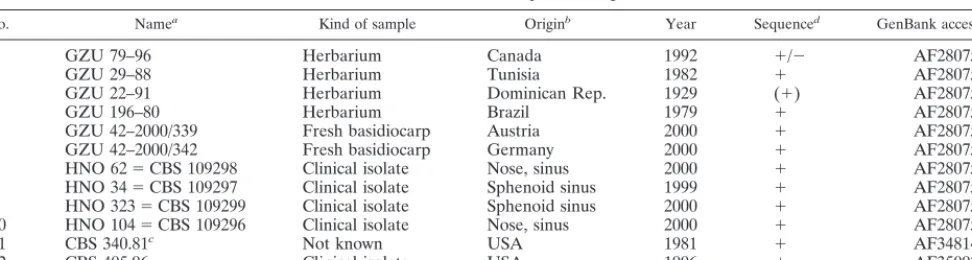
Fungal strains and cultivation.Altogether, 12 isolates ofS. communewere examined for this study (Table 1). Four strains were isolated from patients suffering from chronic sinusitis: samples HNO 34 and HNO 104 were obtained by flushing both nostrils of the patients with sterile 0.9% NaCl solution as described by Ponikau et al. (21), and samples HNO 62 and HNO 323 were obtained by sinus surgery. The mucous material was treated with mucolytic dithiothreitol to release the fungal elements, and these were sedimented by centrifugation. Thereafter, the samples were incubated at 20 and 30°C, respec-tively, on Sabouraud’s glucose agar (SGA), Czapek Dox agar, and malt extract agar. To induce formation of dikaryotic mycelium, monokaryotic mycelia of the clinical isolates HNO 34 and HNO 62 were inoculated together on one petri dish (SGA) about 3 cm apart and incubated at room temperature in daylight for 1 week. Mycelium of the confluence zone was examined by microscopy. Two samples were isolated from living basidiocarps growing on broken branches of a European hornbeam (Carpinus betulus) in a forest in Graz, Austria, and on a dead branch of a tree of heaven (Ailanthus altissima) in the Tiergarten in Berlin, Germany, respectively. Four isolates were obtained from desiccated specimens from the herbarium of the Institute of Botany, Karl-Franzens-University Graz, Graz, Austria. These herbarium specimens were up to 71 years old and origi-nated from North and South America and Africa. To compare the samples with known strains of this fungus, two strains (CBS 340.81 [ATCC 44201] and CBS 405.96) from the Centraalbureau voor Schimmelcultures (CBS), Utrecht, The Netherlands, were examined as well. Cultures of all clinical isolates were depos-ited at the CBS with the accession no. CBS 109296 to CBS 109299 (Table 1).
DNA isolation.DNA was extracted from fungal cells as described previously (15). In this procedure, cultivated material (clinical samples and reference strains) or fragments of basidiocarps (herbarium and fresh samples) were ho-mogenized under liquid nitrogen, suspended in lysis buffer (1.4% N
-cetyl-N,N,N,-trimethylammonium bromide [CTAB], 1 M NaCl, 7 mM Tris, 30 mM EDTA), and incubated at 65°C for 1 h. Proteins were removed by extraction with chloroform-isoamyl alcohol (24:1), and DNA was precipitated with precipitation buffer (0.5% CTAB, 40 mM NaCl) and pelleted by centrifugation. Afterwards, the pellet was resuspended in 1.2 M NaCl, and the DNA was reprecipitated with isopropanol (60% final concentration) at⫺20°C, washed with 70% ethanol, air dried, and resuspended in sterile bidistilled water.
PCR.The 5.8S rDNA and the flanking ITS regions (ITS1 and ITS2) were amplified using the primers ITS5 (GGAAGTAAAAGTCGTAACAAGG) and ITS4 (TCCTCCGCTTATTGATATGC) (30). All primers were prepared com-mercially by Metabion, Planegg-Martinsried, Germany. PCR was carried out in a reaction mixture containing 14.8l of sterile bidistilled water, 5.0l of buffer (10 mM Tris-HCl, 1.5 mM MgCl2, 50 mM KCl, 0.1% Triton X-100), 5.0l of deoxynucleoside triphosphates (10 mM), 1 U of DNA polymerase (DyNAzyme II; Finnzymes Oy, Turku, Finland), 2.5l of each primer (10M), and 20l of fungal DNA (10 to 50 ng/l). To prevent evaporation of the mixture during amplification, it was overlaid with 2 to 3 drops of mineral oil (Sigma; M3516). One reaction mixture containing water in place of DNA template was used as the contamination control. For PCR in the thermocycler (TC480; PE Biosystems), the following parameters were chosen: 30 cycles of 1 min at 95°C, 1 min at 50°C,
and 2 min at 72°C, with a final extension at 72°C for 10 min. The amplification products (1l each) were visualized after gel electrophoresis and staining in ethidium bromide under UV light in a transilluminator.
Cycle sequencing.Excess primers and deoxynucleoside triphosphates were removed with chromatography columns (Microspin S-300 HR; Pharmacia). For sequencing the entire ITS region with the enclosed 5.8S rDNA, we used the primers ITS2 (GCTGCGTTCTTTCATCGATGC) and ITS3 (GCATCGATGA AGAACGCAGC) (30) in addition to the primers ITS5 and ITS4 at a 1.6M concentration. Sequencing was carried out with the ABI Prism BigDye Termi-nator Cycle Sequencing Kit (PE Biosystems) according to the manufacturer’s recommendations. The parameters for cycle sequencing in the GeneAmp 2400 thermocycler (PE Biosystems) were 18 s of delay at 96°C, followed by 25 cycles of 18 s at 96°C, 5 s at 50°C, and 4 min at 60°C.
Analysis of sequences.Sequence analysis was performed using an automated sequence analyzer (ABI Prism 310; PE Biosystems) in conjunction with the ABI Prism Auto Assembler software (version 1.4.0, 1995; Applied Biosystems Divi-sion, PE Biosystems) and aligned using Pileup of the Wisconsin Package software (version 9.0-open VMS, 1993; Genetics Computer Group).
RFLP.For fast and reliable identification of isolates ofS. communewithout the need of sequencing, a map of cutting positions for the restriction enzymes
EcoRI andAvaI was created. Therefore, the sequence data were cut virtually with the program Restriction Enzyme Analysis, which is an online freeware program (http://darwin.bio.geneseo.edu/⬃yin/WebGene/RE.html). This restric-tion map was verified by RFLP analysis of the amplified ITS regions. For this, 10
l of PCR product from medical samples was mixed with 7l of bidistilled water and 3l of 1:2 enzyme buffer (Boehringer, Mannheim, Germany) and incubated at 37°C for 4 h. Restriction fragments were separated on a 2% agarose gel and visualized as described above.
Species-specific primers.Highly variable regions of both ITS1 and ITS2 were screened for possible priming sites. The selected sequences were checked with the program Primer Designer (version 2.0; Scientific & Educational Software) for their applicability as primers, as were GC content, melting temperature, and the lack of hairpins and possible dimers. These priming sites were also examined for their specificity forS. communeby comparing the sequences with entries in gene data banks (http://www2.ebi.ac.uk/fasta3/).
Nucleotide sequence accession numbers.All sequence data were submitted to GenBank (http://www2.ncbi.nlm.nih.gov/) and were registered with the accession no. AF280750 through AF280759, AF348142, and AF350925 (Table 1).
RESULTS
All four clinical isolates were cultivated successfully on all media provided (SGA, Czapek Dox agar, and malt extract agar) at 20 and 30°C after 5 to 10 days of incubation. The cultures showed a cottony white mycelium with small knots of compressed hyphae 5 to 10 mm in diameter, described as “haploid fruiting” by Raper and Krongelb (22) (Fig. 1a). Older cultures produced yellowish droplets of exudation on the sur-TABLE 1. List of samples investigated
No. Namea Kind of sample Originb Year Sequenced GenBank accession no.
1 GZU 79–96 Herbarium Canada 1992 ⫹/⫺ AF280755
2 GZU 29–88 Herbarium Tunisia 1982 ⫹ AF280752
3 GZU 22–91 Herbarium Dominican Rep. 1929 (⫹) AF280751
4 GZU 196–80 Herbarium Brazil 1979 ⫹ AF280759
5 GZU 42–2000/339 Fresh basidiocarp Austria 2000 ⫹ AF280753
6 GZU 42–2000/342 Fresh basidiocarp Germany 2000 ⫹ AF280754
7 HNO 62⫽CBS 109298 Clinical isolate Nose, sinus 2000 ⫹ AF280750
8 HNO 34⫽CBS 109297 Clinical isolate Sphenoid sinus 1999 ⫹ AF280758
9 HNO 323⫽CBS 109299 Clinical isolate Sphenoid sinus 2000 ⫹ AF280757
10 HNO 104⫽CBS 109296 Clinical isolate Nose, sinus 2000 ⫹ AF280756
11 CBS 340.81c Not known USA 1981 ⫹ AF348142
12 CBS 405.96 Clinical isolate USA 1996 ⫹ AF350925
aGZU samples are from the Herbarium of the Institute of Botany, University Graz. HNO samples are from the ENT-Hospital,
Karl-Franzens-University Graz.
bRep., Republic; USA, United States of America. cStrain CBS 340.81 is the same as ATCC 44201. d⫹, all;⫹/⫺, partial; (⫹), small part of ITS.
on May 15, 2020 by guest
http://jcm.asm.org/
[image:2.612.63.549.83.213.2]face, released a yellow to brown pigment into the medium, and had a pronounced odor. Microscopy showed hyaline hyphae of diverse structures with few spicules. Clamp connections were absent in the cultures of all clinical isolates. To produce dikary-otic mycelium, monokarydikary-otic hyphae of two different strains were inoculated together on one petri dish and incubated at room temperature for 3 weeks. The hyphae fused in the con-fluence zone and formed clearly visible clamp connections (Fig. 1b). Nevertheless, no basidiocarps were produced.
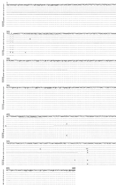
Gel electrophoresis after PCR of the ITS region showed that DNA of all clinical isolates as well as from both fresh and desiccated basidiocarps had been amplified successfully with the primers ITS5 and ITS4 (data not shown). The lengths of all amplicons were identical and lay in the range of 660 bp. The whole ITS region and the enclosed 5.8S rDNA of the CBS strains, the four clinical isolates, and the two samples from the fresh fruiting bodies, as well as the herbarium specimens GZU 29-88 and GZU 196-80, were sequenced successfully (Table 1). DNA of the other two desiccated samples (GZU 22-91 and GZU 79-96) was too degraded to obtain good sequencing results for the entire region. However, some fragments within the 660-bp region could be sequenced. Alignment of the se-quence data showed a similarity of 99.4 to 100% for all four clinical isolates, the samples from the living basidiocarps, the herbarium specimens from Tunisia (GZU 29-88) and Brazil (GZU 196-80), and the CBS strains at a length of 660 nucle-otides with a GC content of 46.4% (Fig. 2). The sequence fragments of the desiccated samples from Canada (553 bp, 99.6% homology) and the Dominican Republic (286 bp, 97.2% homology) fitted well into the aligned sequences.
Analysis of the amplified ITS region showed various cutting sites for restriction enzymes. Two of them were chosen and drawn in a restriction map:EcoRI (281 and 379 bp) andAvaI (391, 125, and 144 bp) (Fig. 3a). Amplicons of all four clinical samples were digested with the chosen restriction enzymes. All
fragments of both restriction enzymes were found to be within the estimated size range, and there was no observable differ-ence between the samples.
Both ITS1 and ITS2 contained segments sufficiently differ-ent from comparable regions in the genomes of other fungi to be specific for S. commune, qualifying them as targets for species-specific PCR primers or DNA probes. The newly de-signed primersscom1 (GTTGACTACGTCTACCTCAC) and
scom2r (GTTAGGCTCCAGCAGACCTC) were both 20 nu-cleotides in length; had GC contents of 50 and 60%, respec-tively; formed no hairpins or dimers; and were in about the same range of melting temperatures. The primers were checked for their specificity by comparing the sequences with entries in gene data banks, and there was no analogy with any other fungi, except for different strains of S. commune. All samples ofS. communeexamined were amplified successfully with the primersscom1 andscom2r, and the resulting 305-bp amplicon was visualized on conventional agarose gels after electrophoresis (Fig. 4). To examine the reproducibility of the primer set, DNA of more than 20 strains of S. commune, collected in the United States and Europe, was extracted and amplified successfully in repeated experiments (data not shown). The specificity of thescom1 andscom2r primer set was confirmed by the unsuccessful amplification of DNA from a variety of fungi under the same conditions as described above (data not shown). The species examined, which included some established human pathogens as well as strains from interna-tionally known culture collections, wereTrichophyton rubrum,
Microsporum gypseum(CBS 161.69),Candida albicans(ATCC 90028), Cryptococcus neoformans (ATCC 90112), Alternaria alternata, Curvularia pallescens (CBS 102694), Aspergillus fu-migatus,Aspergillus ustus(CBS 209.92),Penicillium commune,
Ustilago maydis,Fusarium solani,Beauveria bassiana,and Pseu-dallescheria boidii.
FIG. 1. (a) Three-week-old culture ofS. communeon SGA. (b) Clamp connections (indicated by arrows) on dikaryotic hyphae, derived through mating two monokaryotic isolates (HNO 34 and HNO 62).
on May 15, 2020 by guest
http://jcm.asm.org/
[image:3.612.67.539.71.291.2]FIG. 2. Sequences ofS. communeITS region. ITS1 and ITS2 are in uppercase; small-subunit, 5.8S, and large-subunit rDNA are in lowercase. Priming sites forscom1 (positions 126 to 145) andscom2r (positions 412 to 431) are underlined. Dots represent similarity with the first sequence, dashes represent unknown characters, and underlined spaces represent gaps introduced for alignment. Sequences: 1, GZU 42-2000/339; 2, GZU 42-2000/342; 3, GZU 29-88; 4, GZU 196-80; 5, GZU 79-96; 6, GZU 22-91; 7, HNO 34; 8, HNO 62; 9, HNO 323; 10, HNO 104; 11, CBS 340.81; 12, CBS 405.96.
on May 15, 2020 by guest
http://jcm.asm.org/
DISCUSSION
In the last fifty years, there have been only 16 reported cases of diseases caused byS. commune, most of them concerning the upper respiratory system. We found four isolates of S. communefrom patients with sinusitis within only 6 months, suggesting that this fungus is a much more common human pathogen than expected. The period in whichS. communewas isolated (December to May) correlates well with the main fruiting season of this fungus in central Europe, which is win-tertime. All four patients of this study were female, and this corresponds with previous findings thatS. communeseems to be a gynecotropic agent (13, 24). The age of the patients varied from 51 to 63 years (mean, 59.3⫾5.7 years). All cultures of the clinical isolates showed neither clamp connections on the hy-phae nor fruiting bodies, except for so-called haploid (mono-karyotic) fruiting (22). The macroscopic features, such as rapid growth, cottony white surface, and some droplets of yellowish exudation, made an identification based on morphological characteristics impossible. As has been observed previously,S.
communeoften appears as a monokaryotic isolate in clinical samples and is thus unable to form the characteristic clamp connections and fruiting bodies (27). Kamei et al. (13), when reviewing the cases in which this basidiomycete was isolated from clinical samples, found 58% of the isolates to be mono-karyotic. It can be assumed, therefore, that many cases of infections caused byS. communewere and are misdiagnosed (12). The formation of dikaryotic hyphae through mating ex-periments using a known strain ofS. communemostly leads to clamp connections and spicules (27, 29). Nevertheless, Raper and Krongelb (22) reported successful fruiting in only 80% of mating experiments because of incompatibility between some mating strains. Incubation in daylight, which is normally not possible with clinical laboratory incubators, may support the formation of basidiocarps. However, this procedure may take too much time for identification of the pathogen in most cases. Comparison of the ITS sequences from fungi isolated from four patients with sinusitis, on the one hand, and with speci-mens both from desiccated and from living basidiocarps from three continents (North and South America, Africa, and Eu-rope), on the other, showed a very high similarity in the 660-bp-long rDNA fragment investigated. The test strains CBS 340.81 and CBS 405.96 (the latter was isolated by L. Sigler et al. [27] from a patient in California) fitted well into the aligned sequences. This can be seen as evidence for a highly conserved ITS region within the species ofS. commune, independent of where and when the fungus was found. The samples under investigation were from three continents, and their age ranged from some days to 71 years. Such a high intraspecific conser-vation, but with sufficient difference with regard to other med-ically relevant fungi, offers a great tool for identifying this fungus.
Although sequencing the ITS region of the rDNA is the most accurate method to identify monokaryotic nonclamped strains ofS. commune, it is too costly and time-consuming for the clinical routine. Above all, most clinical laboratories do not have the ability to perform sequence analysis. PCR of the ITS region followed by an analysis of the RFLP can be a valuable tool for a fast and reliable identification ofS. commune, as is described frequently for other medically important fungi (e.g., see references 3, 10, 11, 17, 19, and 31). Another possibility is to perform a PCR with the newly designed species-specific primersscom1 (GTTGACTACGTCTACCTCAC) andscom2r (GTTAGGCTCCAGCAGACCTC). This set of primers cre-ates a specific amplicon of 305 bp within the ITS region and can thus also be used in a nested PCR together with the primers ITS5 and ITS4 when only a very small amount of sample is available. No other fungus (or any other organism) in the gene data banks was found to contain these priming sites. Using the set of PCR primers to amplify 13 of the most com-mon pathogenic and nonpathogenic fungi was unsuccessful. Therefore, it can be assumed that positive amplification with this set of primers is highly specific forS. commune. Repeated amplifications of more than 30 strains ofS. communeon dif-ferent PCR machines showed 100% reproducibility of the primer setscom1-scom2r (data not shown). The sites of both primers,scom1 andscom2r, are also convenient as targets for labeled DNA probes. There are various protocols for hybrid-ization and detection of DNA probes to identify other human FIG. 3. (a) Restriction map of the ITS region for the restriction
enzymesEcoRI andAvaI. (b) RFLP pattern of the clinical isolates of
[image:5.612.53.295.72.280.2]S. commune. Lane 0, 100-bp DNA ladder; lanes 1 and 5, HNO 62; lanes 2 and 6, HNO 34; lanes 3 and 7, HNO 323; lanes 4 and 8, HNO 104.
FIG. 4. Agarose gel showing the amplicons of 12 isolates of S. commune, amplified with the species-specific primers scom1 and
scom2r. Lane 0, 100-bp DNA ladder; lane 13, open tube control. The numbering of samples 1 to 12 is the same as in Table 1.
on May 15, 2020 by guest
http://jcm.asm.org/
pathogenic fungi for which this primer pair may be applicable (e.g., see references 7, 8, 9, 20, and 26).
Our data suggest that whenever a white, cottony, rapidly growing culture is obtained from clinical samples and fails to show distinct microscopic features for a clear identification, one should think ofS. commune,and this possibility may be confirmed by one of the above techniques. This may show that the occurrence ofS. communeas a human pathogenic fungus is much more frequent than assumed previously and will lead to a better understanding of its role in human health.
REFERENCES
1.Amitani, R., K. Nishimura, A. Niimi, H. Kobayashi, R. Nawada, T. Mu-rayama, H. Taguchi, and F. Kuze.1996. Bronchial mucoid impaction due to the monokaryotic mycelium ofSchizophyllum commune. Clin. Infect. Dis.
22:146–148.
2.Batista, A. C., J. A. Maia, and R. Sigler.1955. Basidioneuromycosis on man. An. Soc. Pernambuco13:52–60.
3.Buzina, W.1999. Molecular examinations on human pathogenic dermato-phytes. Ph.D. thesis. Karl-Franzens-University Graz, Graz, Austria. 4.Catalano, P., W. Lawson, E. Bottone, and J. Lebenger.1990.
Basidiomyce-tous (mushroom) infection of the maxillary sinus. Otolaryngol. Head Neck Surg.102:183–185.
5.Cifferi, R., A. C. Batista, and S. Campos.1956. Isolation ofSchizophyllum communefrom a sputum. Atti Ist. Bot. Lab. Crittogam. Univ. Pavia14:3–5. 6.Clark, S., C. K. Campbell, A. Sandison, and D. I. Choa.1996.Schizophyllum commune: an unusual isolate from a patient with allergic fungal sinusitis. J. Infect.32:147–150.
7.Einsele, H., H. Hebart, G. Roller, J. Lo¨ffler, I. Rothenho¨fer, C. A. Mu¨ller, R. A. Bowden, J.-A. van Burik, D. Engelhard, L. Kanz, and U. Schumacher.
1997. Detection and identification of fungal pathogens in blood by using molecular probes. J. Clin. Microbiol.35:1353–1360.
8.Enns, R. K.1988. DNA probes: an overview and comparison with current methods. Lab. Med.19:295–300.
9.Hall, G. S.1993. Probe technology for the clinical microbiology laboratory. Arch. Pathol. Lab. Med.117:578–583.
10. Howell, S. A., R. J. Barnard, and F. Humphreys. 1999. Application of molecular typing methods to dermatophyte species that cause skin and nail infections. J. Med. Microbiol.48:33–40.
11. Jackson, C. J., R. C. Barton, and E. G. V. Evans.1999. Species identification and strain differentiation of dermatophyte fungi by analysis of ribosomal-DNA intergenic spacer regions. J. Clin. Microbiol.37:931–936.
12. Kamei, K., H. Unno, K. Nagao, T. Kuriyama, K. Nishimura, and M. Miyaji.
1994. Allergic bronchopulmonary mycosis caused by the basidiomycetous fungusSchizophyllum commune. Clin. Infect. Dis.18:305–309.
13. Kamei, K., H. Unno, J. Ito, K. Nishimura, and M. Miyaji.1999. Analysis of the cases in whichSchizophyllum communewas isolated. Nippon Ishinkin Gakkai Zasshi40:175–181.
14. Kern, M. E., and F. A. Uecker.1986. Maxillary sinusitis infection caused by the homobasidiomycetous fungusSchizophyllum commune. J. Clin. Micro-biol.23:1001–1005.
15. Kielstein, P., H. Wolf, Y. Gra¨ser, W. Buzina, and P. Blanz.1998. On the variability ofTrichophyton verrucosumisolates from vaccinated herds with ringworm of cattle. Mycoses41(Suppl. 2):58–64.
16. Kligman, A. M.1950. A. basidiomycete probably causing onychomycosis. J. Investig. Dermatol.14:67–70.
17. Lin, D., P. F. Lehmann, B. H. Hamory, A. A. Padhye, E. Durry, R. W. Pinner, and Lasker.1995. Comparison of three typing methods for clinical and environmental isolates ofAspergillus fumigatus. J. Clin. Microbiol.33:1596– 1601.
18. Marlier, S., J. P. de Jaureguiberry, P. Aguilon, E. Carloz, J. L. Duval, and D. Jaubert.1993. Sinusite chronique due aSchizophyllum communeau cours du SIDA. Press Med.22:1107.
19. Mitchell, T. G., T. J. White, and J. W. Taylor.1992. Comparison of 5.8S ribosomal DNA sequences among the basidiomycetous yeast genera Cysto-filobasidium,FilobasidiumandFilobasidiella. J. Med. Vet. Mycol.30:207– 218.
20. Mitchell, T. G., R. L. Sandin, B. H. Bowman, W. Meyer, and W. G. Merz.
1994. Molecular mycology: DNA probes and applications of PCR technol-ogy. J. Med. Vet. Mycol.32:351–366.
21. Ponikau, J. U., D. A. Sherris, E. B. Kern, H. A. Homburger, E. Frigas, T. A. Gaffey, and G. D. Roberts.1999. The diagnosis and incidence of allergic fungal sinusitis. Mayo Clin. Proc.74:877–884.
22. Raper, J. R., and G. S. Krongelb.1958. Genetic and environmental aspects of fruiting inSchizophyllum commune. Mycologia50:707–740.
23. Restrepo, A., D. L. Greer, M. Robledo, O. Osorio, and H. Mondragon.1973. Ulceration of the palate caused by a basidiomyceteSchizophyllum commune. Sabouraudia9:201–204.
24. Rihs, J. D., A. A. Padhye, and C. B. Good.1996. Brain abscess caused by
Schizophyllum commune: an emerging basidiomycete pathogen. J. Clin. Mi-crobiol.34:1628–1632.
25. Rosenthal, J., R. Katz, D. B. Dubois, A. Morrisey, and A. Machicao.1992. Chronic maxillary sinusitis associated with the mushroomSchizophyllum communein a patient with AIDS. Clin. Infect. Dis.14:46–48.
26. Shin, J. H., F. S. Nolte, and C. J. Morrison.1997. Rapid identification of
Candidaspecies in blood cultures by a clinically useful PCR method. J. Clin. Microbiol.35:1454–1459.
27. Sigler, L., L. M. de la Maza, G. Tan, K. N. Egger, and R. K. Sherburne.1995. Diagnostic difficulties caused by nonclampedSchizophyllum commune iso-late. J. Clin. Microbiol.33:1979–1983.
28. Sigler, L., S. Estrada, N. A. Montealegre, E. Jaramillo, M. Arango, C. De Bedout, and A. Restrepo.1997. Maxillary sinusitis caused bySchizophyllum communeand experience with treatment. J. Med. Vet. Mycol.35:365–370. 29. Sigler, L., J. R. Bartley, D. H. Parr, and A. J. Morris.1999. Maxillary sinusitis
caused by medusoid form ofSchizophyllum commune. J. Clin. Microbiol.
37:3395–3398.
30. White, T. J., T. Bruns, S. Lee, and J. W. Taylor.1990. Amplification and direct sequencing of fungal ribosomal genes for phylogenetics, p. 315–322.In
M. A. Innis, D. H. Gelfand, J. J. Sninsky, and T. J. White (ed.), PCR protocols: a guide to methods and applications. Academic Press Inc., San Diego, Calif.
31. Williams, D. W., M. J. Wilson, M. A. O. Lewis, and A. J. C. Potts.1995. Identification ofCandidaspecies by PCR and restriction fragment length polymorphism analysis of intergenic spacer regions of ribosomal DNA. J. Clin. Microbiol.33:2476–2479.