0095-1137/04/$08.00⫹0 DOI: 10.1128/JCM.42.12.5904–5908.2004
Copyright © 2004, American Society for Microbiology. All Rights Reserved.
Characterization of Hemolytic
Escherichia coli
Strains in Ferrets:
Recognition of Candidate Virulence Factor CNF1
Robert P. Marini,
1Nancy S. Taylor,
1Alvin Y. Liang,
2Kimberly A. Knox,
1,2Jeremy Andrew Pen
˜a,
3David B. Schauer,
1,2and James G. Fox
1,2*
Division of Comparative Medicine1and Biological Engineering Division,2Massachusetts Institute of Technology,
Cambridge, Massachusetts, and Department of Pathology, Texas Children’s Hospital, Baylor College of
Medicine, Houston, Texas3
Received 22 January 2004/Returned for modification 6 May 2004/Accepted 31 July 2004
Diseases associated with Escherichia coli infection are the subject of renewed interest due to emerging
conditions such as hemolytic uremia syndrome. A collection of 15 strains of beta-hemolyticE. coliwas isolated
from diarrheic feces and diseased tissues of ferrets. All 15 strains were positive in specific PCR assays for the
presence ofhlyA,pap1, andcnf1. Seven of thecnf1-positive isolates were tested and shown to have a cytopathic
effect on HeLa cell monolayers. The pathogenesis of these strains warrants future study.
Cytotoxic necrotizing factor 1 (CNF1) is a 115-kDa protein toxin produced by uropathogenic and some otherEscherichia colistrains which have been designated necrotoxigenicE. coli
(NTEC) strains (2, 7, 23). In humans, CNF1-producingE. coli
isolates have been implicated in extraintestinal infections, es-pecially urinary tract disease and meningitis. NTEC strains cause at least 80% of uncomplicated urinary tract infections and most nosocomial urinary tract infections (18). A distinct set of virulence determinants distinguish these strains from commensal strains of E. coli found in the colon and feces; additional factors such asibeA(invasion of brain endothelium) may be required to produce meningitis (16). Strains belonging to a limited number of O serogroups possess P fimbriae, iron acquisition systems, toxins such as hemolysin (Hly) and CNF1, capsule formation, and serum resistance (1, 14). These factors presumably allow NTEC to evade host urinary tract defense mechanisms. In animals,cnf1-positiveE. colistrains have been isolated from dogs with diarrhea and urinary tract infections (17, 28), cats with urinary tract infections (11), piglets with diarrhea and edema disease (12), cattle with both extraintes-tinal infections and diarrhea (3, 6), and healthy animals of the aforementioned species. In cattle, the related toxin CNF2 is especially prevalent amongE. coliisolates (6).
CNF1 is a member of a family of bacterial toxins that in-cludes the dermonecrotizing toxin ofBordetella bronchiseptica
and CNF1 of Yersinia pseudotuberculosis. It interferes with cellular signaling by causing the constitutive activation of Rho-GTPases. Activation leads to actin cytoskeleton changes which manifest as stress fiber formation, an increased number of focal adhesions, membrane ruffling with subsequent macropi-nocytosis, and the formation of multinucleate cells in cell cul-ture systems (9, 21, 26). Later intoxication events include the activation of cell motility, filopodium formation, and enhanced internalization of bacteria (8, 10, 15, 29).
Many E. coli-associated extraintestinal diseases, including
gangrenous mastitis, pyometra, vaginitis, pyelonephritis, omphalophlebitis, and septicemia, have been reported for the ferret (13). Most common among these is a severe mastitis characterized histopathologically by coagulative and liquefac-tive necrosis of the mammary tissues and adjacent fat, muscle, and skin (20). During the course of clinical investigations, our laboratory archived E. coli isolates from diarrheic feces and diseased tissues (mammary gland, uterus, and brain) from fer-rets over a 10-year period. The purposes of this study were to characterize these E. coli isolates and to identify candidate virulence determinants as a necessary first step toward eluci-dating the pathogenesis of these infections.
Animals. Ferrets (Mustela putorius furo) were purchased
from a commercial vendor (Marshall Farms, North Rose, N.Y.) as time-pregnant jills (i.e., jills ordered at a specific time during gestation) or were born at our institution from such jills. Vendor jills were vaccinated for canine distemper and rabies and received type C botulism toxoid and bacterins of
Pseudo-monas aeruginosa and B. bronchiseptica. Jills born in-house
were vaccinated for distemper. Animals were housed in an AAALAC International accredited animal facility in modified rabbit caging made of stainless steel (24 by 30 by 27 in.) or plastic (27 by 26 by 17.5 in.). Jills with litters were provided a nest box and nesting material; all ferrets were provided a cloth sleeping tube. Cage board was provided within the cage and was changed daily. Ferrets were fed an admixture of ferret chow (high-density chow 5L14; PMI, St. Louis, Mo.) and canned cat food (Science Diet feline kitten growth; Hill’s, Topeka, Kans.); water was provided ad libitum. Pregnant and postparturient jills were maintained with 14 h of light and 10 h of dark per day; the temperature and humidity were controlled (68 to 74°F and 40 to 65%, respectively), and there were 12 nonrecirculated air changes per h.
Microbiology.Fifteen isolates were collected from each
an-imal from the following sites: milk (n ⫽ 5), feces (n ⫽ 5), vagina (n⫽2), urine (n⫽1), blood (n⫽1), and brain (n⫽
1). TheseE. coliisolates from clinical cases were isolated by plating on MacConkey lactose agar (Remel, Lenexa, Kans.). Lactose-positive colonies were then plated on sheep blood agar (Remel). The pattern of hemolysis was determined by
* Corresponding author. Mailing address: Division of Comparative Medicine, Massachusetts Institute of Technology, Cambridge, MA 02139. Phone: (617) 253-3050. Fax: (617) 258-5708. E-mail: jgfox@mit .edu.
5904
on May 15, 2020 by guest
http://jcm.asm.org/
0.5M concentration of each primer (Integrated DNA Tech-nologies Inc., Coralville, Iowa), a 0.18 mM concentration of each deoxynucleoside triphosphate, 3 mM MgCl2, 0.4 U ofTaq
DNA polymerase (PGC Scientific, Gaithersburg, Md.), 50 mM Tris (pH 8.3), 250 mg of bovine serum albumin/ml, 2% sucrose, and 0.1 mM Cresol Red. PCRs were performed in a Rapid-Cycler instrument (Idaho Technologies Inc., Salt Lake City, Utah). The cycling conditions consisted of initial denaturing at 94°C followed by 30 cycles of denaturation at 94°C, annealing at 55 or 48°C, and extension at 74°C. The amplified products were electrophoresed in 1% agarose gels at 200 V for 1 h, stained with ethidium bromide, and visualized under UV light. Positive samples were identified based on the presence of bands of appropriate sizes compared to positive control strains (strain B41 forsta, strain 80–2575 for heat-labile enterotoxin andstb, and strain 43895 forstx1andstx2[Gastroenteric Dis-ease Center of Pennsylvania State University]). The primer sequences and thermocycling conditions used were from pub-lished methods forstaandstb(25), for heat-labile enterotoxin (27), and for stx1 and stx2 (31). Examinations of additional virulence factors were performed via PCRs with previously reported conditions and primers (4, 18).
Cytopathic effect on HeLa cell monolayers.Bacterial
soni-cates or supernatants of seven ferretE. coliisolates and one nonpathogenic laboratory strain ofE. coliwithout detectable
cnfDNA homology or CNF activity, DH5␣(Invitrogen, Carls-bad, Calif.), were evaluated for cytopathic effects on HeLa cell (CCL-2) monolayers. Five of the isolates were from clinical cases of mastitis (97–1214, 92–834, 93–1626, 98–1362, and 94– 2593), one was from a urine culture (92–336), and one was from a rectal culture (02–0042). After overnight growth in Lennox broth (American Bioanalytical, Natick, Mass.), the bacteria were pelleted for 10 min at 2,040⫻gat room tem-perature. The supernatants were filtered through a 0.2- m-pore-size syringe filter (Pall Corporation, Ann Arbor, Mich.) and stored at⫺80°C. The pellets were resuspended in 1 ml of phosphate-buffered saline and disrupted by six 30-s pulses on ice with a Virsonic 50 sonicator (Virtis, Gardiner, N.Y.). Soni-cates were cleared of debris by centrifugation for 3 min at 16,000 ⫻g at room temperature and were frozen at⫺80°C until use.
For assays of cytopathic effect, 6 ⫻ 103 HeLa cells were
plated on 13-mm-diameter glass coverslips in 24-well plates in Dulbecco’s modified Eagle’s medium (Mediatech, Inc., Hern-don, Va.). The cells were incubated at 37°C in 5% CO2for 3 h.
Twelve microliters of either supernatant or sonicate was added to the HeLa cell monolayers, which were then incubated at 37°C in 5% CO2for 72 h. The coverslips were stained with an
eosin-azure B-based stain (Diff-Quik staining kit; Dade Behring, Newark, Del.) and mounted for visualization by light micros-copy.
High-resolution genotyping.The genomic DNA of each
fer-retE. colistrain was evaluated by high-resolution genotyping
according to the method of Versalovic et al. (30). Briefly, repetitive element-PCR (Rep-PCR) takes advantage of inter-spersed repetitive elements that are found throughout the bac-terial genome. Rep-PCR amplification patterns can provide strain-level discrimination. REP1R and REP2 (30) were used as primers with puReTaqReady-To-Go PCR beads (Amer-sham Biosciences, Piscataway, N.J.) and with 100 ng of genomic DNA as a template. Samples were amplified under cycling conditions of 95°C for 1 min, 40°C for 1 min, and 65°C for 8 min, with a final extension time of 16 min. The products were separated in 1% agarose gels, stained with ethidium bro-mide, and visualized with an Image Station 1000 (Eastman Kodak Co., Rochester, N.Y.). DNA profiles were quantita-tively analyzed with GelCompar II software, v. 2 (Applied Maths, Kortrijk, Belgium). Similarity coefficients were calcu-lated by use of the Pearson correlation and were clustered by the unweighted pair group method with arithmetic means.
Microbiology results.The results of serotyping and the
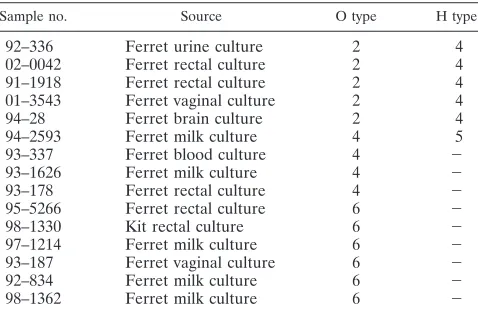
iden-tification of virulence factors are summarized in Table 1. All 15 isolates were beta-hemolyticE. coliand contained␣-hemolysin (hlyA),cnf1, and the P pilis (pap1). Isolates were negative for several other virulence factors, including heat-labile entero-toxin, st, cnf2, and eae. Serotypes included O2:H4 (five iso-lates), O6:H⫺(six isolates), O4:H⫺(three isolates), and O4:H5 (one isolate).
Ferret NTEC strains have CNF1 activity.In order to
deter-mine if cnf1 sequence homology was correlated with CNF1 activity, we treated subconfluent HeLa cell monolayers with sonicates and culture supernatants from seven ferret NTEC strains. After 72 h of incubation, HeLa cells that were treated with sonicates exhibited changes that were characteristic of CNF1 activity. Treated cells were larger than untreated control cells, and⬎80% of the treated cells had multiple nuclei (Fig. 1). In addition, evidence of nuclear fragmentation was appar-ent in some of the treated cells. Sonicates of all seven ferret
97–1214 Ferret milk culture 6 ⫺
93–187 Ferret vaginal culture 6 ⫺
92–834 Ferret milk culture 6 ⫺
98–1362 Ferret milk culture 6 ⫺
a
Serotyping was performed at Pennsylvania State University. All samples were positive forcnf1,hly, andpap1, and all samples were negative forcnf2, heat-labile enterotoxin,sta,stb,stx1,stx2, andeae. All samples exhibited beta-hemolysis.
on May 15, 2020 by guest
http://jcm.asm.org/
[image:2.585.302.541.80.237.2]NTEC strains had comparable cytopathic effects on HeLa cell monolayers 72 h after treatment. Weaker activities were ob-served when HeLa cells were treated with culture supernatants (data not shown).
High-resolution genotyping confirms serogroup-based
rela-tionship of ferret NTEC strains.To confirm the relationship of
ferret NTEC strains based on O and H serotyping, we per-formed PCR amplification of genomic DNAs, using primers that were complementary to interspersed repetitive elements (Rep-PCR). Three groups of ferret NTEC strains could be distinguished based on their Rep-PCR amplification patterns (Fig. 2). The human O4:K6 strain J96, three ferret O4:H⫺
strains, one ferret O4:H5 strain, and two ferret O6:H⫺strains made up the first group (top seven strains). The second group (middle four strains) were all O6:H⫺strains. The third group (bottom five strains) were all O2:H4 strains. Despite the lim-ited genetic diversity among the ferret NTEC strains, the strains did not stratify as a function of the date of isolation. Strains comprising the first genotype, made up of O4 and O6 strains, were isolated between 1993 and 1997. The O6:H⫺ strains comprising the second genotype were isolated between 1992 and 1998. The O2:H4 strains comprising the third geno-type were isolated between 1991 and 2002. Interestingly, the O6:H⫺ strains 93–187 and 97–1214 appeared to be more
FIG. 1. Ferret NTEC strains exhibit CNF1-mediated cytopathic effects. Subconfluent monolayers of HeLa cells were treated with sonicates of ferret NTEC strains. After 72 h, the cells exhibited distention and⬎80% were multinucleated. Nuclear fragmentation was also apparent in some cells. Representative monolayers of HeLa cells treated with a sonicate of ferret NTEC strain 02–0042 (A) and a sonicate of the nonpathologic strain DH5␣(B) and an untreated control monolayer (C) are shown at the same magnification after being stained with Diff-Quik.
FIG. 2. Dendrogram showing similarities of Rep-PCR genotypes of ferret NTEC strains, representing five O4 strains, six O6 strains, and five O2 strains. Three groups of strains could be distinguished. From top to bottom, the strains are grouped as follows: three ferret O4:H⫺strains, one ferret O4:H5 strain, two ferret O6:H⫺strains, and the human O4:K6 strain J96 (top); four ferret O6:H⫺strains (middle); and five ferret O2:H4 strains (bottom).
on May 15, 2020 by guest
http://jcm.asm.org/
necrotoxigenicE. coli(NTEC). NTEC may be the most appro-priate term for these E. coli strains. Extraintestinal diseases associated with cnf1-positive E. coli have also been demon-strated in other species.Uropathogenic organisms contain many virulence factors, including bacterial adhesins, toxins, iron acquisition systems, and host defense avoidance mecha-nisms (1, 24). In the study reported here, representatives of all of these categories of virulence factors (with the exception of iron acquisition systems) were identified. All 15 ferret isolates were positive for P pili by PCRs using a pair of primers de-signed to amplify an internal fragment ofpapC(19) The hu-man O4:K6 strain J96 contains unlinked loci encoding two distinct P pili, pyelonephritis-associated pili (pap) and Pap-related sequence (prs) (22). pap and prs cannot be distin-guished by primers because the two loci are identical except for the gene encoding the adhesin (papGandprsG). Further char-acterization of P pili in ferret NTEC strains is needed, but a preliminary characterization of O6 strains suggested that they express Prs, while O2:H4 strains may express Pap (data not shown). The existence ofpap,hly, andcnf1in urosepsis strains is evidence of the presence of pathogenicity islands (1). Addi-tional characterizations of papand hly, along with challenge studies using isogenic mutants, will provide insight into the contribution of these virulence factors to disease.The ferret may be a promising model for evaluations of the contribution ofcnf1and other genetic factors toE. coli-associated virulence. Ferrets are carnivores, are easily maintained and manipulated in a vivarium, and are of an adequate size (approximately 1 kg) to perform longitudinal studies with the ability to obtain mul-tiple samples. Another advantage over existing models is that the ferret is the natural host for several coliform diseases. In the clinical realm, extraintestinalE. coli-associated diseases of the ferret are major causes of morbidity and mortality in post-parturient jills and in neonates (5, 20).cnf1or other virulence genes of theseE. coliisolates may also be appropriate targets for vaccine development. Finally, comparisons of fecal and extraintestinal strains of pap-, hly-, and cnf1-positive E. coli
from ferrets may provide insights into the critical genetic and proteomic differences required by these organisms for select-ing ecological niches and their subsequent ability to elicit dis-ease.
REFERENCES
1.Bingen-Bidois, M., O. Clermont, S. Bonacorsi, M. Terki, N. Brahimi, C. Loukil, D. Barraud, and E. Bingen.2002.Escherichia colipathogenicity island-like domains. Infect. Immun.70:7185–7186.
2.Bingen-Bidois, M., O. Clermont, S. Bonacorsi, M. Terki, N. Brahimi, C. Loukil, D. Barraud, and E. Bingen.2002. Phylogenetic analysis and preva-lence of urosepsis strains ofEscherichia colibearing pathogenicity island-like domains. Infect. Immun.70:3216–3226.
L. Gagnoux, M. Piechaczyk, P. Boquet, and E. Lemichez.2002. CNF1 ex-ploits the ubiquitin-proteasome machinery to restrict Rho GTPase activation for bacterial host cell invasion. Cell111:553–564.
9.Fabbri, A., L. Falzano, S. Travaglione, A. Stringaro, W. Malorni, S. Fais, and C. Fiorentini.2002. Rho-activating Escherichia colicytotoxic necrotizing factor 1: macropinocytosis of apoptotic bodies in human epithelial cells. Int. J. Med. Microbiol.291:551–554.
10.Falzano, L., R. Rivabene, M. T. Sanini, A. Fabgri, and C. Fiorentini.2001. AnEscherichia colicytotoxin increases superoxide anion generation via rac in epithelial cells. Biochem. Biophys. Res. Commun.283:1026–1030. 11.Feria, C., J. Machado, J. D. Correia, J. Goncalves, and W. Gaastra.2001.
Virulence genes and P fimbriae PapA subunit diversity in canine and feline uropathogenicEscherichia coli.Vet. Microbiol.82:81–89.
12.Fournout, S., C. M. Dozois, M. Odin, C. Desautels, S. Peres, F. Herault, F. Daigle, C. Segafredo, J. Laffitte, E. Oswald, J. M. Fairbrother, and I. P. Oswald.2000. Lack of a role of cytotoxic necrotizing factor 1 toxin from
Escherichia coliin bacterial pathogenicity and host cytokine response in infected germfree piglets. Infect. Immun.68:839–847.
13.Fox, J. G. (ed.).1998. Biology and diseases of the ferret, 2nd ed., p. 321–354. Williams & Wilkins, Baltimore, Md.
14.Guyer, D. M., J.-S. Kao, and H. L. T. Mobley.1998. Genomic analysis of a pathogenicity island in uropathogenicEscherichia coliCFT073: distribution of homologous sequences among isolates from patients with pyelonephritis, cystitis, and catheter-associated bacteriuria and from fecal samples. Infect. Immun.66:4411–4417.
15.Guzman-Verri, C., E. Chaves-Olarte, C. von Eichel-Streiber, I. Lopez-Goni, M. Thelestam, S. Arvidson, J. P. Gorvel, and E. Moreno.2001. GTPases of the Rho subfamily are required forBrucella abortusinternalization in non-professional phagocytes: direct activation of Cdc42. J. Biol. Chem.276:
44435–44443.
16.Johnson, J. R., P. Delavari, and T. T. O’Bryan.2001.Escherichia coliO18: K1H7 isolates from patients with acute cystitis and neonatal meningitis exhibit common phylogenetic origins and virulence factor profiles. J. Infect. Dis.183:1546.
17.Johnson, J. R., N. Kaster, M. A. Kuskowski, and G. V. Ling.2003. Identi-fication of urovirulence traits inEscherichia coliby comparison of urinary and rectalE. coliisolates from dogs with urinary tract infection. J. Clin. Microbiol.41:337–345.
18.Landraud, L., M. Gauthier, T. Fosse, and P. Boquet.2000. Frequency of
Escherichia colistrains producing the cytotoxic necrotizing factor (CNF1) in nosocomial urinary tract infections. Lett. Appl. Microbiol.30:213–216. 19.Le Bouguenec, C., M. Archambaud, and A. Labigne.1992. Rapid and specific
detection of thepap,afa, andsfaadhesin-encoding operon in uropathogenic
Escherichia colistrains by polymerase chain reaction. J. Clin. Microbiol.
30:1189–1193.
20.Liberson, A. J., C. E. Newcomer, J. I. Ackerman, J. C. Murphy, and J. G. Fox.
1983. Mastitis caused by hemolyticEscherichia coliin the ferret. J. Am. Vet. Med. Assoc.183:1179–1181.
21.Lockman, H. A., R. A. Gillespie, B. D. Baker, and E. Shakhnovich.2002.
Yersinia pseudotuberculosisproduces a cytotoxic necrotizing factor. Infect. Immun.70:2708–2714.
22.Lund, B., B.-I. Marklund, N. Stro¨mberg, F. Lindberg, K.-A. Karlsson, and S. Normark. 1988. UropathogenicEscherichia coli can express serologically identical pili of different receptor specificities. Mol. Microbiol.2:255–263. 23.Mainil, J. G., E. Jacquemin, F. Heraul, and E. Oswald.1997. Presence of
pap-, sfa-, and afa-related sequences in necrotoxigenicEscherichia coli iso-lates from cattle: evidence for new variants of the AFA family. Can. J. Vet. Res.61:193–199.
24.Mulvey, M. A., J. D. Schilling, J. J. Martinez, and S. J. Hultgren.2000. Bad bugs and beleaguered bladders: interplay between uropathogenic Esche-richia coliand innate host defenses. Proc. Natl. Acad. Sci. USA16:8829– 8835.
25.Ojeniyi, B., P. Ahrens, and A. Meyling.1994. Detection of fimbrial and toxin genes inEscherichia coliand their prevalence in piglets with diarrhea. The application of colony hybridization assay, polymerase chain reaction and phenotypic assays. Zentbl. Vet. Med. B41:49–59.
on May 15, 2020 by guest
http://jcm.asm.org/
26.Schmitt, C. K., K. C. Meysick, and A. D. O’Brien.1999. Bacterial toxins: friends or foes? Emerg. Infect. Dis.15:224–234.
27.Schultsz, C., G. J. Pool, R. van Ketel, B. De wevek, P. Speelman, and J. Dankert.1994. Detection of enterotoxigenicEscherichia coliin stool samples by using nonradioactively labeled oligonucleotide DNA probes and PCR. J. Clin. Microbiol.32:2393–2397.
28.Starc˘ic˘, M., J. R. Johnson, A. L. Stell, J. van der Goot, H. G. Hendriks, C. van Vorsenbosch, L. van Dijk, and W. Gaastra.2002. HaemolyticEscherichia coliisolated from dogs with diarrhea have characteristics of both uropatho-genic and necrotoxiuropatho-genic strains. Vet. Microbiol.85:361–377.
29.Travaglione, S., L. Falzano, A. Fabbre, A. Stringaro, S. Fais, and C. Fioren-tini.2002. Epithelial cells and expression of the phagocytic marker CD68: scavenging of apoptotic bodies following Rho activation. Toxicol. In Vitro
16:405–411.
30.Versalovic, J., M. Schnieder, F. J. de Bruijn, and J. R. Lupski. 1994. Genomic fingerprinting of bacteria using repetitive sequence-based polymer-ase chain reaction. Methods Mol. Cell. Biol.5:25–40.
31.Witham, P. K., C. T. Yamashiro, K. J. Livak, and C. A. Bat. 1996. A PCR-based assay for the detection ofEscherichia coliShiga-like toxin gene in ground beef. Appl. Environ. Microbiol.62:1347–1353.