0095-1137/05/$08.00⫹0 doi:10.1128/JCM.43.4.1575–1580.2005
Copyright © 2005, American Society for Microbiology. All Rights Reserved.
Clonal Spread of a Vancomycin-Resistant
Enterococcus faecium
Strain
among Bloodstream-Infecting Isolates in Italy
Lucia Stampone,
1Maria Del Grosso,
1Delia Boccia,
2† and Annalisa Pantosti
1*
Dipartimento di Malattie Infettive, Parassitarie ed Immunomediate1and Centro Nazionale di Epidemiologia, Sorveglienza e
Promozione della Salute,2Istituto Superiore di Sanita`, Rome, Italy
Received 25 August 2004/Returned for modification 7 October 2004/Accepted 7 December 2004
Recent data indicated that the rate of vancomycin resistance in bloodstream-infecting enterococcal isolates in Italy is one of the highest in Europe. The aims of this study were to characterize bloodstream-infecting vancomycin-resistant enterococci (VRE) obtained from various Italian hospitals and to establish whether the isolates were clonally related. During the years 2001 to 2003, a total of 39 VRE isolates were obtained from 19 hospital laboratories in various areas of Italy. Species identification and resistance genotypes of the isolates were obtained by multiplex PCR. Further characterization included antibiotic susceptibility testing, pulsed-field gel electrophoresis (PFGE) of SmaI-digested genomic DNA, detection of virulence genes (espandhyl), and multilocus sequence typing (MLST) of selected isolates. VRE were identified as 31 Enterococcus faecium
(VREfm) isolates and 8E.faecalisisolates. All but one isolate carried thevanAgene; one VREfm isolate carried thevanBgene. Analysis of the PFGE profiles showed that 28 VREfm isolates shared a similar electrophoretic profile, designed type 1, and were clonally related. All type 1 isolates were resistant to ampicillin, streptomycin, gentamicin, and rifampin and were positive for the esp gene. MLST identified an allelic profile (ST78) comprisingpurKallele 1, belonging to the C1 clonal lineage, characteristic of human infection and hospital outbreak isolates. ThevanB-carrying VREfm isolate, of PFGE type 2, was shown to be a single-locus variant of ST78. Our data indicate that the recent increase in the number of bloodstream infections caused by VRE in Italy is due to the spread of a hospital-adapted, multidrug-resistant VREfm clone belonging to an interna-tionally disseminated lineage.
Since the first report of vancomycin-resistant enterococci (VRE) in 1988, VRE have emerged as an important cause of hospital-acquired infections, particularly in the United States. According to the National Nosocomial Infection Surveillance System of the Centers for Disease Control and Prevention, over a 10-year time period the percentage of VRE has in-creased from 0.4 to 25% among enterococcal isolates from patients in intensive care units (ICUs) (17). Conversely, infec-tions with VRE are still relatively uncommon in European hospitals, although a large pool of VRE strains is present in Europe, especially among the intestinal flora of farm animals and healthy humans. This large pool is likely due to the use of the glycopeptide avoparcin as a growth promoter in animal husbandry until its ban in 1997 (17).
In European countries, including Italy, VRE have been demonstrated in the feces of farm animals and in animal-derived foods, such as raw meat products (7). After discontin-uation of the use of avoparcin, the frequency of VRE in these reservoirs decreased significantly (1; A. Pantosti, M. Del Grosso, S. Tagliabue, A. Macrı`, and A. Caprioli, Letter, Lancet
354:741–742, 1999).
In the past decade, two hospital outbreaks were reported in
Italy; one was due to VRE belonging to the species
Enterococ-cus faecalis(VREfl) in a neurosurgical ICU (E. Manso, G. De
Sio, F. Biavasco, P. E. Varaldo, G. Sambo, and C. Maffei, Letter, Lancet 342:616–617, 1993), and another was due to VRE belonging to the speciesE. faecium(VREfm) in a he-matology department (21). The latter outbreak was followed by a long period of endemic circulation of genetically related strains in the same hospital. In addition, sporadic cases of infections caused by VRE were observed. In a multicenter study carried out from 1993 to 1995, the proportion of VRE among enterococcal isolates from infected sites varied between 0 and 36% in the 20 hospitals surveyed. The highest proportion was associated with the VREfm outbreak described above (10).
According to the European Antibiotic Resistance Surveil-lance System, an international network which collects data on antibiotic resistance of bloodstream-infecting isolates in 28 European countries (http://www.earss.rivm.nl/), in Italy the proportion of VRE of higher than 10% in 2001 and 2002. In particular, the proportion of VRE among
bloodstream-infect-ingE.faeciumisolates in 2002 (19%) was one of the highest in
Europe. In contrast, the proportion of VRE among blood-stream-infectingE.faecalisisolates (4%) was within the aver-age for the other European countries. As in Italy, in the United Kingdom the vancomycin resistance rate among E. faecium
from bacteremia increased sharply, from 6.3% in 1995 to 24% in 1998 (18). The high prevalence of VRE in Italy and the United Kingdom was also confirmed by another European study carried out in 2001 in ICUs and other at-risk hospital wards; in that study, the United Kingdom and Italy were the countries with the highest rates of VRE, 10.4 and 19.6%,
* Corresponding author. Mailing address: Dipartimento di Malattie Infettive, Parassitarie ed Immunomediate, Istituto Superiore di Sanita`, Viale Regina Elena 299, 00161 Rome, Italy. Phone: (39) 064990 2852. Fax: (39) 0649387112. E-mail: [email protected].
† Present address: Health Protection Agency (HPA), Communica-ble Disease Surveillance Centre, 61 Colindale Ave., London NW9 5EQ, United Kingdom. Phone: (44) 208 2006868. Fax: (44) 208 2007868. E-mail: [email protected].
1575
on May 16, 2020 by guest
http://jcm.asm.org/
respectively (11). The high rate in Italy was ascribed to the circulation of a multidrug-resistant VREfm clone in two hos-pital centers.
The aims of this study were to characterize bloodstream-infecting VRE isolates obtained from various Italian hospitals during the years 2001 to 2003 and to establish whether the isolates were clonally related.
MATERIALS AND METHODS
The laboratories collaborating in the Italian nationwide antibiotic resis-tance surveillance system (AR-ISS), started in May 2001 (http://www.simi.iss.it /antibiotico_resistenza.htm), were asked to send data on the antibiotic suscepti-bilities of all bloodstream-infectingE. faecalisandE. faeciumisolates and to send VRE (the first isolate from each patient) for further testing.
The species identification and the genotypes of glycopeptide resistance were obtained by a previously described multiplex PCR assay (7).
Susceptibilities to ampicillin, aminoglycosides (streptomycin and gentamicin for high-level aminoglycoside resistance), glycopeptides (vancomycin and teico-planin), tetracycline, chloramphenicol, rifampin, quinupristin-dalfopristin, and linezolid were assayed by the microdilution method according to NCCLS guide-lines (16) with prepared Sensititre panels (Biomedical s.r.l., Venice, Italy).
The presence of putative virulence genes, such as the enterococcal surface protein geneesp(29) and the hyaluronidase genehylEfm(20), in VRE isolates was investigated by PCR with the primer pairs suggested by Vankerckhoven et al. (25).
To analyze clonal relatedness among the isolates, total genomic DNAs were digested with SmaI and separated by pulsed-field gel electrophoresis (PFGE) by previously described methods (7). Isolates whose profiles differed by one to six bands were assigned to different subtypes of the same type. Isolates that differed by more than six bands were considered unrelated and were assigned to different types (23). A computer-assisted dendrogram of fragment patterns was con-structed by using Diversity Database Fingerprinting software, version 2 (Bio-Rad Laboratories). Clustering was obtained by the unweighted-pair group method using average linkages (UPGMA) with the Dice similarity coefficient.
To better characterize the principal VREfm clone, multilocus sequence typing (MLST) was carried out with isolates showing different PFGE subtype profiles. For each isolate, the sequences of seven housekeeping genes (adk,atpA,ddl,gdh,
gyd,purK, andpstS) were obtained by previously published methods (13) and compared with those of the alleles recorded in the database available at the MLST website (http://efaecium.mlst.net).
The sequence of thepurKgene was obtained for VREfm isolates not belong-ing to the main clone as well.
RESULTS
Identification and antibiotic susceptibility of VRE. In the years 2001 to 2003, 918 bloodstream-infectingE. faecalis iso-lates and 345E. faecium isolates were reported by hospital laboratories participating to the AR-ISS project. In particular, 1.2% of the E. faecalis isolates and 20% of the E. faecium
isolates were VRE. The isolation of VRE was reported by 26 of 62 hospital laboratories. Only 39 VRE isolates, approxi-mately half of all those reported, from 19 laboratories through-out Italy were available for further testing: 13 isolates in 2001, 15 isolates in 2002, and 11 isolates in 2003. Each laboratory sent one to three isolates. All patients were adults (median age, 68.5 years). They were admitted to hematology (14 patients), ICU (10 patients), surgical (7 patients), medical (5 patients), and other (3 patients) wards, mainly in large hospitals (⬎600 beds).
A total of 31 isolates belonged to the speciesE.faecium, and 8 belonged to the speciesE. faecalis. ThevanAgene was the glycopeptide resistance determinant found in all but one of the isolates; the exception was a VREfm isolate carrying thevanB
gene.
The resistance patterns for the isolates are shown in Table 1. All VREfm isolates were resistant to ampicillin, erythromycin, and rifampin. All but two isolates showed high-level resistance to both streptomycin and gentamicin. In addition, four isolates were resistant to tetracycline, three isolates were resistant to chloramphenicol, and one isolate was resistant to quinupristin-dalfopristin. All VREfl isolates were susceptible to ampicillin and resistant to tetracycline. All but one isolate were resistant to erythromycin, four isolates were resistant to chloramphen-icol, four isolates were resistant to both of the aminoglycosides tested, one isolate was resistant to streptomycin only, and three isolates were resistant to gentamicin only. All of the VRE isolates tested, bothE.faeciumandE.faecalis, were suscepti-ble to linezolid.
[image:2.585.44.544.82.226.2]The results of the glycopeptide susceptibility tests were in agreement with the resistance genotypes. For all of the isolates except thevanB-carrying isolate, the MIC of vancomycin was
TABLE 1. PFGE profiles of bloodstream-infecting VRE isolates
Species PFGE
type
No. of
isolates Yr
No. of hospitals
Area of Italya
Presence (⫹) or absence (⫺) of
espgene
Resistance patternb
E. faecium 1 28 2001–2003 18 N, C, S ⫹ AMP, STR, GEN, ERY, RIFc
2 1d 2002 1 N ⫹ AMP, ERY, TET, RIF
3 1 2001 1 N ⫺ AMP, STR, ERY, TET, RIF
4 1 2002 1 N ⫺ AMP, STR, GEN, ERY, TET, RIF, CHL
E. faecalis A 1 2001 1 S ⫺ GEN, ERY, TET, CHL
B 1 2001 1 S ⫹ STR, TET, CHL
C 1 2001 1 S ⫺ STR, GEN, ERY, TET
D 1 2002 1 C ⫺ STR, GEN, ERY, TET, CHL
E 2 2002, 2003 1 C, S ⫺ GEN, ERY, TET
F 1 2003 1 N ⫹ STR, GEN, ERY, TET
G 1 2003 1 S ⫺ STR, GEN, ERY, TET, CHL
aN, northern; C, central; S, southern.
bAMP, ampicillin; STR, streptomycin; GEN, gentamicin; ERY, erythromycin; RIF, rifampin; TET, tetracycline; CHL, chloramphenicol. cOne isolate was also resistant to CHL, and one isolate was also resistant to TET, CHL, and quinupristin-dalfopristin.
dvanB-carrying isolate.
on May 16, 2020 by guest
http://jcm.asm.org/
ⱖ32 g/ml, and the MIC of teicoplanin wasⱖ64g/ml. For the vanB-carrying isolate, the MIC of vancomycin was ⱖ32
g/ml, and the MIC of teicoplanin wasⱕ0.5g/ml.
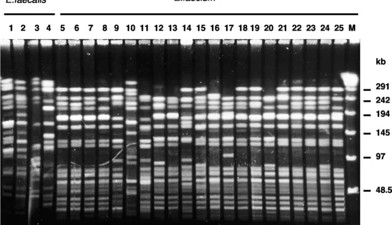
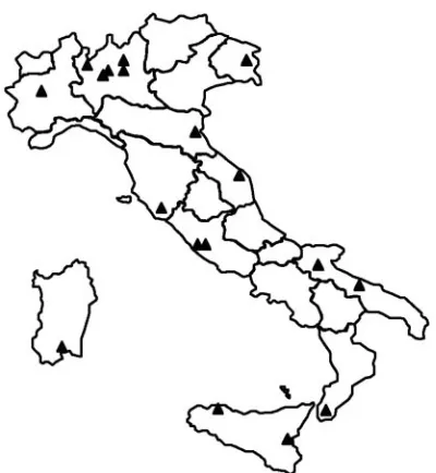
Molecular typing and clonal characteristics of VRE. Anal-ysis of VREfm isolates by PFGE showed that 28 isolates had identical or similar profiles, differing by six or fewer bands; they were assigned to PFGE type 1 (Fig. 1). The computer-assisted clustering of VREfm based on the Dice similarity coefficient is shown in Fig. 2. PFGE type 1 encompassed 14 different types (1.1 to 1.14). Subtype 1.1 contained nine isolates, sub-types 1.2 and 1.10 contained two isolates each, and subsub-types 1.4 and 1.7 contained three isolates each. The other subtypes were represented by single isolates. Type 1 isolates were obtained over a span of 3 years from 18 hospitals in various geographical areas of Italy (Fig. 3). An apparent cluster of isolates was observed in hospitals in the Lombardia region (Milan area), but this finding could be a consequence of the number of large hospitals in the area participating in the surveillance.
The other three VREfm isolates had PFGE profiles that differed by seven or more bands from that of type 1 (Fig. 1) and were assigned to three different PFGE types; type 2 included the onlyvanB-carrying isolate. The dendrogram showed that the isolates belonging to types 2 and 3 were not genetically distant from type 1 isolates, as their coefficient of similarity was 0.8, at the limit for clonal relatedness (Fig. 2).
For the eight VREfl isolates, heterogeneity of PFGE profiles was observed (Fig. 1). Seven different types (A to G) were recognized, with only one type containing two isolates that had identical profiles but were obtained from different hospitals (Table 1).
The esp gene was amplified by PCR from 29 of the 31 VREfm isolates and from 2 of the 8 VREfl isolates. The two
esp-negative VREfm isolates did not belong to PFGE type 1.
The hly gene was not detected in any of the VRE isolates examined.
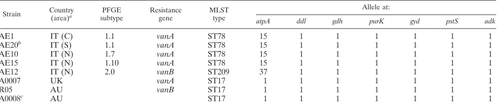
MLST was performed for four VREfm isolates belonging to PFGE type 1 and obtained from different hospitals. Strain AE1, subtype 1.1, was considered the reference strain for the group. Strain AE20 was chosen because it was resistant to quinupristin-dalfopristin, and strains AE10 and AE15 were chosen because their PFGE profiles showed a difference of six bands from the profile for the reference subtype 1.1 strain. In addition, strain AE12, PFGE type 2, was analyzed by MLST, as it was the onlyvanB-carrying isolate.
The four type 1 isolates were found to share an identical allelic profile, to which ST78 was assigned at the MLST web-site. ThevanB-carrying isolate was found to be a single-locus variant of ST78 bearing a newly discovered allele of theatpA
gene, to which ST209 was assigned (Table 2). These findings confirm the partial relatedness obtained by computer-assisted clustering of strain AE12 and type 1 isolates.
Both ST78 and ST209 were single-locus variants of ST17, found in isolates from hospital outbreaks in different countries, including the United Kingdom, Australia, and the United States, and comprising either vancomycin-susceptible isolates or those carryingvanAorvanB(Table 2). In addition, ST78 shared five or more alleles out of seven with 30 other STs retrievable from the MLST website (last accessed 1 December 2004).
The sequence of thepurK gene was obtained for the two VREfm isolates belonging to PFGE types 3 and 4 as well; both carriedpurKallele 1.
DISCUSSION
The use of molecular methods such as AFLP and PFGE has led to major advancements in the understanding of the
epide-FIG. 1. PFGE profiles of bloodstream-infecting VRE isolates. Lanes 1 to 4, VREfl isolates of different PFGE types. Lanes 5 to 25, VREfm isolates; all of the profiles shown belong to different subtypes of type 1, with the exception of the profiles in lane 10 (PFGE type 4), lane 11 (PFGE type 3), and lane 14 (PFGE type 2). M, molecular size markers.
on May 16, 2020 by guest
http://jcm.asm.org/
[image:3.585.94.489.77.304.2]miology of VRE, allowing comparisons of isolates from differ-ent sources, especially animal and human isolates (3, 8, 30), and from hospital outbreaks (14, 19, 21, 24). However, these methods often have proven too discriminatory to establish genetic relationships in a broader setting and to gain general insight about isolates from diverse areas and typed by different laboratories. Recently, an MLST scheme originally devised for
Streptococcus pneumoniae (9) was proposed for E. faecium
(13). The different sequenced alleles of seven housekeeping genes constitute an unambiguous database that is available through the World Wide Web and that allows comparisons of isolates from different environments and different laboratories. Although we studied a limited number of VRE isolates, they represent clinically significant isolates obtained from blood-stream infections. All VREfm isolates were multidrug resistant and carriedpurKallele 1, which has been demonstrated to be typical of isolates of human origin (13). While all of the VREfl isolates seemed to retain their susceptibility to ampicillin, ther-apeutic options for the VREfm isolates appeared to be very limited, with only quinupristin-dalfopristin and linezolid being active.
[image:4.585.95.485.68.390.2]By using PFGE we demonstrated that the majority of the VREfm isolates are clonally related. We are not aware of epidemiological links among the patients infected, as the iso-lates were obtained from hospitals scattered throughout the country over a span of 3 years. VREfl isolates appeared to be
FIG. 2. Dendrogram based on PFGE SmaI restriction pattern analysis of 31 VREfm isolates. Similarity analysis was performed with the Dice coefficient, and clustering was performed by UPGMA.
FIG. 3. Map of Italy. Triangles indicate the sites of hospital labo-ratories at which PFGE type 1 VREfm isolates were obtained.
on May 16, 2020 by guest
http://jcm.asm.org/
[image:4.585.64.264.485.702.2]more heterogeneous on the basis of their PFGE profiles and susceptibility patterns.
The findings obtained by PFGE regarding the clonality of VREfm isolates were confirmed by MLST typing. The five representative isolates analyzed by this method belonged to ST78 or to a single-locus variant of this ST. PFGE appears to be more discriminatory than MLST for the typing ofE. fae-cium, since thevanB-positive strain, considered different from the main clonal group by the former method, was shown by MLST to be a single-locus variant of the same clone bearing a novelatpAallele. This discordance might be due to the con-tribution of genetic mobile elements, such as plasmids, to the number and size of DNA bands in PFGE profiles (28).
According to the published MLST-based classification (13), ST78 is encompassed by clonal group C1, a genetic lineage with a worldwide distribution and including mainly isolates from human infections and hospital outbreaks. Although spe-cific information on isolates belonging to ST78 is not available to date at the MLST website, Bonora and coworkers reported that recent VRE outbreaks in various hospitals in northern Italy were due to VREfm isolates belonging to ST78 (3). Com-mon features of the ST78 clone appear to be multidrug resis-tance and the presence of theespgene, a marker of hospital-adapted isolates that may be linked to the ability to persist in the hospital environment (29).
Intrahospital transmission of the same clone of VREfm has been demonstrated frequently (5, 21, 27). Conversely, inter-hospital dissemination of isolates has been rarely studied, al-though it has been shown to occur when investigated (3, 6, 15, 19, 24). In this study, we found a widespread distribution of a single clone involving the entire country. Since only isolates originating from bloodstream infections were studied—likely representing only a small portion of all of the VRE isolates in health care facilities, which are usually recovered from less serious infections or from colonized patients (12)—the spread of the VREfm clone is likely to be wider and to involve addi-tional hospitals.
In the past decade in Italy, most vancomycin-susceptibleE.
faeciumisolates from clinical infections were found to be
re-sistant to ampicillin (10) and to bear theespgene (L. Baldas-sarri, L. Bertuccini, M. Ammendolia, G. Gherardi, and R. Creti, Letter, Lancet357:1802, 2001), representing a fit genetic background for the acquisition of vancomycin resistance
de-terminants. An ampicillin- and streptomycin-resistant, vanco-mycin-susceptible,esp-positiveE.faeciumisolate from our col-lection, obtained in 1997 from a human infection in Rome, was found to belong to ST78 (data not shown). It can be speculated that a similar hospital-adapted isolate acquired thevanA de-terminant, generating the widespread ST78 VREfm clone. This occurrence has been demonstrated in outbreaks described in France, Poland, and Germany (14, 22, 27). We did not examine thevanAresistance determinants of the VREfm iso-lates to ascertain whether they have similar or different struc-tures and reside on the chromosome or on a plasmid.
As for a possible role of the animal reservoir in the spread of VRE isolates in Italian hospitals, we recently demonstrated that animal VREfm isolates are different from VREfm isolates from human infections on the basis of their antibiotic suscep-tibility profiles; notably, they are generally susceptible to am-picillin, while the human isolates are not (4). Using various typing methods, several investigators have demonstrated that animal and human VREfm isolates are genetically distinct (2, 13, 30) and that animal isolates do not carry theespgene (29). All of these observations suggest host specificity for VRE iso-lates, although the genetic elements carrying vanA can be shared between human and animal isolates (26). Therefore, it is possible that the animal VRE reservoir that is still present in Italy several years since the withdrawal of avoparcin (A. Ricci and A. Battisti, personal communication) can contribute to the spread of VRE through the sharing ofvanAresistance deter-minants with human and hospital-adapted isolates.
In conclusion, our data indicate that the recent increase in the number of VRE infections in Italy might be due to the appearance and spread of a hospital-adapted, multidrug-resis-tant VREfm clone belonging to an internationally dissemi-nated lineage. It is possible that both horizontal gene transfer and clonal spread have contributed to the high rate of VRE infections in Italy.
ACKNOWLEDGMENTS
[image:5.585.43.542.90.197.2]We are indebted to Stefania Salmaso, Paolo Fortunato D’Ancona, and Stefano Fokas for scientific organization and data management of the AR-ISS project and to the laboratories participating in the AR-ISS project. We are especially indebted to the colleagues who provided strains: V. Amato, Catania; L. Biagianti, Orbetello, Grosetto; G. Boli-gnano, Reggio Calabria; C. Bonato, Milan; S. Bramati, Monza; F. Dell’Anno, Ravenna; D. De Vito, Bari; A. Giammanco, Palermo; A. TABLE 2. Allelic profiles (MLST), PFGE types, and vancomycin-resistance genes for selected bloodstream-infectingE. faeciumisolates
from Italy
Strain Country
(area)a
PFGE subtype
Resistance gene
MLST type
Allele at:
atpA ddl gdh purK gyd pstS adk
AE1 IT (C) 1.1 vanA ST78 15 1 1 1 1 1 1
AE20b IT (S) 1.1 vanA ST78 15 1 1 1 1 1 1
AE10 IT (N) 1.7 vanA ST78 15 1 1 1 1 1 1
AE15 IT (N) 1.10 vanA ST78 15 1 1 1 1 1 1
AE12 IT (N) 2.0 vanB ST209 37 1 1 1 1 1 1
A0007 UK vanA ST17 1 1 1 1 1 1 1
R05 AU vanB ST17 1 1 1 1 1 1 1
A0008c AU ST17 1 1 1 1 1 1 1
aIT, Italy; UK, United Kingdom; AU, Australia; C, central; S, southern; N, northern. bResistant to quinupristin-dalfopristin.
cSusceptible to vancomycin.
on May 16, 2020 by guest
http://jcm.asm.org/
Grossi, Treviglio, Bergamo; A. Ingianni, Monserrato (CA); M. Liber-goli, San Giovanni Rotondo, Foggie; F. Luzzaro, Varese; E. Manso, Ancona; R. Minniti, Rome; M. P. Molinari, Novara; G. Parisi, Rome; A. Piscina, Rimini; R. Trevisan, Udine; and F. Vailati, Bergamo. We thank Fabio D’Ambrosio for assistance with the image analysis soft-ware.
This study was supported in part by a grant from Istituto Zoopro-filattico Sperimentale delle Venezie, Ricerca Finalizzata 2000.
REFERENCES
1.Aarestrup, F., A. M. Seyfarth, H.-D. Emborg, K. Pedersen, S. Hendriksen, and F. Bager.2001. Effect of abolishment of the use of antimicrobial agents for growth promotion on occurrence of antimicrobial resistance in fecal enterococci from food animals in Denmark. Antimicrob. Agents Chemother.
45:2054–2059.
2.Bonora, M. G., C. Bordin, L. Bragagnolo, L. Cirelli, M. De Fatima, A. Grossato, M. Ligozzi, G. Lo Cascio, and R. Fontana.2001. Molecular anal-ysis ofvanAenterococci isolated from humans and animals in northeastern Italy. Microb. Drug Resist.7:247–256.
3.Bonora, M. G., M. Ligozzi, M. De Fatima, L. Bragagnolo, A. Goglio, G. C. Guazzotti, and R. Fontana.2004. Vancomycin-resistantEnterococcus fae-ciumisolates causing hospital outbreaks in northern Italy belong to the multilocus sequence typing C1 lineage. Microb. Drug Resist.10:114–123. 4.Busani, L., M. Del Grosso, C. Paladini, C. Graziani, A. Pantosti, F. Biavasco,
and A. Caprioli.2004. Antimicrobial susceptibility of vancomycin-susceptible and -resistant enterococci isolated in Italy from raw meat products, farm animals, and human infections. Int. J. Food Microbiol.97:17–22. 5.Christiansen, K. J., P. A. Tibbett, W. Beresford, J. W. Pearman, R. C. Lee,
G. W. Coombs, I. D. Kay, F. G. O’Brien, S. Palladino, C. R. Douglas, P. D. Montgomery, T. Orrell, A. M. Peterson, F. P. Kosaras, J. P. Flexman, C. H. Heath, and C. A. McCullough.2004. Eradication of a large outbreak of a single strain ofvanBvancomycin-resistantEnterococcus faeciumat a major Australian teaching hospital. Infect. Control Hosp. Epidemiol.25:384–390. 6.D’Agata, E. M. C., L. H. C. Goulding, and Y.-W. Tang.2001. Clinical and molecular characterization of vancomycin-resistant Enterococcus faecium
strains during establishment of endemicity. Clin. Infect. Dis.33:511–516. 7.Del Grosso, M., A. Caprioli, P. Chinzari, M. Fontana, G. Pezzotti, A.
Man-frin, E. Giannatale, E. Goffredo, and A. Pantosti.2000. Detection and char-acterization of vancomycin-resistant enterococci in farm animals and raw meat products in Italy. Microb. Drug Resist.6:313–318.
8.Dicuonzo, G., G. Gherardi, G. Lorino, S. Angeletti, F. Battistoni, L. Bertuc-cini, R. Creti, R. Di Rosa, M. Venditti, and L. Baldassarri.2001. Antibiotic resistance and genotypic characterization by PFGE of clinical and environ-mental isolates of enterococci. FEMS Microbiol. Lett.201:205–211. 9.Enright, M. C., and B. G. Spratt.1998. A multilocus sequence typing scheme
forStreptococcus pneumoniae: identification of clones associated with serious invasive disease. Microbiology144:3049–3060.
10.Fontana, R., M. Ligozzi, A. Mazzariol, G. Veneri, and G. Cornaglia.1998. Resistance of enterococci to ampicillin and glycopeptide antibiotics in Italy. Clin. Infect. Dis.27(Suppl. 1):S84–S86.
11.Goossens, H., D. Jabes, R. Rossi, C. Lammens, G. Privitera, and P. Cour-valin.2003. European survey of vancomycin-resistant enterococci in at-risk hospital wards and in vitro susceptibility testing of ramoplanin against these isolates. J. Antimicrob. Chemother.51:5–12.
12.Hayden, M. K.2000. Insights into the epidemiology and control of infection with vancomycin-resistant enterococci. Clin. Infect. Dis.31:1058–1065. 13.Homan, W., D. Tribe, S. Poznanski, M. Li, G. Hogg, E. Spalburg, J. van
Embden, and R. Willems.2002. Multilocus sequence typing scheme for
Enterococcus faecium. J. Clin. Microbiol.40:1963–1971.
14.Kawalec, M., M. Gniadkowski, M. Zaleska, T. Ozorowski, L. Konopka, and W. Hryniewicz.2001. Outbreak of vancomycin-resistantEnterococcus fae-ciumof the phenotype VanB in a hospital in Warsaw, Poland: probable
transmission of the resistance determinants into an endemic vancomycin-susceptible strain. J. Clin. Microbiol.39:1781–1787.
15.Klare, I., C. Konstabel, D. Badstubner, G. Werner, and W. Witte.2003. Occurrence and spread of antibiotic resistances inEnterococcus faecium. Int. J. Food Microbiol.88:269–290.
16.National Committee for Clinical Laboratory Standards.2002. Performance standards for antimicrobial susceptibility testing; 12th informational supple-ment. National Committee for Clinical Laboratory Standards, Wayne, Pa. 17.Patel, R.2003. Clinical impact of vancomycin-resistant enterococci. J.
Anti-microb. Chemother.51(Suppl. 3):13–21.
18.Reacher, M. H., A. Shah, D. M. Livermore, M. C. J. Wale, C. Graham, A. P. Johnson, H. Heine, M. A. Monnickendam, K. F. Barker, D. James, and R. C. George.2000. Bacteraemia and antibiotic resistance of its pathogens re-ported in England and Wales between 1990 and 1998: trend analysis. Br. Med. J.320:213–216.
19.Reinert, R. R., G. Conrads, J. J. Schlaeger, G. Werner, W. Witte, and R. Lutticken.1999. Survey of antibiotic resistance among enterococci in North Rhine-Westphalia, Germany. J. Clin. Microbiol.37:1638–1641.
20.Rice, L., L. Carias, S. Rudin, C. Vael, H. Goossens, C. Konstabel, I. Klare, S. R. Nallapareddy, W. Huang, and B. Murray.2003. A potential virulence gene,hylEfm, predominates inEnterococcus faeciumof clinical origin. J. In-fect. Dis.187:508–512.
21.Scagnelli, M., G. Pellizer, F. De Lalla, A. D’Emilio, M. Rassu, L. Bragagnolo, P. Reatto, G. Veneri, M. Ligozzi, and R. Fontana.2001. Epidemiological analysis of vancomycin-resistant enterococci in a large tertiary-care hospital in northern Italy. Eur. J. Clin. Microbiol. Infect. Dis.20:609–616. 22.Suppola, J., E. Kolho, S. Salmenlinna, E. Tarkka, J. Vuopio-Varkila, and M.
Vaara.1999.vanAandvanBincorporate into an endemic ampicillin-resistant vancomycin-sensitiveEnterococcus faeciumstrain: effect on interpretation of clonality. J. Clin. Microbiol.37:3934–3939.
23.Tenover, F. C., R. D. Arbeit, R. V. Goering, P. A. Mickelsen, B. E. Murray, D. H. Persing, and B. Swaminathan.1995. Interpreting chromosomal DNA restriction patterns produced by pulsed-field gel electrophoresis: criteria for bacterial strain typing. J. Clin. Microbiol.33:2233–2239.
24.Thal, L., S. Donabedian, B. Robinson-Dunn, J. Chow, L. Dembry, D. Clewell, D. Alshab, and M. Zervos.1998. Molecular analysis of glycopeptide-resistant
Enterococcus faecium isolates collected from Michigan hospitals over a 6-year period. J. Clin. Microbiol.36:3303–3308.
25.Vankerckhoven, V., T. Van Autgaerden, C. Vael, C. Lammens, S. Chapelle, R. Rossi, D. Jabes, and H. Goossens.2004. Development of a multiplex PCR for the detection ofasa1,gelE,cylA,esp, andhylgenes in enterococci and survey for virulence determinants among European hospital isolates of En-terococcus faecium. J. Clin. Microbiol.42:4473–4479.
26.Wegener, H. C.2003. Antibiotics in animal feed and their role in resistance development. Curr. Opin. Microbiol.6:439–445.
27.Werner, G., I. Klare, F.-B. Spencker, and W. Witte.2003. Intra-hospital dissemination of quinupristin/dalfopristin- and vancomycin-resistant Entero-coccus faeciumin a paediatric ward of a German hospital. J. Antimicrob. Chemother.52:113–115.
28.Werner, G., R. Willems, B. Hildebrandt, I. Klare, and W. Witte.2003. Influence of transferable genetic determinants on the outcome of typing methods commonly used forEnterococcus faecium. J. Clin. Microbiol.41:
1499–1506.
29.Willems, R., W. Homan, J. Top, M. van Santen-Verheuvel, D. Tribe, X. Manzioros, C. Gaillard, C. Vandenbroucke-Grauls, E. Mascini, E. van Kregten, J. van Embden, and M. Bonten.2001. Variantespgene as a marker of a distinct genetic lineage of vancomycin-resistantEnterococcus faecium
spreading in hospitals. Lancet357:853–855.
30.Willems, R., J. Top, N. van den Braak, A. van Belkum, H. P. Endtz, D. Mevius, E. E. Stobberingh, A. E. van den Bogaard, and J. D. A. van Embden.
2000. Host specificity of vancomycin-resistantEnterococcus faecium. J. In-fect. Dis.182:816–823.