Georgia State University Georgia State University
ScholarWorks @ Georgia State University
ScholarWorks @ Georgia State University
Public Health Theses School of Public Health
Fall 12-20-2012
A Historical Perspective and Review of the Evidence to Support
A Historical Perspective and Review of the Evidence to Support
Fruit Bats as the Natural Reservoir for Ebola Viruses
Fruit Bats as the Natural Reservoir for Ebola Viruses
Zachary Reed
Follow this and additional works at: https://scholarworks.gsu.edu/iph_theses
Recommended Citation Recommended Citation
Reed, Zachary, "A Historical Perspective and Review of the Evidence to Support Fruit Bats as the Natural Reservoir for Ebola Viruses." Thesis, Georgia State University, 2012.
https://scholarworks.gsu.edu/iph_theses/241
A Historical Perspective and Review of the Evidence to Support Fruit Bats as the
Natural Reservoir for Ebola Viruses
By
Zachary D. Reed
B.S. University of Georgia
A Capstone Project Submitted to the Graduate Faculty of Georgia State University in Partial Fulfillment
of the Requirement for the Degree
MASTER OF PUBLIC HEALTH
ATLANTA, GEORGIA
Abstract
The Ebola viruses cause sporadic outbreaks of Ebola hemorrhagic fever (EHF) where origins
have been traced to the continent of Africa and the Philippines. Since the initial discovery of Zaire and
Sudanebolavirus in 1976, the Ebola viruses have been responsible for severe hemorrhagic fever
outbreaks in Africa with case fatality rates between 40-90%. The natural reservoir(s) of the Ebola
viruses is currently unknown, but there is mounting evidence that fruit bats may play a key role. The
goal of the current study is to screen a large variety of bat species from Africa and Asia where Ebola is
known to be endemic for the presence of IgG specific antibody to Ebola virus in order to see which bat
species may show evidence of past Ebola virus infection. Ebola virus would not be expected to cause
lethal disease in its natural reservoir; therefore the presence of IgG antibody would be present.
Identifying the species of bats that have been infected will allow researchers to hopefully isolate Ebola
virus from bats adding to the evidence that bats are a reservoir species. The knowledge gained may also
provide clues to new species of bats yet to be identified as possible natural reservoir(s) as well as expand
the known geographical range of known Ebola virus outbreaks. Knowing which species of bats as well
as their geographic range may help prevent future Ebola outbreaks by minimizing human-reservoir
contact.
Historical Perspective and Review of the Evidence to Support Fruit Bats as the
Natural Reservoir for Ebola Viruses
By
Zachary D. Reed
Approved:
Dr. Richard Rothenberg_________________ Committee Chair
Dr. Jonathan Towner____________________ Committee Member
Authors Statement
In presenting this thesis as a partial fulfillment of the requirements for an advanced degree from Georgia State University, I agree that the Library of the University shall make it available for inspection and circulation in accordance with its regulations governing materials of this type. I agree that
permission to quote from, to copy from, or to publish this thesis may be granted by the author or, in his/her absence, by the professor under whose direction it was written, or in his/her absence, by the Associate Dean, College of Health and Human Sciences. Such quoting, copying, or publishing must be solely for scholarly purposes and will not involve potential financial gain. It is understood that any copying from or publication of this dissertation which involves potential financial gain will not be allowed without written permission of the author.
Notice to Borrowers Page
All theses deposited in the Georgia State University Library must be used in accordance with the stipulations prescribed by the author in the preceding statement.
The author of this thesis is:
Student’s Name: Zachary D. Reed
Users of this capstone who are not enrolled at Georgia State University are required to attest acceptance of the preceding stipulation by signing below. Libraries borrowing this capstone for the use of their patrons are required to see that each user records below the information requested.
Name of User Address Date Type of Use
Table of Contents
Chapter 1: Introduction………..…7
Chapter 2: Literature Review………...9
Chapter 3: Materials and Methods………...25
Chapter 4: Results………....29
Chapter 5: Discussion and Conclusion………....30
Chapter I: Introduction
Background:
The Ebola viruses cause sporadic outbreaks of Ebola hemorrhagic fever (EHF) where origins
have been traced to the continent of Africa and the Philippines. The Ebola viruses belong to the family
Filoviridae, order Mononegavirales which are characterized by a filamentous shape with a
nonsegmented, single strand, negative sense RNA genome. The family Filoviridae currently includes
three genera, Ebolavirus, Marburgvirus, and “Cuevavirus” (proposed). The genus Ebolavirus includes
five species: Zaire ebolavirus (EBOV), Sudan ebolavirus (SUDV), Reston ebolavirus (RESTV),
Bundibugyo ebolavirus (BDBV), and Taï Forest ebolavirus (TAFV)formerly Côte d’Ivoire ebolavirus
(CIEBOV) (International Committee on Taxonomy of Viruses. & King, 2012).
Since the initial discovery of the first two species, Zaire and Sudanebolavirus in 1976, the Ebola
viruses have been responsible for severe hemorrhagic fever outbreaks in Africa with high case fatality
rates between 40-90% (World Health Organization, 2009). Reston ebolavirus is the only species that
has been found outside the African continent and has not been found to be pathogenic to humans
(Miranda et al., 1991). There has only been one non-fatal human case of Côte d’Ivoire ebolavirus
reported (Le Guenno et al., 1995).
Outbreak investigations of Ebola virus have shown that introductions into the human population
result from either a single introduction into the human population followed by human-to-human
transmission, or through multiple introductions into the human population from a zoonotic source
(Newman & Food and Agriculture Organization of the United Nations., 2011). After introduction into
the human population, human-to-human transmission of Ebola viruses occurs through contact with
infectious bodily fluids particularly in healthcare settings where improper barrier nursing techniques are
used or during traditional African burials that include communal washing of bodies.
which a fruit bat has been identified (Amman et al., 2012; Leroy et al., 2009; Leroy et al., 2005; Pourrut
et al., 2009). The goal of the current study is to screen a large variety of bat species from Africa and
Asia where Ebola is known to be endemic for the presence of IgG specific antibody to Ebola virus in
order to see which bat species may show evidence of past Ebola virus infection. Ebola virus would not
be expected to cause lethal disease in its natural reservoir; therefore the presence of IgG antibody would
be present. Identifying the species of bats that have been infected will allow researchers to hopefully
isolate Ebola virus from bats adding to the evidence that bats are a reservoir species. The knowledge
gained may also provide clues to new species of bats yet to be identified as possible natural reservoir(s)
as well as expand the known geographical range of known Ebola virus outbreaks. Knowing which
species of bats as well as their geographic range may help prevent future Ebola outbreaks by minimizing
Chapter II: Literature Review
Introduction to Filoviridae: Ebolavirus, Marburgvirus, and “Cuevavirus” (proposed):
The family Filoviridae contains the genera Ebolavirus,Marburgvirus, and the recently
discovered “Cuevavirus” (proposed) (International Committee on Taxonomy of Viruses. & King, 2012).
There are five species within the genus Ebolavirus. Zaire ebolavirus, Sudan ebolavirus, Bundibugyo
ebolavirus, and Taï Forest ebolavirus are found on the African continent. Reston ebolavirus, originating
from the Philippines,is the only one that has been found outside of the African continent. Marburg
marburgvirus is the only species within the genus Marburgvirus, but there are two distinct viruses
Marburg and Ravn (International Committee on Taxonomy of Viruses. & King, 2012). Lloviu virus is a
new Ebola-like filovirus that was recently discovered in Spain (Negredo et al., 2011).
Transmission Patterns and Epidemiology of Ebola Virus:
With the exception of the recognized outbreaks, the epidemiology of Ebola virus infections in
humans is unknown (Sanchez A, 2007). After infection from the initial source, infection is spread by
person-to-person contact through infected bodily fluids such as blood, excrement, oral secretions, and
tissue. Healthcare facilities can be a major source of virus transmission. Improper barrier nursing
techniques as well as the reuse of infected needles help spread the virus into the community and to the
healthcare workers. For example, in the 1995 Zaire ebolavirus outbreak in Zaire, one-fourth of the cases
were among healthcare workers (Sanchez A, 2007).
Clinical Presentation and Symptoms:
The incubation period of EHF is between 2 and 21 days (Hartman, Towner, & Nichol, 2010).
Symptoms manifest abruptly and are often non-specific flu-like and may include chills, fever, myalgia,
and malaise followed by lethargy, nausea, vomiting, abdominal pain, anorexia, diarrhea, coughing,
headache, and hypotension (Hartman, et al., 2010). Hemorrhaging occurs in less than 50 percent of
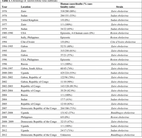
Ebola Hemorrhagic Fever Outbreaks:
[image:11.612.58.558.118.601.2]Table 1 summarizes the known Ebola virus outbreaks through September 2012.
Table 1. Chronology of known Ebola virus outbreaks
Year Location
Human cases/deaths (%
case-fatality ratio) Strain
1976 Zaire 318/280 (88%) Zaire ebolavirus
1976 Sudan 284/151 (53%) Sudan ebolavirus
1976 United Kingdom 1/0 (0%) Sudan ebolavirus
1977 Zaire 1/1 (100%) Zaire ebolavirus
1979 Sudan 34/22 (65%) Sudan ebolavirus
1989-1990 USA Epizootic, 4-6 human cases (0%) Reston ebolavirus
1992 Itally, Philippines Epizootic Reston ebolavirus
1994 Côte d’Ivoire 1/0 (0%) Côte d’Ivoire ebolavirus
1994-1995 Gabon 52/31 (60%) Zaire ebolavirus
1995 Zaire 315/250 (81%) Zaire ebolavirus
1996 Gabon 37/21 (57%) Zaire ebolavirus
1996 USA, Philippines Epizootic Reston ebolavirus
1996 Russia 1/1 (100%) Zaire ebolavirus
1996-1997 Gabon, South Africa 60/45 (74%) Zaire ebolavirus
2000-2001 Uganda 425/224 (53%) Sudan ebolavirus
2001-2002 Gabon, Republic of 122/96 (78%) Zaire ebolavirus
2002 Gabon, Republic of Congo 11/10 (90%) Zaire ebolavirus
2002-2003 Republic of Congo 143/128 (89.5%) Zaire ebolavirus
2003-2004 Republic of Congo 35/29 (82.9%) Zaire ebolavirus
2004 Russia 1/1 (100%) Zaire ebolavirus
2004 Sudan 17/7 (41%) Sudan ebolavirus
2005 Republic of Congo 12/10 (83%) Zaire ebolavirus
2007 Democratic Republic of the Congo 264/186 (71%) Zaire ebolavirus
2007-2008 Uganda 131/42 (37%) Bundibugyo ebolavirus
2008 Philippines 6/0 (0%) Reston ebolavirus
2008- 2009 Democratic Republic of the Congo 32/15 (47%) Zaire ebolavirus
2011 Uganda 1/1 (100%) Sudan ebolavirus
2012 Uganda 24/17 (71%) Sudan ebolavirus
Zaire ebolavirus:
1976 Zaire
Ebola hemorrhagic fever (EHF) was first recognized in 1976 during two simultaneous but
independent outbreaks in Sudan and Zaire. Between September 1, 1976 and October 24, 1976, an
outbreak of Zaire ebolavirus occurred in Zaire, which centered around Yambuku Missionary Hospital
(YMH) in the Bumba Zone of the Equateur Region ("Ebola haemorrhagic fever in Zaire, 1976," 1978).
The first known case was a 44-year-old male instructor at the Mission School who reported to the
outpatient clinic at YMH on August 26, 1976 with what was thought to be malaria ("Ebola
haemorrhagic fever in Zaire, 1976," 1978). The man had been touring the northern Equateur Region and
had purchased smoked antelope and monkey meat. The man indicated that he and his family had eaten
the antelope, but not the monkey meat. He was admitted to YMH on September 5, 1976 with
gastrointestinal bleeding and died September 8 ("Ebola haemorrhagic fever in Zaire, 1976," 1978).
There were 318 cases, 280 deaths and a case fatality ratio of 88 percent ("Ebola haemorrhagic
fever in Zaire, 1976," 1978). YMH was the main source of transmission during the outbreak where the
reuse of needles and close body contact with infected individuals helped spread the virus ("Ebola
haemorrhagic fever in Zaire, 1976," 1978). Of the 288 cases where the means of transmission could be
determined, 85 received one or more injections at YMH. Another 149 cases acquired the disease after
being in contact with patients, usually in their home village. Forty three cases had a history of both
injections at YMH and contact with patients ("Ebola haemorrhagic fever in Zaire, 1976," 1978).
Thirteen of the seventeen staff members became infected with EHF of whom eleven died ("Ebola
haemorrhagic fever in Zaire, 1976," 1978). The outbreak eventually ended with the closing of YMH and
1977 Zaire
Approximately one year after the original EBOV outbreak, Zaire ebolavirus reemerged in the
Tandala region of Zaire in 1977 marking the first time the virus had been seen since its discovery. A
nine-year-old girl from the village of Bonduni, presented to Tandala Mission Hospital in June 1977 with
a three day fever, abdominal pain, and hematemesis (Heymann et al., 1980). Twenty-eight hours after
hospitalization, the child died. Ebola virus was isolated from guinea pigs that had been inoculated with
the girl’s clinical specimens (Heymann, et al., 1980).
1995 Zaire
In April of 1995 an epidemic of dysentery broke out in Kikwit II Maternity Hospital in Kikwit,
Zaire (Khan et al., 1999). Later that month, in Kikwit’s other large hospital, Kikwit General Hospital, a
similar cluster of dysentery was identified among the operating room staff in which two Italian
missionary nurses died after caring for a patient who had a laparotomy performed (Khan, et al., 1999).
The epidemic dysentery was originally misdiagnosed and later confirmed as Zaire ebolavirus after
testing by the Centers for Disease Control and Prevention in Atlanta, Georgia. Fourteen samples were
tested, which confirmed that the 2 Italian nurses as well as twelve additional members of the surgical
team and their contacts had recent infection. There were a total of 315 cases, 250 deaths and a case
fatality ratio of 81 percent (Khan, et al., 1999).
December 1994- January/February 1995 Gabon
There were five independent outbreaks of EBOV between November 1994 and February 1995 in
Gabon. The first epidemic involved two waves of patients who were from the gold-panning
communities of Mekouka, Andock, and Minkebe (Georges et al., 1999). The first wave of patients
consisted of 32 patients who went to the hospital in Makokou for treatment. The second wave originated
occurred in the second wave (Georges, et al., 1999). There were 52 cases, 31 deaths, and a case fatality
ratio of 60 percent (Georges, et al., 1999).
January - April 1996 Gabon
In February 1996, the second epidemic of Zaire ebolavirus began in the village of Mayibout 2,
Gabon. The epidemic began with 18 people who had skinned a chimpanzee cadaver and became ill with
fever, headache, and bloody diarrhea (Georges, et al., 1999). All patients were admitted to Makokou
General Hospital where four died within 48 hours. The bodies were returned to their villages for burial
where traditional burial practices were performed without proper precautions (Georges, et al., 1999).
There were 37 cases, 21 deaths and a case fatality ratio of 57 percent (Georges, et al., 1999).
July 1996 - January 1997 Gabon
The third epidemic in Zaire ebolavirus in Gabon occurred in Booue area. Virus was isolated
from two of six samples from patients hospitalized at Booue (Georges, et al., 1999). A retrospective
study of the epidemic traced the index case back to a 39-year-old hunter in a logging camp near Booue
in July who had typical symptoms of viral hemorrhagic fever. Furthermore, dead chimpanzees were
found in the area of which one was confirmed to be positive for Ebola (Georges, et al., 1999). At the end
of August, a second hunter died at the same logging camp. A third hunter became ill twelve days later,
and died in the village of Balimba.
At the end of November 1996, another wave of transmission occurred in three locations: Lolo,
SHM, a timber company, and Balimba, a logging camp (Georges, et al., 1999). The epidemic also
spread to Libreville, the capital of Gabon, Lastourville, and South Africa. A Gabonese doctor performed
an endoscopy in Libreville on an infected patient and went to Johannesburg for treatment (Georges, et
al., 1999). A nurse who treated the infected doctor became ill and died. There were 60 cases, 45 deaths
and a case fatality ratio of 74 percent (Georges, et al., 1999).
An outbreak of Zaire ebolavirus was reported between October 2001 and July 2002 in Gabon
and Republic of Congo. This was the first reported instance of Ebola being in the Republic of Congo
("Outbreak(s) of Ebola haemorrhagic fever, Congo and Gabon, October 2001-July 2002," 2003). In
November 2001, medical personnel at Mekambo Medical Centre in La Zadie health district of Gabon
reported five deaths to the regional health authorities. At the same time there had been a large number of
dead non-human primates found in the rainforest and reported to the authorities ("Outbreak(s) of Ebola
haemorrhagic fever, Congo and Gabon, October 2001-July 2002," 2003). On November 30, 2001 blood
samples from two suspected cases were sent to Centre International de Recherches Médicales de
Franceville (CIRMF) for testing. Ebola virus was confirmed in both samples. In Gabon, there were 65
cases, 53 deaths and a case fatality ratio of 82 percent ("Outbreak(s) of Ebola haemorrhagic fever,
Congo and Gabon, October 2001-July 2002," 2003). In the Republic of the Congo, there were 57 cases,
43 deaths and a case fatality ratio of 75 percent ("Outbreak(s) of Ebola haemorrhagic fever, Congo and
Gabon, October 2001-July 2002," 2003).
2002-2003 Republic of Congo
On June 28, 2003, ten deaths and five hospitalized cases were reported by the Kéllé Health
Center in Kéllé District, Cuvette-Ouest Region of the Republic of Congo (Kuhn, 2008). This was
preceded by a die off chimpanzees, gorillas, and duikers in which Zaire ebolavirus had been isolated
from their dead carcasses. Human infections were also recorded in workers of a gold mine in Mbomo
District (Formenty et al., 2003). There were a total of 143 cases, 128 deaths and a case fatality ratio of
89 percent (Formenty, et al., 2003).
2003-2004 Republic of Congo
An outbreak of Zaire ebolavirus occurred in the Mbomo district, Cuvette Ouest Department
during November and December of 2003 in the Republic of the Congo. Testing performed by the
(World Health Organization, 2004). There were 35 cases, 29 deaths, and a case fatality ratio of 83
percent (World Health Organization, 2004).
2005 Republic of Congo
Between April 25 and June 16, 2005, there were twelve cases of Zaire ebolavirus in Etoumbi
District, Cuvette Ouest Region, Republic of the Congo. The outbreak began with two men who were
poaching for elephants in Parc d’Odzala forest (Nkoghe, Kone, Yada, & Leroy, 2011). They returned to
the village of Etoumbi to get help with the elephant and was hospitalized with fever and hemorrhaging.
They died shortly after hospitalization. There were ten contacts epidemiologically linked to the index
cases in which eight died (Nkoghe, et al., 2011). The case fatality ratio was 83 percent (Nkoghe, et al.,
2011).
2007 Democratic Republic of the Congo
A large outbreak of Zaire ebolavirus occurred in Luebo, Democratic Republic of the Congo in
2007 that may have started from the direct exposure to fruit bats (Leroy, et al., 2009). The Kampungu
agglomeration of villages where the index case, a 55-year-old woman, lived was once located in a
forest/savanna transition zone along the Lulua River. Bats were reported to have migrated to the islands
of Ndongo and Koumulele to feed on the fruit trees (Leroy, et al., 2009). Villagers routinely hunted the
bats for food and sold them at the village market (Leroy, et al., 2009). There were 264 cases and 186
deaths with a case fatality ratio of 71 percent (Leroy, et al., 2009).
2008-2009 Democratic Republic of the Congo
The last known outbreak of Zaire ebolavirus occurred in 2008-2009 in the Mweka and Luebo
health zones of the Democratic Republic of the Congo. The outbreak was confirmed by laboratory tests
at the Institut National de Recherches Biologiques (INRB) in Kinshasa, the CIRMF in Gabon, and the
2009). There were 32 cases reported, 15 fatalities with a case fatality ratio of 47 percent (World Health
Organization, 2009).
Sudan ebolavirus:
1976 Sudan
Between June and November of 1976, an outbreak of Sudan ebolavirus occurred simultaneously
in Sudan as the Zaire ebolavirus outbreak was occurring in Yambuku, Zaire. Three employees of a
cotton factory in Nzara township became ill with a severe febrile illness with profuse bleeding ("Ebola
haemorrhagic fever in Sudan, 1976. Report of a WHO/International Study Team," 1978). The outbreak
later spread to Maridi 128km from Nzara, where it was able to amplify in Maridi hospital. There were
284 cases, 151 fatalities with a case fatality ratio of 53 percent ("Ebola haemorrhagic fever in Sudan,
1976. Report of a WHO/International Study Team," 1978).
1979 Sudan
In 1979, thirty four cases of Sudan ebolavirus occurred in Nzara, Sudan (Baron, McCormick, &
Zubeir, 1983). The outbreak began in August with a 45-year-old man entering Nzara hospital with 3
days of fever, a recent onset of vomiting and diarrhea, and eventually developing gastrointestinal
bleeding (Baron, et al., 1983). He passed away three days later. Neither the hospital staff nor his family
taking care of him practiced barrier-nursing procedures which led to the outbreak. There were 34 cases,
22 deaths, and a case fatality ratio of 65 percent (Baron, et al., 1983).
August 2000- January 2001 Uganda
The largest outbreak of Ebola hemorrhagic fever to occur was an outbreak of Sudan ebolavirus
in Gulu, Masindi, and Mbarara districts of Uganda. On October 8, 2000, an unusual febrile illness with
hemorrhage and significant mortality was reported to the Ministry of Health in Kampala, Uganda
(Centers for Disease Control, 2001). There were 425 presumptive cases of EHF with 224 deaths with a
attending funerals of Ebola hemorrhagic fever case-patients, having contact with case-patients in one's
family, and providing medical care to Ebola case-patients without using adequate personal protective
measures were all identified as the most important means of transmission during the outbreak (Centers
for Disease Control, 2001).
2004 Sudan
In April 2004 Sudan ebolavirus reemerged in Yambio, Sudan. The medical staff at Yambio
County Health Department and the coordinator of the South Sudan Early Warning and Response
Network (EWARN) reported 7 suspected cases of hemorrhagic fever, which included two deaths to the
EWARN leader in Lokichoggio, Kenya (Onyango et al., 2007; "Outbreak of Ebola haemorrhagic fever
in Yambio, south Sudan, April - June 2004," 2005). Five of the suspected cases were from the same
family and two were hospital staff. Symptoms manifested over a three-week period and included fever,
vomiting, and bloody diarrhea (Onyango, et al., 2007). Overall, there were 17 confirmed cases, seven
deaths and case fatality ratio of 41 percent (Onyango, et al., 2007). The outbreak coincided with an
outbreak of measles, which made cases of Ebola difficult to diagnose.
2011 Uganda
In 2011, a single case of Sudan ebolavirus occurred in Luwero District, Uganda. On May 6,
2011, a 12 year-old girl presented to Bombo Military Hospital with fever, jaundice, and hemorrhagic
signs that included vaginal bleeding (Shoemaker et al., 2012). Her condition worsened and she died
three hours after admission. She was isolated from the general hospital population and the hospital staff
used proper protective equipment, which limited the outbreak to the one case.
2012 Uganda
An outbreak of Sudan ebolavirus was confirmed on July 28, 2012 in Kibaale District, Uganda. A
outbreak. The outbreak was declared over on October 4, 2012 with 24 cases, 17 deaths, and a case
fatality ratio of 71 percent.
Bundibugyo ebolavirus:
2007-2008 Uganda
The most recently discovered species of Ebola virus, Bundibugyo ebolavirus, was identified five
years ago in Budibugyo District, Uganda. In late November of 2007, 29 blood samples were sent to the
Centers for Disease Control and Prevention in Atlanta, Georgia for filovirus testing (Towner et al.,
2008). Eight of the samples were acutely positive using an antigen capture ELISA assay and IgM
capture assay for Zaire ebolavirus (Towner, et al., 2008). Once an outbreak of Ebola virus was
identified, an international team of scientists was deployed to Uganda to assist with the outbreak. There
were a total of 131 suspect, probable, and confirmed cases of identified (Chowell, Hengartner,
Castillo-Chavez, Fenimore, & Hyman, 2004). There were 56 laboratory confirmed cases, 43 of which were acute
phase specimens (MacNeil et al., 2010). Out of the 43 acute specimens, there were 17 deaths with a case
fatality ratio of 40 percent (MacNeil, et al., 2010).
Bundibugyo ebolavirus is thought to have first appeared in August 2007 in the village of
Kabango in Bundibugyo District ("Outbreak news. Ebola virus haemorrhagic fever, Democratic
Republic of the Congo," 2007). A 26-year-old woman developed fever and weakness and was
hospitalized. She was pregnant and gave birth to a preterm infant that later died and the mother died on
August 4th ("Outbreak news. Ebola virus haemorrhagic fever, Democratic Republic of the Congo,"
2007). The mother and sister of the index patient were involved in handling her remains and did not use
proper barrier nursing techniques. A total of nine cases and 6 deaths from the cluster were reported in
which two of the survivors later tested positive for Bundibugyo ebolavirus specific IgG ("Outbreak
news. Ebola virus haemorrhagic fever, Democratic Republic of the Congo," 2007).
On August 17, 2012 the Ministry of Health reported 10 suspected cases and 6 deaths of Ebola
hemorrhagic fever to the World Health Organization ("Outbreak news. Ebola haemorrhagic fever,
Democratic Republic of the Congo," 2012). The outbreak is currently ongoing in Isiro and Dungu
Health Zones of Province Orientale. As of October 24, 2012 there have been 52 reported cases and 25
deaths ("Outbreak news. Ebola, Democratic Republic of Congo - update," 2012).
Cote d’Ivoire ebolavirus:
1994 Cote d’Ivoire
The only known case of Cote-d’Ivoire ebolavirus occurred in the Taï National Forrest of
Cote-d’Ivoire. A troop of chimpanzees was being studied in which several members were found dead with
hemorrhages (Le Guenno, et al., 1995). In November 1994, a field autopsy was performed on a dead
chimpanzee to collect samples in order to find a cause of death. A 34-year old female who autopsied the
chimpanzee developed dengue-like symptoms on the 24th of November and was hospitalized on the 26th
of November (Le Guenno, et al., 1995). Sera was drawn from the patient and virus isolation attempts
were made using Vero E6 monkey kidney cells and AP61 Aedes pseudosutellaris mosquito cells.
Electron microscopy was used to identify Ebola virus in the Vero E6 cells (Le Guenno, et al., 1995). The
patient eventually recovered from the infection.
Reston ebolavirus:
1989-1990 USA
The third species of Ebola to be identified was Reston ebolavirus in 1989 and is the only
non-African species identified. In October 1989, 100 cynomolgus monkeys (Macaca fascicularis), were
flown from Manila, Philippine, to New York and taken by truck to Hazleton Research Products in
Reston, Virginia (Jahrling et al., 1990). They were placed into quarantine where two animals died and
two others became ill. Necropsy findings were consistent with simian hemorrhagic fever (SHF)
filovirus was isolated from four monkeys, which was later named Reston ebolavirus. There were five
confirmed cased of Reston ebolavirus inmonkeys and no reports of disease in humans (Jahrling, et al.,
1990).
1989-1990 Philippines
After the initial importation of infected cynomalgus monkeys into the United States from the
Philippines in 1989, studies were conducted at the export facilities in the Philippines in order to
document transmission of the virus. In March of 1990, dead monkeys were tested from the two export
facilities in the Philippines where the shipments to the United States had originated (Hayes et al., 1992).
In facility A, 85 of 161 dead monkeys tested positive for Reston ebolavirus antigen (Hayes, et al., 1992).
A serosurvey showed that 89 out of 343 monkeys had antibody to the virus. The second facility, facility
B, had no dead monkeys that were positive for Reston ebolavirus but 53 of 958 were antibody positive.
1992 Italy
In March of 1992, 55 cynomologus monkeys were imported into Siena, Italy from the same
monkey import facility in the Philippines implicated in the original 1989-1990 Reston, Virginia Reston
ebolavirus outbreak. Eight monkeys died from the shipment and Reston ebolavirus was isolated and
identified by electron microscopy ("Viral haemorrhagic fever in imported monkeys," 1992). There were
16 human contacts with the monkeys but none of them showed signs of infection or seroconverted
("Viral haemorrhagic fever in imported monkeys," 1992).
1996 USA/ Philippines
Reston ebolavirus was once again imported into the United States in April 1996. One hundred
cynomologus monkeys arrived from the Philippines to a quarantine facility in Alice, Texas where they
were split into two cohorts of 50 animals. One monkey died three days after arrival after exhibiting
anorexia and lethargy and was tested for Reston ebolavirus antigen (Rollin et al., 1999). Federal
quarantine (Centers for Disease Control, 1990; Pourrut, et al., 2009). A second monkey had similar signs
in the same cohort and was euthanized, tested and found positive for Reston ebolavirus (Rollin, et al.,
1999). The remaining 48 monkeys were euthanized and tested in which two additional monkeys were
found positive for Reston ebolavirus. Simian hemorrhagic fever was also isolated from some of the
animals (Rollin, et al., 1999). The monkeys were exported from the same facility that exported monkeys
infected with Reston ebolavirus to Reston Virginia in 1989-1990, and to Italy in 1992 (Miranda et al.,
1999; "Viral haemorrhagic fever in imported monkeys," 1992).
2008 Philippines
Although Reston Reston ebolavirus had been shown to be in the Philippines since 1989, it had
only been found in Cynomolgus monkeys. In 2008, Reston ebolavirus was discovered in swine further
expanding the host range of the virus. In July 2008, assistance from the Foreign Animal Disease
Diagnostic Laboratory (FADDL) of the United States Department of Agriculture (USDA) was requested
in investigating outbreaks of a respiratory and abortion disease syndrome that had been spreading
through Asia (Barrette et al., 2009). Sera and tissue samples were tested for the presence of Porcine
Reproductive and Respiratory Syndrome Virus (PRRSV) as well as other common swine viruses.
PRRSV was discovered to be infecting the swine simultaneously with Reston ebolavirus (Barrette, et al.,
2009). Interestingly, Reston ebolavirus was only found in samples that were also infected with PRRSV.
Laboratory Accidents:
1976 England, Sudan ebolavirus
Specimens from the 1976 Sudan ebolavirus outbreak were sent to investigators at the
Microbiological Research Establishment, Porton Down, England to assist in the identification of the new
agent responsible for the outbreak (Emond, Evans, Bowen, & Lloyd, 1977). On November 5, 1976, an
investigator accidently pricked his thumb through a protective rubber glove while transferring a
complained of anorexia, nausea, and constant central abdominal pain. He recovered after treatment with
interferon and convalescent serum from patients of the recent African outbreak and recovered. Blood
collected 14 hours after the patient became feverish was confirmed to have virus particles similar to
those of Ebola (Emond, et al., 1977).
2004 Russia, Zaire ebolavirus
On May 5, 2004, a Russian scientist working at the State Research Center for Virology and
Biotechnology (VECTOR) in Siberia, accidently punctured her double-gloved hand and pricked her left
palm with blood from a guinea pig infected with Zaire ebolavirus ("Fatal Laboratory Accident, Siberia,"
2004). She was isolated at the hospital of VECTOR’s department of dangerous infections where she was
under medical observation. She died 14 days later.
Fruit Bats as the Natural Reservoir of Filoviridae:
Isolation of Marburgvirus from Rousettus aegyptiacus Fruit Bats:
The first recognition of Marburg hemorrhagic fever was in 1967 when monkeys from Uganda
were imported into Marburg, Germany. Following the original discovery of marburgvirus the natural
reservoir had been unknown until recently. In 2007, following an outbreak of Marburg hemorrhagic
fever involving miners in Kitaka Cave, Uganda, an ecological investigation was launched to try and
identify the natural reservoir of Marburg virus (Towner et al., 2009). Of the 611 R. aegyptiacus bats
collected from Kitaka mine in August 2007 and April-May of 2008, 32 were positive for Marburg virus
by Q-RT-PCR and there were five virus isolates obtained (Towner, et al., 2009).
Experimental Infection of Fruit Bats with Zaire ebolavirus:
Following the Zaire ebolavirus outbreak in Kikwit in 1995, pathogenicity experiments were
undertaken at the National Institute for Virology in South Africa in representative plants and vertebrates
that were collected from a previous visit. Among the animals infected with Ebola virus were
Serological Evidence for Fruit Bats as the Natural Reservoir of Ebolavirus:
In Gabon and the Republic of Congo where known Zaire ebolavirus (ZEBOV) outbreaks have
occurred, screening of 1,030 animals collected between 2001 and 2005 including 679 bats, were
screened for the presence of IgG antibody specific to ZEBOV (Leroy, et al., 2005). IgG antibody to
ZEBOV was detected by ELISA in 8/117 Epomops franqueti bats, 4/17 Hypsignathus monstrosus, and
4/58 Myonycteris torquata (Leroy, et al., 2005). Furthermore, 13 bats of these three species had RNA
sequences that matched ZEBOV, but none of the bats that were IgG positive for ZEBOV had RNA
sequences. The authors suggested that these species of fruit bats are possible reservoirs of ZEBOV
(Leroy, et al., 2005).
Since the discovery of ZEBOV in fruit bats in Gabon and Republic of Gabon a larger serological
survey was performed between 2003 and 2008 that also included the Democratic Republic of Congo.
There were 2,147 bats belonging to nine species tested for the presence of Marburg virus and ZEBOV
(Pourrut, et al., 2009). In addition to confirming the presence of ZEBOV in E. franqueti, H. monstrosus,
and M. torquata, antibody was found in 4/197 Micropteropus pusillus, 24/307 R. aegyptiacus, and 3/24
Microchiroptera (Pourrut, et al., 2009).
Fruit bats of the Greater Accra Region have also been found to be positive for IgG antibody to
ZEBOV (Hayman et al., 2010; Hayman et al., 2012). In 2008, 1/262 Eidolon helvum bats screened using
indirect fluorescent tests was positive for IgG antibody to ZEBOV (Hayman, et al., 2010). In order to
see if Ebola virus is circulating in the Greater Accra Region, 88 bats from five species were screened
following the initial finding of the IgG positive E. helvum (Hayman, et al., 2012). Of those, 32 were
found IgG positive for Ebola virus. IgG antibody specific to Ebola virus was found in 10/27 E.
franqueti, 14/37 Epomophorus gambianus, 7/16 H. monstrosus, and 1/4 Nanonycteris veldkampii
(Hayman, et al., 2012). Some of the bats reacted with ZEBOV specific antigen while others reacted with
increase the ability to detect Ebola virus in Ghana if Taï Forest ebolavirus is the species of Ebola virus
circulating in the area (Hayman, et al., 2012).
Reston ebolavirus (REBOV) epidemics have occurred in the Philippines since 1989, but the
discovery of REBOV in domestic swine in 2008 put more attention on finding the natural reservoir.
During 2008 and 2009, 141 wild bats were caught and tested for REBOV specific IgG antibody using
ELISA (Taniguchi et al., 2011). Only one species, Rousettus amplexicaudatus, had positive results for
Chapter III: Materials and Methods
Study Description:
The CDC’s Viral Special Pathogens Branch (VSPB) in Atlanta, Georgia investigated many of
the Ebola outbreaks detailed in the previous section. Additionally, bat specimens from Nipah virus
outbreaks from Southeast Asia were available to screen for REBOV that VSPB had participated.
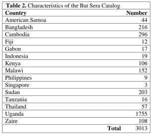
Characteristics of the Bat Sera Collection:
The collection of bat specimens screened for antibody evidence of Ebola virus consisted of 3,013
sera or blood specimens. The bat sera or blood was collected in the years 1995 through 2012 from seven
African countries and eight Asian countries. Some of the bats screened were originally collected during
the Zaire ebolavirus outbreak in 1995, Sudan ebolavirus outbreak in 2001, and the Sudan ebolavirus
outbreak in 2011. Fifty genera of bats are represented in the collection. Table 2 shows the distribution of
[image:26.612.152.459.380.655.2]the bat specimens screened by country.
Table 2. Characteristics of the Bat Sera Catalog
Country Number
American Samoa 44
Bangladesh 216 Cambodia 296
Fiji 12 Gabon 17 Indonesia 19 Kenya 106 Malawi 152 Philippines 9 Singapore 3 Sudan 203 Tanzania 16 Thailand 57 Uganda 1755 Zaire 108
[image:26.612.157.452.384.652.2]Total 3013
Table 3. Distribution by Species of the Bat sera Collection
Genus and Species Sera Collection Location(s) Number Screened
Casinycteris argynnis Zaire 2
Chaerephon plicata Cambodia 108
Chaerephon species unknown Sudan 4
Epomops franqueti* Gabon, Zaire, Uganda 10
Eidolon helvum* Kenya, Malawi, Uganda 179
Epomophorus labiatus Kenya, Sudan, Uganda 590
Eonycteris spelaea Bangladesh, Indonesia, Philippines, Thailand 77
Epomophorus wahlbergi Kenya 14
Emballonuridae Sudan 1
Epomophorus species unknown Malawi, Sudan, Uganda 108
Epomops species unknown Gabon, Malawi, 40
Glauconycteris species unknown Sudan 1
Hypsignathus monstrosus* Gabon 1
Hipposideros cafer Uganda 596
Hipposideros species unknown Bangladesh, Sudan, Uganda 28
Laephotis species unknown Tanzania 1
Megaderma lyra Bangladesh 10
Myotis mystacinus Cambodia 5
Micropteropus pusillus* Sudan, Zaire 83
Myonycteris torquata Gabon 1
Megaloglossus woermanni Zaire 26
Megaderma species unknown Bangladesh 1
Micropteropus species unknown Sudan 6
Molossidae Sudan 21
Notopteris macdonaldi Fiji 7
Neoromicia species unknown Tanzania 10
Nycteris species unknown Sudan 6
Pteropus giganteus Bangladesh 153
Pteropus lylei Cambodia, Thailand 95
Pteropus samoensis American Samoa, Fiji 10
Pteropus tonganus American Samoa, Fiji 39
Pipistrellus species unknown Malawi, Uganda 2
Pteropodidae Zaire 1
Pteropus species unknown Cambodia 8
Rousettus amplexicaudatus* Philippines 1
Rousettus angolensis Gabon 6
Rousettus lanosus Sudan, Tanzania 3
Rousettus leschenaulti Bangladesh 49
Rousettus aegyptiacus* Uganda 549
Rhinolophus species unknown Sudan, Tanzania, Uganda 26
Scotoecus albofuscus Sudan 1
Scotophilus kuhlii Cambodia, Philippines, 36
Scotoecus species unknown Sudan 11
Scotophilus species unknown Sudan, Tanzania 26
Stenonycteris species unknown Malawi 3
Taphozous melanopogon Cambodia 36
Taphozous theobaldi Cambodia 11
Tadarida species unknown Cambodia 6
Taphozous species unknown Uganda 2
Unknown Singapore 3
Laboratory Methods:
Enzyme-Linked Immunoassay for the Detection of Ebola Virus IgG:
Using an established protocol adapted to bats, 3,013 bats were screened for the presence of Ebola
virus specific immunoglobulin G (IgG) antibody (Ksiazek, West, Rollin, Jahrling, & Peters, 1999).
Briefly, Ebola virus antigen and negative control antigen was diluted 1:1,000 in 0.01M, PBS, pH 7.2 and
100 ul per well was absorbed to 96-well plates at 4°C overnight (BD Falcon Cat No. 353910). The
plates were then washed three times with 200 ul of a 0.01M PBS and 0.1% Tween-20 wash buffer
solution and 100 ul of serum diluent (0.01M PBS pH 7.4, 0.5% skim milk, and 0.1% Tween-20) added
to each well. The bat serum was added to the plates in 4-fold serial dilutions beginning with an initial
dilution of 1:100. The sera was incubated at 37°C for 1 hr and allowed to bind to the antigen. The plates
were again washed three times with 200 ul of wash buffer and 100 ul of anti-bat IgG conjugate (Bethyl
laboratories) diluted 1:2,000 in serum diluent was applied to each well and incubated at 37°C for 1 hr.
After the incubation, the plates were washed, and 100 ul per well of ABTS substrate (Kirkgard and
Perry Laboratories) was added and incubated at 37°C for 30 minutes. The plates were read at 405 nm
and 495 nm absorbance on a Biotek platereader. A positive sample was defined as having a titer greater
than or equal to 400 and having a sum optical density (OD) greater than 0.95.
Virus Isolation Techniques:
A pool of spleen and liver tissue from 50 Epomophoruslabiatus bats and 14 insectivorous bats
from the 2011 Sudan ebolavirus outbreak were setup for virus isolation to screen for the presence of
Ebola virus. Approximately 100 mg of tissue was ground using a Genogrinder in 2 mls of Hanks
Balanced Salt Solution (HBSS). The resulting suspension was centrifuged and the supernatant was used
to inoculate Vero E6 cells in 25 cm2
flasks and incubated at 37°C for 1 hr. After the 1 hr incubation,
flasks were re-fed with Eagles Minimum Essential Medium (EMEM) with 2% fetal bovine serum and
2x10^6 rads of gamma radiation, and fixed to slides with acetone in order to examine for Ebola virus
Chapter IV: Results
Ebola Virus Seroprevalence Patterns in the Bat Sera Collection:
Overall, 3 of the 3,014 (0.09%) bats were positive for Ebola virus specific IgG. There was 1
Chaerephon plicata from the October 2001 Cambodia collection that reacted with all five species of
Ebola virus. There were two Epomophorus labiatus batsthat were positive for Sudan ebolavirus IgG
only. One was collected during August 2010 in Sudan and one was collected in April of 2012 in
Uganda.
Limitations of the Study:
One potential limitation of this study is that many of the bat specimens used to screen for Ebola
virus specific IgG were collected as early as 1995 during the Zaire ebolavirus outbreak in Kikwit. The
handling and storage conditions over the years are unknown. The integrity of the specimens may have
Chapter V: Discussion and Conclusion
Interpretation of the Ebola Virus Seroprevalence in the Bat Sera Collection:
The discovery of an Ebola virus IgG positive Chaerephon plicata bat from Cambodia expands
the currently known geographical range of Ebola virus in Southeast Asia as well as indicates another
possible reservoir. Currently, Reston ebolavirus is known to be in the Philippines. Recent research has
indicated that Rousettus amplexicaudatus from the Philippines may be a possible reservoir (Taniguchi,
et al., 2011). It is possible that Reston ebolavirus is also in Cambodia and R. amplexicaudatus is a
possible reservoir species in that country.
There have been 3 Ebola outbreaks in Sudan. Although finding 1 Epomophorus labiatus bat
from Sudan IgG positive to Sudan ebolavirus is not surprising, it is novel in that this species of bat has
not been implicated as a possible reservoir species in the literature. The other positive E. labiatus bat
was collected in Uganda in close proximity to the 2011 Sudan virus outbreak. Since each of these bats
only reacted with Sudan ebolavirus and were collected where known outbreaks of Sudan ebolavirus
have occurred, it makes this bat species a prime candidate for further exploration into the natural
reservoir of Sudan ebolavirus.
The Impact of Ebola Virus Outbreaks on African Communities
The impact of Ebola outbreaks on African communities has varied in the past in size and severity
depending on the strain of virus and how quick the detection and response can be put forth by the
international community. This is in part due to a poor understanding of what Ebola virus is and how it is
spread. For example, the Zaire ebolavirus outbreak in the Republic of the Congo in 2002-2003was
thought to be caused by sorcery by certain ethnic groups while other ethnic groups contributed it to the
pygmy population who were thought to be “dirty disease spreaders” (Hewlett, Epelboin, Hewlett, &
Formenty, 2005). Efforts have been made by public health officials to educate the affected African
the native language of the people and in English. Furthermore, enlisting village elders to cooperate in
helping isolate ill patients and calming fears have led to less severe outbreaks.
The countries in Africa where previous Ebola outbreaks have occurred are among some of the
poorest in the world. The healthcare facilities in countries such as the Democratic Republic of the Congo
may not have a sufficient supply of sterile needles, which leads to the reuse of needles and amplification
of the outbreak in the healthcare setting. In many of the hospitals it is expected that family members
provided much of the basic supplies such as bed linens and provide supportive care while in the hospital.
The close contact between patients and family members taking care of them gives the virus an
opportunity to spread between members of an entire family, thus leading to many family members
becoming victims of Ebola virus. Additionally, if family members are able to leave the hospital freely
after providing care to a relative suspected of having Ebola virus, they may unknowingly spread the
virus throughout the community making it more difficult to contain.
An Ebola outbreak can have a devastating effect on an already fragile healthcare system
especially in a small village where there may be only one hospital. For example in 1995, during the
Zaire ebolavirus outbreak, there were 80 healthcare workers who were infected and Kikwit General
Hospital had to be closed in order to end the outbreak (Khan, et al., 1999). The loss of healthcare
workers and closure of an entire hospital may have devastating effects on an African community
especially when it comes to treating other endemic diseases.
Ebola outbreaks have damaging psychological effects throughout the affected communities.
Ebola victims are often stigmatized or face rejection from their local communities (De Roo et al., 1998;
MacNeil & Rollin, 2012). During the 1995 Zaire ebolavirus outbreak in Kikwit, patients who were
convalescing felt rejected by society including their family members. Furthermore, Ebola virus survivors
Uganda’s Efforts to Enhance Filovirus Surveillance
After the discovery of Marburg virus in R. aegyptiacus fruit bats and the discovery of
Bundibugyo ebolavirus in 2007, Uganda has strengthened their filovirus surveillance activity to better
detect and respond to outbreaks. During the Bundibugyo ebolavirus outbreak in 2007, a BSL-4 field
laboratory was setup at the Uganda Virus Research Institute in Entebbe in order to quickly test
specimens without having to send them to the Centers for Disease Control and Prevention in Atlanta.
This allowed infectious patients to be isolated from the general population which was critical in stopping
the chain of transmission. Since the outbreak, the laboratory in Uganda has been renovated with
enhanced security and more storage capacity for handling future outbreaks.
The Centers for Disease Control and Prevention’s, Viral Special Pathogens Branch currently has
an epidemiologist and a laboratorian stationed at the Ugandan Virus Research Institute. Having
personnel stationed in Uganda makes it possible for suspected viral hemorrhagic fever cases to be
quickly responded to and tested before an outbreak can spread. In 2011, the rapid recognition and
isolation of one case of Sudan ebolavirus limited the outbreak to the single patient. Since 2011, the
personnel in Uganda have detected two Ebola virus outbreaks and a Marburg virus outbreak in 2012.
The ability to test specimens in country allowed for a quick diagnosis and information to be
disseminated before the outbreaks could intensify.
Biosafety and Biosecurity Risks of Working with Ebola Virus
The Ebola viruses are among the most dangerous pathogens in the world and classified in the
United States as biosafety level 4 (BSL-4) pathogens (Chosewood, Wilson, Centers for Disease Control
and Prevention (U.S.), & National Institutes of Health (U.S.), 2009). This designation is given to agents
that are frequently fatal, have no treatment, are easily transmitted through aerosols, and pose a high risk
to the individual working with the agent (Chosewood, et al., 2009). The Ebola viruses have the ability to
public health and the environment if they were to be released from the laboratory. All of these
characteristics with the fact there is no licensed vaccine or therapy puts the Ebola viruses in the highest
biosafety category. In addition to the biosafety risks of working with Ebola virus in the laboratory there
is the potential for their use as agents of bioterrorism. The Ebola viruses are classified as Category A
select agents with the United States Department of Health and Human Services (DHHS). Category A
select agents are those that are rarely seen in the United States and pose a security risk because they can
be easily disseminated, have high mortality rates, cause public panic, and require special action for
public health preparedness (Centers for Disease Control and Prevention).
After the bombing of the Alfred E. Murrah Federal Building in Oklahoma City in 1995,
Congress passed the Antiterrorism and Effective Death Penalty Act of 1996 which gives DHHS the
authority to register laboratories and track select agents by the Centers for Disease Control and
Prevention. Following the terrorist attacks of September 11, 2001, Congress passed the Uniting and
Strengthening America by Providing Appropriate Tools Required to Intercept and Obstruct Terrorism
Act Of 2001 (USA PATRIOT Act) and the Public Health Security and Bioterrorism Preparedness and
Response Act of 2002 which set forth the provisions for the possession, use, and transfer of select agents
(Centers for Disease Control and Prevention, 2010). The final rules for possession, use and transfer of
select agents were published in the Federal Register in 2005 by the Departments of Health and Human
Services and Agriculture (42 C.F.R. Part 73, 7 C.F.R. Part 331, and 9 C.F.R. Part 121) (Centers for
Disease Control and Prevention Office of Inspector General Department of Health Human Services,
2005).
In October 2012, the Ebola viruses were classified as Tier 1 select agents (Centers for Disease
Control and Prevention (CDC) Department of Health and Human Services (HHS), 2012). This
designation further enhances the security surrounding who has possession of and the ability to work with
new regulations are implemented. Ebola virus will be stored in a separate location from other non-tier 1
select agents in the BSL-4 laboratory. Furthermore, researchers working with Ebola virus will undergo
suitability assessments to ensure their mental competency.
Conclusion
Ebola virus outbreaks continue to emerge in rural settings where public health surveillance is
lacking or non-existent. Despite popular belief from the media, people living in these communities are
the ones who are most at risk. In order to better protect these communities from future outbreaks, and
rule out other endemic infections, surveillance and rapid diagnostic testing needs to be implemented in
order to ensure an early public health response. Additionally, the importance of preventing healthcare
associated Ebola infections by using proper barrier protection and not reusing contaminated medical
supplies must be championed through educational material and providing appropriate protective
equipment for healthcare staff when possible.
In many of these outbreaks, a zoonotic source has been implemented in the form of hunting for
and eating bushmeat (Leroy et al., 2004; Nkoghe, et al., 2011). For example, in 2005 in the Republic of
Congo the initial cases of Ebola were identified in men who had been poaching elephants and in the
2007 outbreak in the Democratic Republic of the Congo fruit bats were hunted and eaten as a source of
protein which coincided with the outbreak (Leroy, et al., 2009; Nkoghe, et al., 2011). Educational
campaigns about the dangers of contracting Ebola virus from infected wildlife should be undertaken to
prevent possible future outbreaks. Specifically, bats have been implicated as a possible reservoir for
Ebola virus. Efforts should be made to encourage people to be aware of the potential that bats carry
Ebola virus and to avoid roosting areas where bats are known to inhabit particularly mines and caves.
The threat of an Ebola outbreak in endemic areas will always exist. The extent and severity of
future outbreaks will depend on the ability of public health officials to rapidly diagnose and respond.
such as the one in Uganda could limit the size and severity of future outbreaks as well as detect novel
Ebola strains. Ecological studies into potential reservoirs play an important role in preventing future
outbreaks. Once the virus can be definitively linked to a zoonotic source, efforts can be made to prevent
infection from the source. A concerted effort between international governments, healthcare officials,
and the communities affected by Ebola will have to be made in preventing future outbreaks.
References
Amman, B. R., Carroll, S. A., Reed, Z. D., Sealy, T. K., Balinandi, S., Swanepoel, R., et al. (2012).
Seasonal Pulses of Marburg Virus Circulation in Juvenile Rousettus aegyptiacus Bats Coincide
with Periods of Increased Risk of Human Infection. PLoS Pathog, 8(10), e1002877.
Baron, R. C., McCormick, J. B., & Zubeir, O. A. (1983). Ebola virus disease in southern Sudan: hospital
dissemination and intrafamilial spread. Bull World Health Organ, 61(6), 997-1003.
Barrette, R. W., Metwally, S. A., Rowland, J. M., Xu, L., Zaki, S. R., Nichol, S. T., et al. (2009).
Discovery of swine as a host for the Reston ebolavirus. Science, 325(5937), 204-206.
Centers for Disease Control. (1990). Update: Ebola-related filovirus infection in nonhuman primates and
interim guidelines for handling nonhuman primates during transit and quarantine. MMWR Morb
Mortal Wkly Rep, 39(2), 22-24, 29-30.
Centers for Disease Control. (2001). Outbreak of Ebola hemorrhagic fever Uganda, August
2000-January 2001. MMWR Morb Mortal Wkly Rep, 50(5), 73-77.
Centers for Disease Control and Prevention. Bioterrorism Agents/Diseases. Retrieved October 26,
2012, from http://www.bt.cdc.gov/agent/agentlist-category.asp#a
Centers for Disease Control and Prevention. (2010). About Us. . Retrieved October 26, 2012, from
http://www.selectagents.gov/AboutUS.html
Centers for Disease Control and Prevention (CDC) Department of Health and Human Services (HHS).
(2012). Possession, use, and transfer of select agents and toxins; biennial review. Final rule. Fed
Regist, 77(194), 61083-61115.
Centers for Disease Control and Prevention Office of Inspector General Department of Health Human
Services. (2005). Possession, use, and transfer of select agents and toxins. Final rule. Fed Regist,
70(52), 13293-13325.
ed.). Washington, D.C.: U.S. Dept. of Health and Human Services, Public Health Service,
Centers for Disease Control and Prevention, National Institutes of Health.
Chowell, G., Hengartner, N. W., Castillo-Chavez, C., Fenimore, P. W., & Hyman, J. M. (2004). The
basic reproductive number of Ebola and the effects of public health measures: the cases of
Congo and Uganda. J Theor Biol, 229(1), 119-126.
De Roo, A., Ado, B., Rose, B., Guimard, Y., Fonck, K., & Colebunders, R. (1998). Survey among
survivors of the 1995 Ebola epidemic in Kikwit, Democratic Republic of Congo: their feelings
and experiences. Trop Med Int Health, 3(11), 883-885.
Ebola haemorrhagic fever in Sudan, 1976. Report of a WHO/International Study Team. (1978). Bull
World Health Organ, 56(2), 247-270.
Ebola haemorrhagic fever in Zaire, 1976. (1978). Bull World Health Organ, 56(2), 271-293.
Emond, R. T., Evans, B., Bowen, E. T., & Lloyd, G. (1977). A case of Ebola virus infection. [Case
Reports]. Br Med J, 2(6086), 541-544.
Fatal Laboratory Accident, Siberia. (2004). International Journal of Infectious Diseases, 8(4), 199-200.
Formenty, P., Libama, F., Epelboin, A., Allarangar, Y., Leroy, E., Moudzeo, H., et al. (2003). [Outbreak
of Ebola hemorrhagic fever in the Republic of the Congo, 2003: a new strategy?]. Med Trop
(Mars), 63(3), 291-295.
Georges, A. J., Leroy, E. M., Renaut, A. A., Benissan, C. T., Nabias, R. J., Ngoc, M. T., et al. (1999).
Ebola hemorrhagic fever outbreaks in Gabon, 1994-1997: epidemiologic and health control
issues. J Infect Dis, 179 Suppl 1, S65-75.
Hartman, A. L., Towner, J. S., & Nichol, S. T. (2010). Ebola and marburg hemorrhagic fever. Clin Lab
Hayes, C. G., Burans, J. P., Ksiazek, T. G., Del Rosario, R. A., Miranda, M. E., Manaloto, C. R., et al.
(1992). Outbreak of fatal illness among captive macaques in the Philippines caused by an
Ebola-related filovirus. Am J Trop Med Hyg, 46(6), 664-671.
Hayman, D. T., Emmerich, P., Yu, M., Wang, L. F., Suu-Ire, R., Fooks, A. R., et al. (2010). Long-term
survival of an urban fruit bat seropositive for Ebola and Lagos bat viruses. PLoS One, 5(8),
e11978.
Hayman, D. T., Yu, M., Crameri, G., Wang, L. F., Suu-Ire, R., Wood, J. L., et al. (2012). Ebola virus
antibodies in fruit bats, Ghana, West Africa. Emerg Infect Dis, 18(7), 1207-1209.
Hewlett, B. S., Epelboin, A., Hewlett, B. L., & Formenty, P. (2005). Medical anthropology and Ebola in
Congo: cultural models and humanistic care. Bull Soc Pathol Exot, 98(3), 230-236.
Heymann, D. L., Weisfeld, J. S., Webb, P. A., Johnson, K. M., Cairns, T., & Berquist, H. (1980). Ebola
hemorrhagic fever: Tandala, Zaire, 1977-1978. J Infect Dis, 142(3), 372-376.
International Committee on Taxonomy of Viruses., & King, A. M. Q. (2012). Virus taxonomy :
classification and nomenclature of viruses : ninth report of the International Committee on
Taxonomy of Viruses. London ; Waltham, MA: Academic Press.
Jahrling, P. B., Geisbert, T. W., Dalgard, D. W., Johnson, E. D., Ksiazek, T. G., Hall, W. C., et al.
(1990). Preliminary report: isolation of Ebola virus from monkeys imported to USA. Lancet,
335(8688), 502-505.
Khan, A. S., Tshioko, F. K., Heymann, D. L., Le Guenno, B., Nabeth, P., Kerstiens, B., et al. (1999).
The reemergence of Ebola hemorrhagic fever, Democratic Republic of the Congo, 1995.
Commission de Lutte contre les Epidemies a Kikwit. J Infect Dis, 179 Suppl 1, S76-86.
Ksiazek, T. G., West, C. P., Rollin, P. E., Jahrling, P. B., & Peters, C. J. (1999). ELISA for the detection
Kuhn, J. H. (2008). Filoviruses : a compendium of 40 years of epidemiological, clinical, and laboratory
studies. Vienna: Springer.
Le Guenno, B., Formenty, P., Wyers, M., Gounon, P., Walker, F., & Boesch, C. (1995). Isolation and
partial characterisation of a new strain of Ebola virus. Lancet, 345(8960), 1271-1274.
Leroy, E. M., Epelboin, A., Mondonge, V., Pourrut, X., Gonzalez, J. P., Muyembe-Tamfum, J. J., et al.
(2009). Human Ebola outbreak resulting from direct exposure to fruit bats in Luebo, Democratic
Republic of Congo, 2007. Vector Borne Zoonotic Dis, 9(6), 723-728.
Leroy, E. M., Kumulungui, B., Pourrut, X., Rouquet, P., Hassanin, A., Yaba, P., et al. (2005). Fruit bats
as reservoirs of Ebola virus. Nature, 438(7068), 575-576.
Leroy, E. M., Rouquet, P., Formenty, P., Souquiere, S., Kilbourne, A., Froment, J. M., et al. (2004).
Multiple Ebola virus transmission events and rapid decline of central African wildlife. Science,
303(5656), 387-390.
MacNeil, A., Farnon, E. C., Wamala, J., Okware, S., Cannon, D. L., Reed, Z., et al. (2010). Proportion
of deaths and clinical features in Bundibugyo Ebola virus infection, Uganda. Emerg Infect Dis,
16(12), 1969-1972.
MacNeil, A., & Rollin, P. E. (2012). Ebola and Marburg hemorrhagic fevers: neglected tropical
diseases? PLoS Negl Trop Dis, 6(6), e1546.
Miranda, M. E., Ksiazek, T. G., Retuya, T. J., Khan, A. S., Sanchez, A., Fulhorst, C. F., et al. (1999).
Epidemiology of Ebola (subtype Reston) virus in the Philippines, 1996. J Infect Dis, 179 Suppl
1, S115-119.
Miranda, M. E., White, M. E., Dayrit, M. M., Hayes, C. G., Ksiazek, T. G., & Burans, J. P. (1991).
Seroepidemiological study of filovirus related to Ebola in the Philippines. Lancet, 337(8738),
Negredo, A., Palacios, G., Vazquez-Moron, S., Gonzalez, F., Dopazo, H., Molero, F., et al. (2011).
Discovery of an ebolavirus-like filovirus in europe. PLoS Pathog, 7(10), e1002304.
Newman, S. H., & Food and Agriculture Organization of the United Nations. (2011). Investigating the
role of bats in emerging zoonoses : balancing ecology, conservation and public health interest.
Rome: Food and Agriculture Organization of the United Nations.
Nkoghe, D., Kone, M. L., Yada, A., & Leroy, E. (2011). A limited outbreak of Ebola haemorrhagic
fever in Etoumbi, Republic of Congo, 2005. Trans R Soc Trop Med Hyg, 105(8), 466-472.
Onyango, C. O., Opoka, M. L., Ksiazek, T. G., Formenty, P., Ahmed, A., Tukei, P. M., et al. (2007).
Laboratory diagnosis of Ebola hemorrhagic fever during an outbreak in Yambio, Sudan, 2004. J
Infect Dis, 196 Suppl 2, S193-198.
Outbreak news. Ebola haemorrhagic fever, Democratic Republic of the Congo. (2012). Wkly Epidemiol
Rec, 87(36), 338-339.
Outbreak news. Ebola virus haemorrhagic fever, Democratic Republic of the Congo. (2007). Wkly
Epidemiol Rec, 82(38), 329.
Outbreak news. Ebola, Democratic Republic of Congo - update. (2012). Wkly Epidemiol Rec, 87(38),
357.
Outbreak of Ebola haemorrhagic fever in Yambio, south Sudan, April - June 2004. (2005). Wkly
Epidemiol Rec, 80(43), 370-375.
Outbreak(s) of Ebola haemorrhagic fever, Congo and Gabon, October 2001-July 2002. (2003). Wkly
Epidemiol Rec, 78(26), 223-228.
Pourrut, X., Souris, M., Towner, J. S., Rollin, P. E., Nichol, S. T., Gonzalez, J. P., et al. (2009). Large
serological survey showing cocirculation of Ebola and Marburg viruses in Gabonese bat
populations, and a high seroprevalence of both viruses in Rousettus aegyptiacus. BMC Infect Dis,
Rollin, P. E., Williams, R. J., Bressler, D. S., Pearson, S., Cottingham, M., Pucak, G., et al. (1999).
Ebola (subtype Reston) virus among quarantined nonhuman primates recently imported from the
Philippines to the United States. J Infect Dis, 179 Suppl 1, S108-114.
Sanchez A, G. T., Feldmann H. (Ed.). (2007). Filoviridae: Marburg and Ebola Viruses. (5 ed.).
Philadelphia: Lippincott-Raven.
Shoemaker, T., Macneil, A., Balinandi, S., Campbell, S., Wamala, J. F., McMullan, L. K., et al. (2012).
Reemerging Sudan ebola virus disease in Uganda, 2011. Emerg Infect Dis, 18(9), 1480-1483.
Swanepoel, R., Leman, P. A., Burt, F. J., Zachariades, N. A., Braack, L. E., Ksiazek, T. G., et al. (1996).
Experimental inoculation of plants and animals with Ebola virus. Emerg Infect Dis, 2(4),
321-325.
Taniguchi, S., Watanabe, S., Masangkay, J. S., Omatsu, T., Ikegami, T., Alviola, P., et al. (2011).
Reston Ebolavirus antibodies in bats, the Philippines. Emerg Infect Dis, 17(8), 1559-1560.
Towner, J. S., Amman, B. R., Sealy, T. K., Carroll, S. A., Comer, J. A., Kemp, A., et al. (2009).
Isolation of genetically diverse Marburg viruses from Egyptian fruit bats. PLoS Pathog, 5(7),
e1000536.
Towner, J. S., Sealy, T. K., Khristova, M. L., Albarino, C. G., Conlan, S., Reeder, S. A., et al. (2008).
Newly discovered ebola virus associated with hemorrhagic fever outbreak in Uganda. PLoS
Pathog, 4(11), e1000212.
Viral haemorrhagic fever in imported monkeys. (1992). Wkly Epidemiol Rec, 67(19), 142-143.
World Health Organization. (2004, January 6). Ebola haemorrhagic fever in the Republic of the
Congo-update 6. from www.who.int/csr/don/2004_01_06/en/
World Health Organization. (2009, February 17, 2009). End of the Ebola Outbreak in the Democratic