ScholarWorks @ Georgia State University
ScholarWorks @ Georgia State University
Biology Theses Department of Biology
12-17-2014
Phenotypic Effects of Predicted SigI on Virulence in Bacillus
Phenotypic Effects of Predicted SigI on Virulence in Bacillus
anthracis
anthracis
Jenny Gi Yae Kim
Georgia State University
Adam Christopher Wilson
Georgia State University
Follow this and additional works at: https://scholarworks.gsu.edu/biology_theses
Recommended Citation Recommended Citation
Kim, Jenny Gi Yae and Wilson, Adam Christopher, "Phenotypic Effects of Predicted SigI on Virulence in Bacillus anthracis." Thesis, Georgia State University, 2014.
https://scholarworks.gsu.edu/biology_theses/59
This Thesis is brought to you for free and open access by the Department of Biology at ScholarWorks @ Georgia State University. It has been accepted for inclusion in Biology Theses by an authorized administrator of
by
JENNY KIM
Under the Direction of Adam C. Wilson, PhD
ABSTRACT
Alternative sigma factors play a key role in the physiology of Bacillus anthracis by
regulating the transcription of the appropriate genes required for adaptation and survival. Under
specific conditions, alternative sigma factors activate transcription by binding to the promoter of
the genes relevant to the condition and initiate synthesis of RNA. Here we report that the
transcription of predicted sigI gene in B. anthracis, BAS3231, is induced by elevated
temperatures and involved in the regulation of virulence gene expression. We show that
BAS3231 is required for cell viability at elevated temperatures. We have also demonstrated that
mutation in the BAS3231 gene results in a decrease in virulence gene expression. Our study
provides new insight into the role of alternative sigma factors in B. anthracis.
by
JENNY KIM
A Thesis Submitted in Partial Fulfillment of the Requirements for the Degree of
Master of Science
in the College of Arts and Sciences
Georgia State University
Copyright by Jenny Gi Yae Kim
by
JENNY KIM
Committee Chair: Adam C. Wilson
Committee: Zehava Eichenbaum
Parjit Kaur
Electronic Version Approved:
Office of Graduate Studies
College of Arts and Sciences
Georgia State University
DEDICATION
I would like to dedicate this thesis to the love and support from all the family and friends
who encouraged me in this study. I would like to especially thank my parents Steve and Mi
Hyang Kim and my brother Andrew Kim for being such a huge support. They encouraged me to
constantly keep my head up high and to remain the strong person they raised me to be during
times when giving up was the easy way out. It is trite, but there are no words to describe the
gratification I have for the constant support I received during this unforgettable learning
experience from my family and without them I wouldn’t be where I am today. I owe a gratitude
for pursing my Masters in Science to my friends Paul Salazar, Lauren Adel, Sonia Im and Woori
Koh for constantly staying by my side during difficult times, making me laugh and being the best
support group I could have ever ask for.
To my lab members Sam Han and Mila Iakovenko who have generously given their time
and expertise to provide invaluable advice, support and collaboration. My thanks must also go to
my PI, Adam Wilson for giving me the opportunity to join his lab and mature into the scientist I
have grown up to be. He has given me constant encouragement for improvement, the courage to
challenge what is known about science and the strength to stand up for what I believe in.
Finally, I would like to dedicate this work to my late grandmother, Ok Chul Kim who
very recently passed away from cervical cancer. She was the absolute paragon of compassion,
strength, kindness and appreciation. She constantly pushed me to believe that the sky is the limit
and that I will always make her proud regardless of where I end up in life. She consistently
motivated me to live the life she sacrificed her life for and her strength is what kept me going
when I wanted to give up. She was the woman I constantly strive to be and I hope to make the
ACKNOWLEDGEMENTS
I must first acknowledge my committee, Adam Wilson, Zehava Eichenbaum and Parjit
Kaur for not only generously giving their time to revise and better my work but also providing
insights and their expertise to help me with my research and thesis. I strongly believe that the
support I received from my committee members is what made it possible for me to complete my
thesis. I thank them for taking the time to be on my committee and act as the best professional
support group I could have asked for.
I would like to acknowledge the help from my coworkers Sam Han and Mila Iakovenko
for providing samples and their expertise in both experimental design and scientific guidance.
My thanks goes out to the core facilities personnel Debby Walthall, Sonja Young,
Hyuk-Kyu Seoh and Ping Jiang for taking the time to help me with instrumentation and providing other
TABLE OF CONTENTS
ACKNOWLEDGEMENTS ... v
LIST OF TABLES ... vii
LIST OF FIGURES ... viii
1 INTRODUCTION ... 1
2 MATERIALS AND METHODS ... 4
2.1 Strains and Growth ... 4
2.2 β-Galactosidase Assay ... 5
2.3 Heat Survival Assay ... 5
3 RESULTS ... 6
3.1 Characterization of the sigI deletion strain ... 6
3.2 Characterization of rsgI deletion strain ... 9
3.3 Characterization of sigI-rsgI double deletion strain ... 11
3.4 Predicted σI is induced by heat shock ... 15
3.5 Transcription of sigI is induced by heat shock ... 16
4 DISCUSSION ... 19
5 FUTURE DIRECTIONS ... 22
5.1 Indirect or direct regulation of virulence gene expression by σI ... 22
5.2 BAS3228 and its relationship in cell wall synthesis ... 23
LIST OF TABLES
Table 2.1.1 Relevant Strains and Plasmids ... 5
LIST OF FIGURES
Figure 3.1.1 Cell growth of sigI mutant strain. ... 7
Figure 3.1.2 pagA and atxA expression levels in sigI mutant strain. ... 8
Figure 3.1.3 pagA, atxA, cya and lef expression levels in sigI mutant strain. ... 9
Figure 3.2.1. Cell growth of rsgI mutant strain. ... 10
Figure 3.2.2 pagA, atxA, cya and lef expression levels in rsgI mutant strain. ... 11
Figure 3.3.1 Cell growth sigI-rsgI mutant strain. ... 13
Figure 3.3.2 pagA, atxA, cya and lef expression levels in sigI-rsgI mutant strain. ... 14
Figure 3.5.1 Predicted BAS3231 (sigI) operon structure. ... 16
Figure 3.5.2 sigI promoter activity in sigI mutant strain. ... 17
1 INTRODUCTION
Bacillus anthracis is a gram-positive, endo-spore forming bacterium known as the
etiologic agent of anthrax (1,2). Spores of B. anthracis can result in systemic infection through
three different modes of infection according to their method of entry: cutaneous, pulmonary and
gastrointestinal (1). Upon infection, B. anthracis spores germinate into viable vegetative cells
within the host and begin to spread to all tissues of the body. The high increase in vegetative
cells result in the death of the host leading to the sporulation of the bacteria until another host
becomes infected. Because B. anthracis is predominately found in soil, it is frequently exposed
to variable living conditions. The bacteria’s ability to withstand different environmental
conditions such as thermal, oxidative and osmotic stress is imperative to its virulence and
survival (20). As a common disease of livestock and occasionally humans, understanding the
regulation of virulence is a prioritized topic of interest in bacterial pathogenesis of anthrax.
Virulence of B. anthracis is conferred by two virulence plasmids: pXO1 and pXO2. The
pXO1 plasmid carries gene encoding the anthrax toxin subunits (1, 2, 3): pagA, encoding
protective antigen, lef, encoding lethal factor, and cya, encoding edema factor. The master
regulator of toxic gene expression is AtxA (4, 5, 6) which activates toxin gene expression in the
presence of bicarbonate and in response to body temperature (7). The pXO2 virulence plasmid
encodes a 5 gene operon that encodes the genes responsible for the synthesis of the
poly-γ-D-glutamic acid (polyglutamate) capsule (8). Both virulence plasmids are necessary for the lethality
of B. anthracis (4, 5, 6,7).
In order for B. anthracis to survive under different environmental conditions,
sporulation-related genes. One method of regulation of genes necessary for survival under
different conditions is through alternative sigma factors (14). Sigma factors are responsible for
promoter recognition and will bind to the RNA polymerase to initiate the transcription of RNA.
In doing so, the active core RNAP is able to transcribe genes regulated by its cognate sigma
factor such as the Bacillus subtilis sporulation genes that are regulated by its
sporulation-associated sigma factor (14, 9). Under conditions non-specific to the sigma factor sporulation-associated
with environmental stress, the activity of some sigma factors is regulated by a specific inhibitor,
the anti-sigma factor. Anti-sigma factors act as antagonists by negatively regulating the activity
of its cognate sigma factor. The activity of the sigma factor is controlled by the anti-sigma factor
which binds to the sigma factor to inhibit the formation of the sigma factor-containing RNA
polymerase complex (13). One response to changes in environmental conditions is the
organism’s ability to respond to a sudden elevation in the surrounding temperature. Response to
this increase in temperature is through the heat shock response (15). Heat shock response genes
are responsible for coding proteins appropriate for the survival of the bacteria at elevated
temperatures (15). The regulation of the heat shock genes are controlled by transcriptional
regulators such as alternative sigma factors. Sigma factors responsible for the regulation of heat
shock genes have been identified to be associated with cell viability under extreme
environmental conditions such as elevated temperatures (15). Because survival under various
conditions and utilization of complex systems to sense these changes are essential to the survival
of bacteria, it is hypothesized that sigma factors may be linked to the regulation of virulence in
Bacillus species (10, 14, 24).
The alternative sigma factor, σI, is a sigma factor found in B. subtilis to be associated
temperatures(10). In B. subtilis, the expression of the sigI is autoregulated by activated σI and is
regulated by its anti-sigma factor, RsgI (16). Because of the high degree in homology between B.
subtilis and B. anthracis, the predicted sigI gene in B. anthracis, BAS3231, is hypothesized to
have a function similar to σI in B. subtilis. Although σI is required for adaptation to heat stress,
the role of σI in virulence is not yet understood. σI in B. subtilis however, is not limited to the
regulation of the heat shock genes and have been found to play a role in cell wall hydrolase.
LytE, a peptidoglycan hydrolase, is involved in the synthesis and turnover of cell walls. An actin
homolog gene, mreBH, responsible for the localization of autolysin LytE, belongs to the σI
regulon in B. subtilis. Because mreBH has been found to be a target of σI
in Bacillus subtilis, it
is hypothesized that not only does σI in B. anthracis transcriptionally regulate heat shock genes,
but also target genes responsible for cell wall metabolism.
In this study, we investigate the role of σI
on virulence and heat shock response.
Our results suggest that σI
affects toxin gene expression of pagA, cya and lef. We verified that a
predicted anti-sigma factor, RsgI regulates the expression of σI at elevated temperatures. We also
report that the σI
in B. anthracis has a heat shock response function similar to that seen with B.
2 MATERIALS AND METHODS
2.1 Strains and Growth
Relevant strains and plasmids used for this study are listed in Table 2.1.1. B. anthracis
strain, 34F2(pXO1+ pXO2-) was grown in LB broth or BHI (brain heart infusion) broth
supplemented with the appropriate antibiotics according to specific plasmids used with the
appropriate concentrations: 7.5 µg/mL chloramphenicol, 7.5 µg/mL kanamycin. Transformation
of plasmids were performed using competent cells of B. anthracis and were prepared using the
method as described elsewhere (17) and electroporation was performed using a Bio-Rad Gene
Pulse (2500V, 25µF, 400Ω, 4mm electroporation cuvette). Escherichia coli TG1, C600 and
DH5α competent cells were prepared chemically as previously described (18) and clones were
selected using appropriate antibiotics on LB agar: 7.5 µg/mL chloramphenicol, 30 µg/mL
kanamycin. Plasmid construction and markerless deletion in B. anthracis was performed using
methods previously described (19) Transformants were screened for incorporation of the
mutation and plasmid retention of pXO1. LB-Bic was used to induce toxin expression by
Table 1.1.1 Relevant Strains and Plasmids
Strain or plasmid Relevant characteristic(s) B. anthracis strains
34F2 Parental
AW-A059 Insertional disruption of BAS3231 AW-A094 Markerless deletion of BAS3231 AW-A118 Markerless deletion of BAS3230 AW-A125 Markerless deletion of BAS3231-0 Plasmids
pORI-Cm-I-SceI pORI-Cm vector with I-SceI recognition site and (Cmr) pTCV-lac Promoterless vector, transcriptional lacZ fusion (Kanr) pTCVlac-pagA pagA-lacZ transcriptional fusion in pTCV-lac (Kanr) pTCVlac-atxA12 atxA12-lacZ transcriptional fusion in pTCV-lac (Kanr) pAW193 cya-lacZ transcriptional fusion in pTCV-lac (Kanr) pAW194 lef-lacZ transcriptional fusion in pTCV-lac (Kanr)
2.2 β-Galactosidase Assay
B. anthracis lacZ fusion strains were constructed by fusion of predicted promoter on
pTCV-lac replicative vector (21). Strains were grown in LB supplemented with 20 µg/mL
kanamycin under inducing conditions at 37˚C. β-Galactosidase assays were performed as
previously described in measure β-Galactosidase activity expressed in Miller Units (22, 23).
2.3 Heat Survival Assay
Survival of B. anthracis was assayed through a modification of the technique previously
used (10) with the exception that survival temperature was 44˚C for 18 hours. Survival of strains
3 RESULTS
3.1 Characterization of the sigI deletion strain
To investigate the role of sigI in virulence gene expression and cell growth, we made
markerless deletion strains in B. anthracis of the predicted sigI gene, BAS3231. When
characterizing growth of the deletion strain, the sigI mutant strain resulted in a decrease in
growth shown in Figure 3.1.1 compared to the parental strain, 34F2. As studied in B. subtilis, the
sigI mutant strains showed temperature-sensitive growth under elevated temperatures so a
growth deficiency under inducing conditions at 37˚C was not expected (10). However, with the
loss of sigI in B. anthracis, a deficiency in growth is observed at 37˚C. The growth deficiency
seen at non-elevated temperatures in the sigI mutant strain may be due to differential activities of
the sigI gene, for example, sigI in B. anthracis may function as another regulator other than heat
Figure 3.1.1 Cell growth of sigI mutant strain.
Cell growth of parental, 34F2, and the sigI mutant strain grown in LB at 37°C.
In addition to measuring growth, lacZ fusion plasmids were made with the promoter
regions of virulence genes atxA, pagA, cya and lef to monitor virulence gene expression. As
shown in Figure 3.1.2, the loss of the predicted sigI gene, BAS3231, resulted in a decrease of
transcription of pagA, cya and lef during both exponential and stationary phase compared to the
wild type strain. Unlike the change in expression of pagA, transcription of virulence regulatory
gene atxA did not show a significant difference between the mutant and the wild type. This may
suggest that the σI does not directly affect virulence gene expression through the master
regulator, AtxA but through another mechanism to regulate pagA, cya and lef.
0.1 1 10
2 3 4 5 6 7 8
log
10
[O
D600
]
Time (Hours)
34F2
Figure 3.1.2 pagA and atxA expression levels in sigI mutant strain.
Virulence gene expression in the sigI mutant strain. B-Galactosidase activity in pagA and atxA reporter strains grown in LB-Bic supplemented with 7.5 µg/mL kanamycin under inducing conditions at 37°C.
Because change in virulence gene expression in late stationary phase showed the most
evident differences, we focused more carefully at the 8 hour mark into the stationary phase.
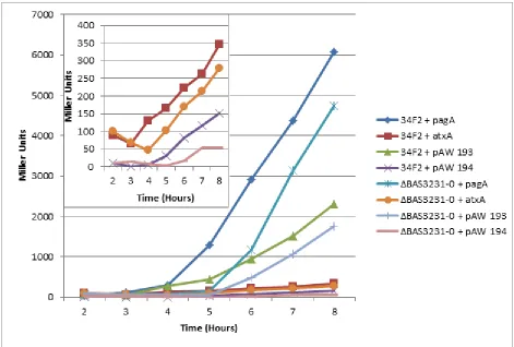
When measuring expression of all 4 virulence genes at 8 hours, the sigI mutant consistently
showed a significant decrease in pagA, cya and lef expression; however, there was no change in
atxA expression as seen in Figure 3.1.3. These results suggest that the loss of sigI is required for
the transcription of toxin genes pagA, cya and lef but does not affect expression of the master
regulator atxA which indicates that σI either directly or indirectly affects pagA, cya and lef
without affecting transcription of the master virulence regulator.
0 1000 2000 3000 4000 5000 6000
2 3 4 5 6 7 8
M ill e r Un its Time (Hours)
34F2 + pagA
34F2 + atxA
A094 + pagA
Figure 3.1.3 pagA, atxA, cya and lef expression levels in sigI mutant strain.
Virulence gene expression in sigI mutant strain at 8hrs. B-Galactosidase activity in pagA and atxA, cya and lef reporter strains grown in LB-Bic supplemented with 7.5 µg/mL kanamycin under inducing conditions at 37°C. ***, p<0.0001
3.2 Characterization of rsgI deletion strain
Although the interaction between σI and its anti-sigma factor, RsgI, in B. subtilis has
already been established, interaction between the two proteins in B. anthracis has yet been
characterized (13). The primary function of an anti-sigma factor is to bind to the sigma factor
and hinder its ability to form an RNAP holoenzyme. Because σI is a known transcription factor
activated by heat, low expression levels of virulence genes at inducing conditions were
unexpected. To determine whether the predicted anti-sigma factor gene, BAS3230, could be
affecting virulence gene expression, deletion strains of BAS3230 were constructed and
characterization of growth and virulence gene expression were done. If the primary role of the
(rsgI) gene would show no change in both growth and virulence gene expression compared to
the parental strain.
Under the same conditions used to perform the previous phenotype characterization, the
same growth curve was done to determine whether loss of BAS3230 alone would affect cell
viability over time as shown in Figure 3.2.1 These results suggest that loss of rsgI has no effect
on cell growth at both exponential and stationary phase, indicating that rsgI has no role in cell
[image:20.612.71.519.263.589.2]growth as was shown with the sigI mutant strain.
Figure 3.2.1. Cell growth of rsgI mutant strain.
Cell growth of parental, 34F2, and the rsgI mutant strain grown in LB at 37˚C.
To determine whether the predicted anti-sigma factor plays a role in virulence gene
expression, β-Galactosidase assays were done using all 4 reporters: pagA, atxA, cya, and lef. As
0.1 1 10
2 3 4 5 6 7 8
log
10
[O
D600
]
Time (Hours)
34F2
seen in Figure 3.2.2, deletion of BAS3230 had no effect on virulence gene expression of all 4
reporter genes. This was anticipated as anti-sigma factors do not function in gene regulation
directly. This may also confirm that σI alone is responsible for virulence gene expression and
[image:21.612.75.525.179.493.2]growth deficiency.
Figure 3.2.2 pagA, atxA, cya and lef expression levels in rsgI mutant strain.
Virulence gene expression in the rsgI mutant strain at 8hrs. β-Galactosidase activity in pagA and atxA, cya and lef reporter strains grown in LB-Bic supplemented with 7.5 µg/mL kanamycin under inducing conditions at 37˚C.
3.3 Characterization of sigI-rsgI double deletion strain
Our earlier analysis of the sigI and rsgI mutant strains shows that the growth deficiency and
lowered virulence gene expression were seen with the single deletion of the predicted sigI gene,
presence of the anti-sigma factor may be causing this change in phenotype, a double gene
deletion of both the sigma factor and its anti-sigma factor was constructed. The double gene
deletion strain was analyzed using the same conditions as the single gene deletion analysis for
both growth and virulence gene expression. The double gene deletion of BAS3231-0 had a
growth deficiency similar to the single gene deletion of BAS3231 under the same conditions as
seen in Figure 3.3.1. This suggests that with the absence of both the sigma factor and its
regulating anti-sigma factor, the phenotype is similar to the single deletion of the sigma factor
alone. This observation was anticipated due to the understandings that without the anti-sigma
factor present to regulate its cognate sigma factor; the sigma factor will consistently be active.
However, with the sigma factor deleted in lieu of the anti-sigma factor, both the transcription
Figure 3.3.1 Cell growth sigI-rsgI mutant strain.
Cell growth of parental, 34F2, and the sigI-rsgI mutant strain grown in LB at 37˚C.
To further characterize the double mutant strain and to verify that the predicted sigI gene
is responsible for the decrease in virulence gene expression of pagA, cya and lef, we performed a
β-Galactosidase assay using the same conditions as mentioned before for the single gene
deletions. When virulence gene expression was analyzed in the double mutant, we observed
decrease in virulence gene expression similar to the phenotype seen with the single sigI deletion
mutant as shown in Figure 3.3.2. The double gene deletion of both predicted sigI and rsgI had a
decrease in pagA, cya and lef. When the same assay was performed at late stationary phase, gene
expression of pagA, cya and lef were lower compared to the parental strain, 34F2.
0.1 1 10
2 3 4 5 6 7 8
lo
g10
[OD
600
]
Time (Hours)
34F2
Figure 3.3.2 pagA, atxA, cya and lef expression levels in sigI-rsgI mutant strain.
Virulence gene expression in the sigI-rsgI mutant strain. β-Galactosidase activity in pagA and atxA, cya and lef reporter strains grown in LB-Bic supplemented with 7.5 µg/mL kanamycin under inducing conditions at 37˚C.
This decrease in virulence gene expression seen with the double gene deletion of both
sigI and rsgI was also observed in the sigI mutant strain. The decrease in pagA, cya and lef in the
sigI-rsgI mutant strain were anticipated considering that the primary role of the anti-sigma factor
is to regulate its cognate sigma-factor and is expected to have no effect on virulence gene
expression. The data suggests that the role of the predicted anti-sigma factor does not play a role
3.4 Predicted σI is induced by heat shock
In B. subtilis, the putative sigma factor σI has been found to be heat-inducible (10).
Because of the high similarity in homology between B. subtilis and B. anthracis, a heat shock
survival assay was done with the sigI mutant strain in B. anthracis. The sigI deletion strain and
the parental strain were grown at an elevated temperature of 44˚C and survival was determined.
Survival rate was quantified by counting individual colonies after the plating of heat shock
cultures followed by growth at 37˚C. Similar to the results seen with the sigI mutant strains in B.
subtilis (10), sigI mutant strains in B. anthracis were unable to survive at elevated temperatures
as seen in Table 3.4.1. The deletion mutant strains had similar growth as the parental at 37˚C;
however, at an elevated temperature the mutant strains had a survival rate of almost 2%
compared to the survival rate of 36% seen with the parental strain. These results suggest that just
like σI in B. subtilis, the predicted σI in B. anthracis is a heat shock sigma factor that is necessary
for the cell’s ability to grow at elevated temperatures.
Table 2.4.1. Heat shock survival of sigI mutant strains at 44°C.
Survival of parental strain and sigI mutant strain after incubation in LB at 44˚C and 37˚C for 20 hours.
CFU after incubation at:
3.5 Transcription of sigI is induced by heat shock
sigI appears to be a part of an operon containing both the sigI predicted gene BAS3231
and the predicted anti-sigma factor, BAS3231. Upstream of the BAS3231 gene is a large
intergenic region of 778bp between the genes BAS3231 and BAS3232 as seen in Figure 3.5.1.
Although the neighboring gene BAS3232 lies in the same direction as the predicted sigI and rsgI
gene, this intergenic region is a fairly large space between two neighboring genes. This allowed
us to predict the presence of a possible sigI promoter within the intergenic space. Hypothesizing
the presence of a promoter, we created a plasmid to measure promoter activity of BAS3231
[image:26.612.77.505.343.423.2]during elevated temperatures.
Figure 3.5.1 Predicted BAS3231 (sigI) operon structure.
To measure promoter activity, a lacZ fusion plasmid was constructed using the entire
778bp intergenic region between the genes of BAS3231 and BAS3232. Promoter activity was
measured using β-Galactosidase assay of both the parental strain and the sigI mutant strains at
both 37˚C and at an elevated temperature of 44˚C as seen in Figure 3.5.2. At normal
physiological conditions, promoter activity of both the parental strain and the sigI mutant strain
falls to very low levels during late exponential phase, however, at approximately 5 hours of
growth, promoter activity for both strains increases to levels up to 6 times higher. Interestingly,
parental strain. When both strains were grown at an elevated temperature of 44˚C, promoter
activity increased at an earlier time around 4 hours of growth, but activity in the mutant strain
was significantly lower than the parental. These results not only provide evidence that σI is
involved in heat shock but also that because promoter activity of sigI still increases at lower
levels in the mutant strain during elevated temperature, regulation of the heat shock sigma factor
[image:27.612.70.512.262.569.2]could be induced by another promoter.
Figure 3.5.2 sigI promoter activity in sigI mutant strain.
BAS3231 promoter activity in the sigI mutant strain. β-Galactosidase activity in parental and sigI mutant strains grown in LB supplemented with 7.5 µg/mL kanamycin at 37˚C and 44˚C.
Promoter activity of the predicted sigI promoter was also measured in rsgI mutant strains
using the same β-Galactosidase assay mentioned. Activity was measured in both the parental
0 10 20 30 40 50 60 70 80 90
2 3 4 5 6 7 8
M ill er Uni ts Time (Hours)
34F2 + pAW399 @ 37⁰C
ΔBAS3231 + pAW399 @ 37⁰C
34F2 + pAW399 @ 44⁰C
strain and the rsgI mutant strains at 37˚C seen in Figure 3.5.3. As expected, when the cognate
regulator of the sigma factor is removed, transcription of the regulated gene will exceed normal
levels as seen in the parental strain. Activity of the promoter was measured in both LB and
inducing LB media containing bicarbonate to determine whether the promoter is inducible.
Miller units for BAS3231 promoter activity in the rsgI mutant strain remained the same
regardless of whether cultures were assayed in non-inducing LB or inducing LB. Interestingly,
promoter activity in the rsgI mutants were significantly higher than the parental strain. As
expected, an increase in promoter activity of sigI in the rsgI mutant strain was observed due to
the absence of its regulator with a 30-fold increase promoter activity. These results suggest that
without the presence of the anti-sigma factor to regulate levels of its cognate sigma factor,
promoter activity of the predicted sigI levels increase to extremely high levels even without
Figure 3.5.3 sigI promoter activity in rsgI mutant strain.
BAS3231 promoter activity in the rsgI mutant strain β-Galactosidase activity of predicted in parental and rsgI mutant strains grown in LB and LB-Bic supplemented with 7.5 µg/mL kanamycin under inducing conditions at 37˚C.
4 DISCUSSION
Heat shock sigma factors contribute to cell viability at elevated temperatures by initiating
the transcription of appropriate heat shock genes by activating the inactive RNAP. Due to the
homology between the two species B. subtilis and B. anthracis, the effect of deleting sigI in B.
anthracis was done to test whether the function of σI is conserved between the two similar
species. In B. subtilis, σI
is induced by heat shock and mutant strains of the predicted sigI gene
are unable to grow at elevated temperatures. Very similar to B. subtilis, the σI
in B. anthracis is
also induced by elevated temperatures. In addition to induction of σI by elevated temperatures,
0 500 1000 1500 2000 2500
1 3 5 7 9
sigI mutants in B. anthracis have decreased toxin gene expression and a deficiency in growth in
both non-inducing and inducing conditions supplemented with CO2/bicarbonate.
Mutant sigI strains in B. anthracis show a decrease in pagA, cya and lef toxin gene
expression; however, levels of atxA remain the same compared to the parental 34F2 strain.
Although σI is a heat shock sigma factor, virulence gene regulation mechanisms have not been
found for the heat shock sigma factor and its correlation to virulence gene expression. sigI in B.
subtilis has been linked to the regulation of cell wall synthesis pathways such as the WalRK two
component system in growing and stressed cells (24, 25, 26). In B. subtilis, the alternative sigma
factor, σI
, is a target of the WalRK two component system and is targeted by the WalR regulon
under heat stress (25). With the regulation of sigI and other genes associated with cell
morphogenesis and cell wall hydrolysis by the WalR regulon, transcription of sigI has been
found to be heat-inducible(25, 26). Whether the regulation of virulence gene expression by sigI
in B. anthracis is direct or indirect, toxin gene expression appears to require the presence of sigI.
This report suggests that σI is involved in heat shock. In the taxonomically similar species,
B. subtilis, sigI mutant strains showed temperature-sensitive growth at elevated temperatures
resulting in the conclusion that transcription of sigma factor σI in B. subtilis is induced by heat
shock (10). When the mutation strain of the putative sigI gene in B. anthracis was grown at
elevated temperatures, temperature-sensitive growth was also seen. This provides evidence that,
without the sigI gene, the strain is unable to withstand elevated temperatures. Mutant sigI strains
in B. anthracis also had a decreased level of sigI promoter activity at elevated temperature.
However, at 37˚C, promoter activity of the sigI mutant strain was higher than the parental at not
only early exponential phase but more importantly the late stationary phase. Our anticipated
activity as the parental strain or slightly lower due to the previous data suggesting that sigI is
induced by heat shock. However, the elevated promoter activity in the mutant strain at normal
conditions may suggest the presence of a secondary transcriptional regulator. This suggests that,
similar to the sigI operon seen in B. subtilis, a σA
promoter may be involved in the regulation of
sigI(26). If a second promoter is located in proximity to the sigI promoter, deletion of the sigI
gene may increase the activity of this promoter. We hypothesize that the lower promoter activity
seen in the parental strain at normal conditions is low due to the idea that the presence of both σI
and σA
could act as an interference with one another resulting in a lower level of promoter
activity. If this is true, in the mutant strain, sigI is deleted which results in a decrease in
interference between σI and σA
which may cause the increase in promoter activity of sigI.
We also showed that the transcription of sigI is tightly regulated by its cognate anti-sigma
factor, RsgI. When promoter activity was measured in strains missing the anti-sigma factor gene
rsgI, sigI promoter activity increased 100 fold. The function of the anti-sigma factor is known to
act as an antagonist by negatively regulating its sigma factor-dependent transcription by
inhibiting the sigma factor. We found that with the rsgI mutant strain, promoter activity of sigI
was increased significantly which was expected. Given the results of the no change in toxin gene
expression with the rsgI mutant strain, it was hypothesized that the anti-sigma factor had no
effect on virulence phenotypes other than to regulate the sigma factor. However, our findings of
increased promoter activity of sigI in the rsgI mutant strain provides evidence that regulation by
rsgI is incredibly controlled under elevated temperatures and without it , sigI would be strongly
produced.
Although our results suggest that σI
is a heat induced sigma factor, we are uncertain about
possible that pagA is indirectly regulated by σI and that additional mechanisms are used for the
regulation of toxin gene expression. While these observations provide evidentiary support of the
homology found between the two species of B. subtilis and B. anthracis, the understanding of
virulence regulation is still unknown. Additional work is required to identify the regulation
mechanism and relationship between σI and pagA, cya and lef to define their relationship and the
role of a heat shock sigma factor in virulence gene regulation.
5 FUTURE DIRECTIONS
5.1 Indirect or direct regulation of virulence gene expression by σI
Using methods such as in vitro transcription, microarray analysis and β-Galactosidase
reporter assays, we hope to verify our hypothesis that the putative σI of B. anthracis either
indirectly or directly regulates virulence gene expression. To determine whether there is a direct
correlation between pagA, cya and lef expression and the presence of σI, in vitro transcription
will be used. Using core RNA polymerase, recombinant σI
proteins and templates for the toxin
genes pagA, cya and lef, we will be able to determine whether σI
alone is able to initiate the
transcription of the toxin genes by regulating transcription from the virulence gene templates.
To determine whether there is an indirect regulation of toxin gene expression, microarray
analysis will be used to measure fold change of genes affected by the deletion of sigI under
normal physiological conditions and at elevated temperatures. Fold changes of genes expressed
in the parental strain compared to the mutant strains at different conditions will provide
information on the possible genes involved in a mechanistic pathway in virulence gene
virulence gene expression in the mutant strains but also shed light on other affected genes to
make inferences on the possible mechanistic pathway of virulence gene regulation.
5.2 BAS3228 and its relationship in cell wall synthesis
Cell wall hydrolases are critical for bacterial virulence in its involvement with cell wall
turnover and synthesis. In the taxonomically similar species B. subtilis, autolysin LytE is a
peptidoglycan hydrolase that has been found to be driven by a σA
promoter (11) and a two
component system, YycFG (WalRK) (11). It has been found that an actin homolog gene mreBH
belongs to the σI
regulon and is responsible for the localization of LytE in B. subtilis (11).
Because it has been found that the mreBH gene is a target of σI in B. subtilis, we have
investigated neighboring genes upstream and downstream of the sigI gene. Downstream of the
sigI gene, BAS3231, in B. anthracis lies the gene BAS3228 that has been annotated as a
hydroxyl transferase. Because hydroxyl transferases are involved in the synthesis of
polysaccharides that compose the peptidoglycan of gram-positive bacteria, we are hypothesizing
that the transferase gene, BAS3228 plays an active role in not only cell wall synthesis but also an
indirect or direct role in antibiotic resistance to β-lactam antibiotics such as Oxacillin. In our
preliminary study of B. anthracis sigI mutant strains, it has been observed that some sigI mutant
strains are susceptible to β-lactam class antibiotics, specifically Oxacillin. Given our hypothesis
and observations, we hope to identify the role of the predicted transferase gene BAS3228 in the
REFERENCES
1. Dixon, T. C., Meselson, M., Guillemin, J., & Hanna, P. C. (1999). Anthrax. N Engl J
Med, 341(11), 815-826.
2. Koehler, T. M. (2009). Bacillus anthracis physiology and genetics. Mol Aspects Med,
30(6), 386-396.
3. Little, S. F., & Ivins, B. E. (1999). Molecular pathogenesis of Bacillus anthracis
infection. Microbes Infect, 1(2), 131-139.
4. Bongiorni, C., Fukushima, T., Wilson, A. C., Chiang, C., Mansilla, M. C., Hoch, J. A., &
Perego, M. (2008). Dual promoters control expression of the Bacillus anthracis virulence
factor AtxA. J Bacteriol, 190(19), 6483-6492.
5. Fouet, A. (2010). AtxA, a Bacillus anthracis global virulence regulator. Res Microbiol,
161(9), 735-742.
6. Hammerstrom, T. G., Roh, J. H., Nikonowicz, E. P., & Koehler, T. M. (2011). Bacillus
anthracis virulence regulator AtxA: oligomeric state, function and CO(2) -signalling. Mol
Microbiol, 82(3), 634-647.
7. Bertin, M., Chateau, A., & Fouet, A. (2010). Full expression of Bacillus anthracis toxin
gene in the presence of bicarbonate requires a 2.7-kb-long atxA mRNA that contains a
terminator structure. Res Microbiol, 161(4), 249-259.
8. Candela, T., Mock, M., & Fouet, A. (2005). CapE, a 47-amino-acid peptide, is necessary
for Bacillus anthracis polyglutamate capsule synthesis. J Bacteriol, 187(22), 7765-7772.
9. Piggot, P. J., & Hilbert, D. W. (2004). Sporulation of Bacillus subtilis. Curr Opin
10. Zuber, U., Drzewiecki, K., & Hecker, M. (2001). Putative sigma factor SigI (YkoZ) of
Bacillus subtilis is induced by heat shock. J Bacteriol, 183(4), 1472-1475.
11. Margot, P., Wahlen, M., Gholamhoseinian, A., Piggot, P., & Karamata, D. (1998). The
lytE gene of Bacillus subtilis 168 encodes a cell wall hydrolase. J Bacteriol, 180(3),
749-752.
12. Tseng, C. L., & Shaw, G. C. (2008). Genetic evidence for the actin homolog gene mreBH
and the bacitracin resistance gene bcrC as targets of the alternative sigma factor SigI of
Bacillus subtilis. J Bacteriol, 190(5), 1561-1567.
13. Dufour, A., & Haldenwang, W. G. (1994). Interactions between a Bacillus subtilis
anti-sigma factor (RsbW) and its antagonist (RsbV). J Bacteriol, 176(7), 1813-1820.
14. Kazmierczak, M. J., Wiedmann, M., & Boor, K. J. (2005). Alternative sigma factors and
their roles in bacterial virulence. Microbiol Mol Biol Rev, 69(4), 527-543.
15. Schumann, W. (2003). The Bacillus subtilis heat shock stimulon. Cell Stress Chaperones,
8(3), 207-217.
16. Asai, K., Ootsuji, T., Obata, K., Matsumoto, T., Fujita, Y., & Sadaie, Y. (2007).
Regulatory role of RsgI in sigI expression in Bacillus subtilis. Microbiology, 153(Pt 1),
92-101.
17. Koehler, T. M., Dai, Z., & Kaufman-Yarbray, M. (1994). Regulation of the Bacillus
anthracis protective antigen gene: CO2 and a trans-acting element activate transcription
from one of two promoters. J Bacteriol, 176(3), 586-595.
18. Sambrook J., Russell D. W., (2001) Molecular cloning: a laboratory manual, 3rd edition.
19. Han, H., Wilson, A. C. (2013). The two CcdA proteins of Bacillus anthracis differentially
affect virulence gene expression. J Bacteriol, 195(23), 5242-5249.
20. Cybulski, R. J., Jr., Sanz, P., Alem, F., Stibitz, S., Bull, R. L., & O'Brien, A. D. (2009).
Four superoxide dismutases contribute to Bacillus anthracis virulence and provide spores
with redundant protection from oxidative stress. Infect Immun, 77(1), 274-285.
21. Poyart, C., & Trieu-Cuot, P. (1997). A broad-host-range mobilizable shuttle vector for
the construction of transcriptional fusions to beta-galactosidase in gram-positive bacteria.
FEMS Microbiol Lett, 156(2), 193-198.
22. Miller J. H., (1972) Experiments in molecular genetics, p 352-355. Cold Spring Harbor
Laboratory, Cold Spring Harbor, NY
23. Wilson, A. C., Hoch, J. A., & Perego, M. (2008). Virulence gene expression is
independent of ResDE-regulated respiration control in Bacillus anthracis. J Bacteriol,
190(15), 5522-5525.
24. Hadjifrangiskou, M., Chen, Y., & Koehler, T. M. (2007). The alternative sigma factor
sigmaH is required for toxin gene expression by Bacillus anthracis. J Bacteriol, 189(5),
1874-1883.
25. Huang, W. Z., Wang, J. J., Chen, H. J., Chen, J. T., & Shaw, G. C. (2013). The
heat-inducible essential response regulator WalR positively regulates transcription of sigI,
mreBH and lytE in Bacillus subtilis under heat stress. Res Microbiol, 164(10), 998-1008.
26. Salzberg, L. I., Powell, L., Hokamp, K., Botella, E., Noone, D., & Devine, K. M. (2013).
The WalRK (YycFG) and sigma(I) RsgI regulators cooperate to control CwlO and LytE
expression in exponentially growing and stressed Bacillus subtilis cells. Mol Microbiol,