2016 Joint International Conference on Artificial Intelligence and Computer Engineering (AICE 2016) and International Conference on Network and Communication Security (NCS 2016)
ISBN: 978-1-60595-362-5
Bleeding Detection in Wireless Capsule Endoscope
Based on Color Feature Vector
Shang-Bo ZHOU
1,2, Shu-Fang CHEN
1,2, Awudu Karim
1,2,
Jian-Ying BAI
3, Chao-Qiang FAN
31Key Laboratory of Dependable Service Computing in Cyber Physical Society, Ministry of Education, Chongqing University, Chongqing 400030, China
2College of Computer Science, Chongqing University, Chongqing 400030, China
3Department of Gastroenterology, Xinqiao Hospital, Chongqing, 400037, China
Keywords: Bleeding Detection, Wireless Capsule Endoscopy (WCE), CMYK Color Space, CIELAB Color Space, Supported Vector Machine (SVM).
Abstract. Wireless capsule endoscopy (WCE) is an electronic revolutionary technique with no pain, easy to use and provides the capability of inspecting the whole small intestine which traditional ways do not. However, WCE produces so many pictures of a patient that it burdens on the work of doctors detecting diseases. To solve this problem, a highly promising automatic method based on the color feature of CIELAB and CMYK color space is proposed. The first step is image preprocessing, which extracts the region of interest from the image. Then, according to the characteristics of the CIELAB color space, color balance based on gray world assumption is executed and the images’ local contrast is improved. The second step is to generate a binary vector. The third step, we use a support vector machine (SVM) as a classifier to identify images and use RBF kernel function to check the feature vector’s performance. Experimental work shows that the proposed approach improves precision.
Introduction
Wireless Capsule Endoscopy (WCE) is an effective method to examine gastrointestinal (GI) tract with a non-invasive procedure, which is less painful and comfortable than the traditional endoscopy. The earliest developed capsule endoscopy is given by an imaging company in 2000, which released the first capsule endoscopy M2A and conducted clinical experiments in 2001[1]. WCE is a capsule shaped device with a dimension of 26mm in length by 11mm in diameter, consisting of an optical dome, an imaging sensor, a battery, a transmitter and lights [2]. The WCE device is swallowed by a patient who is asked not to eat or drink water before the procedure. The speed of image capture is 2 frames per second. In the end, the doctor needs to examine a total of about 60,000 images or even higher to make diagnosis of diseases [3]. Therefore, it is necessary to have computer aided systems to help doctors efficiently detect bleeding images [4]. This is one of the reasons why efforts have been made to develop automated computer-aided detection and diagnostic systems for bleeding screening in WCE images [5].
method using histogram intersection to distinguish a bleeding image in WCE images. The Authors in [5], presented a two-stage map extraction method to enhance bleeding regions. However, the intermediate steps should be simplified further and the dimension is relatively large. Currently in [11], the authors proposed a binary vector technique which was used to get over the drawbacks of conventional color histogram. However, the images are not preprocessed.
An effective algorithm to detect bleeding in WCE images with increased accuracy is presented in this paper. Firstly, we eliminate irrelevant information of capsule endoscopic images. Secondly, we extract second channel (A) of CIELAB color space and second channel (M) of CMYK color space. Contrast can be enhanced by adjusting A and M gray value range. Then we set the threshold to get a binary vector. Finally, support vector machine is applied as the classifier.
Image Preprocessing
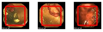
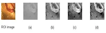
[image:2.612.227.394.316.450.2]The typical bleeding and non-bleeding WCE images are shown in Fig.1 These images in Fig.1 clearly show that the bleeding zones are unlike the non-bleeding zones. We find the most effective region in WCE image as the region of interest (ROI) without the interfering information. The size of ROI is 400×400 from the WCE image of the size 576×576 as shown in Fig.2. It can be seen that ROI images make the feature extraction more accurate and more convenient.
Figure 1. Bleeding and non-bleeding WCE images.
Figure 2. ROI from the WCE images.
The wireless capsule endoscopy is powered by an internal battery which runs out gradually with the movement of the capsule inside the digestive tract and the illumination is gradually decreased. The light intensity slowly decreases as the battery runs out. Therefore, it is essential to execute color balance based on gray world assumption to improve the images’ local contrast. In this paper, we use the LAB color space and set a normal WCE image as a reference to balance the color of the other pictures, as in Eq. (1).
) , ( ) ( ) ,
( I x y
I avg
TH y x
I C
C C
C . (1)
Where avg I c represents the mean of C channel, THcis the threshold of the C channel. As shown in
[image:2.612.221.399.493.549.2]image pixel in the Lab color space channel. In the a and b channels, we can observe that the values are basically concentrated in one place, thresholds (THaandTHb) of a and b channels.
Figure 3. Color distribution for standard image.
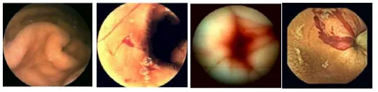
[image:3.612.174.450.289.357.2]After taking the threshold into account to balance the color of WCE images, the tones of the image tend to resemble the original image. As shown in Fig.4, the color is not balanced. After color balance, we can see the results as shown Fig.5.
Figure 4. Wireless capsule endoscopy images from different bands.
Figure 5. Color balancing based on linear transformation.
Feature Extraction
Color feature is important for bleeding detection. Choosing an appropriate color space is a major step for color image analysis. In this paper, the proposed method uses CIELAB and CMYK color space for color feature extraction, because CIELAB and CMYK represent the color with an intuitive value. This property enables us to extract the color feature from A channel of the CIELAB color space and M channel from CMYK color space. In particular, pixels of the bleeding region are significantly greater than the pixels of the normal region in the M and A channels. So, we need to use color histogram to concretely analyze the difference between the bleeding region and the normal region.
Processing Gray Information
[image:3.612.177.447.388.454.2]Figure 6. The gray image.
The Binary Feature Vector
[image:4.612.200.402.265.424.2]Because WCE images only contain the contents of GI tracts, they do not take up the entire spectrum of the color space. Based on extensive experimentations, we find that only red color is useful. As illustrated in Fig. 7, we can find out that the range of A channel is mostly considered between 145 and 185. As shown in Fig. 8, it is obvious that the range of M channel is between 165 and 240. This reduces A and M spaces by 14% to 24% of the original A and M spaces.
[image:4.612.201.401.449.593.2]Figure 7. A channel of WCE images.
Figure 8. M channel of WCE images.
1 ( , )
( , ) .
0 ( , )
AM x y threshold
features x y
AM x y threshold
(2)
In order to facilitate the computation, feature vector (AM) is a normalized histogram in our clipped color space. x and y denote the xth in range_a and yth in range_m respectively. Thresholds are used to eliminate the interference of a small area or a small number of discrete pixels, preventing misjudgment. When threshold is 0.4%, the result is the best through the experiments.
Results and Discussion
The performance of the proposed method is evaluated using the testing set of 1512 WCE images comprising 1122 bleeding images and 390 normal images. The training set is composed of 50 bleeding images and 50 non-bleeding images.In this paper, the classifier used is a support vector machine (SVM). According to this characteristic of SVM, the classification of bleeding images from WCE images is much easier and more convenient. So, we use libSVM [12] to complete the bleeding detection tasks. An RBF kernel function is used for SVM in [12].
Accuracy, sensitivity, and specificity are applied in this experiment in order to assess the performance of the proposed method. These three indicators are calculated as in Eq. (3).
TP Sensitivity TP FN TN Specificity TN FP TP TN Accuracy
TP TN FP FN
. (3)
[image:5.612.145.468.499.579.2]Where true positive (TP) cases are the number of bleeding images that are correctly labeled as the bleeding images. False positive (FP) are the ones incorrectly labeled as the bleeding images. False negative (FN) represents the number of bleeding images which are recognized as non-bleeding images. True negative (TN) represents the number of non-bleeding images that we recognize correctly.
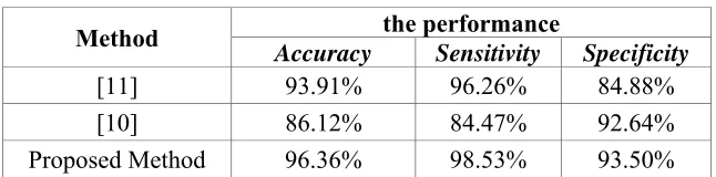
Table 1. Experiment results.
Method the performance
Accuracy Sensitivity Specificity
[11] 93.91% 96.26% 84.88%
[10] 86.12% 84.47% 92.64%
Proposed Method 96.36% 98.53% 93.50%
Figure 9. Experimental results compare.
Figure 9 shows the results of ten tests. The training set and testing set are selected randomly. Our algorithm presents more stable characteristics in sensitivity and accuracy. We have proposed an effective algorithm in bleeding detection of WCE images in the GI tract. In the future, we will find more appropriate methods for the bleeding detection of WCE images to decrease the mistake rate.
Acknowledgment
This work was supported by a grant from Chongqing People’s Livelihood Science and technology innovation projects (Grant No. cstc2015shmszx120010).
References
[1] G. Iddan, G. Meron, A. Glukhovsky, and P. Swain, Wireless capsule endoscopy, Nature, vol. 405, p. 417, 2000.
[2] Yuan Y., Li B., Meng Q. Bleeding Frame and Region Detection in the Wireless Capsule Endoscopy Video, IEEE Journal of Biomedical and Health Informatics, 2015, pp. 1-1.
[3] M. Liedlgruber, A. Uhl, Endoscopic Image Processing - An Overview, Proceedings of the 6th International Symposium on Image and Signal Processing and Analysis, 2009.
[4] G. Pan, G. Yan, X. Qiu, and J. Cui, Bleeding detection in wireless capsule endoscopy based on probabilistic neural network, Journal of medical systems, vol. 35, no. 6, pp. 1477-1484, 2011. Yuan Y, Meng M Q H, Automatic bleeding frame detection in the wireless capsule endoscopy images, Robotics and Automation (ICRA), 2015 IEEE International Conference on. IEEE, 2015, pp. 1310-1315.
[5] J.M. Buscaglia et al. Performance characteristics of the suspected blood indicator feature in capsule endoscopy according to indication for study, Clinical gastroenterology and hepatology: the Official Clinical Practice Journal of the American Gastroenterological Association, vol. 6, no. 3, pp. 298-301, Mar. 2008.
[6] S. Liangpunsakul, L. Mays, and D.K. Rex, Performance of given suspected blood indicator, Americal Journal of Gastroenterology, vol. 98, no. 12, pp. 2676-8, Jan. 2004.
[7] Ghosh T., Fattah S.A., Wahid K. Automatic bleeding detection in wireless capsule endoscopy based on RGB pixel intensity ratio, Electrical Engineering and Information & Communication Technology (ICEEICT), 2014 International Conference on. IEEE, 2014, pp. 1-4.
[9] G. Lv, G. Yan, and Z. Wang, Bleeding detection in wireless capsule endoscopy images based on color invariants and spatial pyramids using support vector machines, in Engineering in Medicine and Biology Society, EMBC, 2011 Annual International Conference of the IEEE. IEEE, 2011, pp. 6643-6646.
[10]Mathew M., Gopi V.P. Transform based bleeding detection technique for endoscopic images, Electronics and Communication Systems (ICECS), 2015 2nd International Conference on. IEEE, 2015, pp. 1730-1734.
[11]Zhou S., Song X., Siddique M.A., et al. Bleeding detection in wireless capsule endoscopy images based on binary feature vector, Intelligent Control and Information Processing (ICICIP), 2014 Fifth International Conference on. IEEE, 2014, pp. 29-33.