Femoral stem wear in cemented total hip replacement
H-Y Zhang1*, L Blunt1, X-Q Jiang1, L Brown1, S Barrans1,andY Zhao21Centre for Precision Technologies, School of Computing and Engineering, University of Huddersfield, Huddersfield,
UK
2School of Management, Tianjin University, Tianjin, People’s Republic of China
The manuscript was received on 24 July 2007 and was accepted after revision for publication on 22 January 2008.
DOI: 10.1243/09544119JEIM346
Abstract: The great success of cemented total hip replacement to treat patients with end-stage osteoarthritis and osteonecrosis has been well documented. However, its long-term survivorship has been compromised by progressive development of aseptic loosening, and few hip prostheses could survive beyond 25 years. Aseptic loosening is mainly attributed to bone resorption which is activated by an in-vivo macrophage response to particulate debris generated by wear of the hip prosthesis. Theoretically, wear can occur not only at the articulating head–cup interface but also at other load-bearing surfaces, such as the stem– cement interface. Recently, great progress has been made in reducing wear at the head–cup interface through the introduction of new materials and improved manufacture; consequently femoral stem wear is considered to be playing an increasingly significant role in the overall wear of cemented total hip replacement. In this review article, the clinical incidences of femoral stem wear are comprehensively introduced, and its significance is highlighted as a source of generation of wear debris and corrosion products. Additionally, the relationship between femoral stem surface finish and femoral stem wear is discussed and the primary attempts to reproduce femoral stem wear throughin-vitrowear testing are summarized. Furthermore, the initiation and propagation processes of femoral stem wear are also proposed and a better understanding of the issue is considered to be essential to reduce femoral stem wear and to improve the functionality of cemented total hip replacement.
Keywords: wear, femoral stem, bone cement, simulation, total hip replacement
1 INTRODUCTION
Cemented total hip replacement (THR) has been performed worldwide to improve the quality of life of patients suffering from debilitating hip disorders, such as end-stage osteoarthritis and osteonecrosis. It is considered to have been a momentous stride forward in orthopaedics since its introduction as a pioneering method to stabilize the femoral stem by Sir John Charnley in the 1960s [1]. The advent of poly(methyl methacrylate) bone cement undoubt-edly promoted the great success of cemented THR, and cemented THR still predominates in terms of the number of operations carried out in the UK in
comparison with uncemented THR [2]. However, controversies with regard to bone cement have prevailed across previously published literature, not only because of its inherent shortcomings such as the generation of exothermic heat released by a redox reaction during polymerization and the for-mation of high residual stresses which could potentially result in initial damage to the bone cement [3], but also owing to a vulnerable interface that has been introduced as a consequence of its application, namely the stem–cement interface. It has been demonstrated in retrieval studies that failure of cemented THR has been initiated at this interface, associated with bone cement fractures and significant areas of osteolysis [4,5]. In fact, the stem– cement interface, which functions as a transitional zone by mechanical bonding between two materials with significantly different mechanical properties,
has consistently been cited as a weak link in cemented THR. Recently, there is an increasing body of evidence which suggests that debonding at the stem–cement interface not only is common but in fact may be inevitable, regardless of femoral stem geometry and surface finish [6–8]. These findings as a consequence imply the occurrence of a low-amplitude oscillatory micromotion at this interface due to the unmatched strain under cyclically physiological loading. Indeed, roentgen stereophoto-grammetric analyses have shown evidence of fem-oral stem movement within the cement mantle in all stem designs that have been studied [9]. Consequently, this movement would lead to poten-tial wear on both components. This paper concen-trates on the wear generated at the stem–cement interface in cemented THR. However, it should be mentioned that femoral stem wear is not exclusively involved in cemented THR; it has also been reported on the stem surface in uncemented THR [10].
2 THE SIGNIFICANCE OF FEMORAL STEM WEAR
Historically, femoral stem wear has received rela-tively little attention in spite of its potential sig-nificance as a reason for the failure of cemented THR. There are several reasons for this. First, the femoral stem has previously been believed to anchor well in the cement mantle, especially for those ‘shape-closed’ stem designs which take advantage of a collar, a flange, a rectangular cross-section, and a matt surface finish to achieve stabilization [11]. Second, femoral stem wear is sometimes impossible to recognize with the naked eye, and it is difficult to detect even with the assistance of conventional light microscopy under certain cases. Third, another articulating interface in cemented THR, namely the head–cup interface which is designed to give the patients more flexibility, has attracted much of the researchers’ attention because this interface is generally considered to experience severe wear in vivo. Recently, with the introduction of cross-linked ultra-high molecular weight polyethylene (UHMWPE) and hard-on-hard bearing systems, wear at the head–cup interface has been greatly reduced [12–14]. Therefore, more research interest has been transferred to the stem–cement interface. In addition, the majority of modern femoral stems are of modular design, usually the head–neck Morse taper junction. The modular design has met with great success, providing the orthopaedic sur-geons with more flexibility in terms of adaptation to the patient’s anatomy and allowing for the best
combination of materials, i.e. the femoral stem obtains excellent mechanical properties of stainless steel and the femoral head benefits from the wear resistance of Co–Cr alloy. However, modular design is often criticized for bringing about another inter-face that is easily damaged by wear, namely the head–neck interface. Indeed, there are reports of wear in this site from both in-vivo retrieval studies and in-vitro wear tests [15, 16]. This further indi-cates that femoral stem wear is now playing an increasingly significant role in the overall wear of cemented THR, although it is for the most part caused by the relative micromotion at the stem– cement interface.
3 THE CLINICAL INCIDENCES OF FEMORAL STEM WEAR
against the bone cement. Likewise in 1992, Bulyet al. [21] reviewed 71 titanium alloy cemented total hip arthroplasties, in which 71 per cent of the femoral stems with aseptic loosening were considered to be abraded by the bone cement. Again in a study carried out by Salvati et al. [22] in 1993, it was indicated that, when the titanium alloy femoral stem became loosenedin vivo, it would abrade against the fragmented bone cement and generate metallic as well as cement debris. In 1997, Shardlow et al. [23] reported ‘fretting wear’ of Charnley low-friction arthroplasties, and they further identified that this wear affected the anterolateral and posteromedial edges of the femoral stems. All these early studies have strengthened the significance of femoral stem wear and have indicated the necessity for further investigation. However, various terms have been used to describe femoral stem wear in these studies, such as ‘polishing’, ‘rub marks’, ‘burnishing’, ‘abra-sion’, and ‘fretting’, which has made comparison between the wear damage a little confusing.
4 THE RELATIONSHIP BETWEEN FEMORAL
STEM SURFACE FINISH AND FEMORAL STEM WEAR
From the 1990s, most studies concerning femoral stem wear were focused on matt femoral stems. There was a dearth of research in this area as to polished femoral stems, and only a few studies have been performed to investigate the relationship between femoral stem surface finish and femoral stem wear. This issue came to the fore because most femoral stem designs experienced much evolution as they were continually developed, and change in stem surface finish proved to be one of the most contentious aspects [24]. It has generally been accepted that matt stems can provide a greater bond strength because of the enhanced bone cement integration, whereas polished stems form a much lower bond strength and potentially suffer from a higher probability of local micromotion. The first attempt to investigate this area was considered to be the study which was carried out by Howell et al. [25] in 1999. They examined 150 explanted femoral stems and characterized femoral stem wear using contact profilometry, interference profilome-try, and SEM associated with energy-dispersive X-ray analysis (EDXA), from which it was considered that a fretting mechanism was responsible for the damage and the wear was influenced by the femoral stem alloy and surface finish. However, a deep insight into the influence of femoral stem surface finish was not
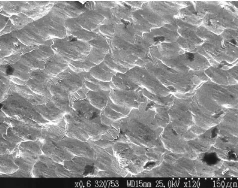
gained at that time. Later, numerous reports were published on failed prostheses that exhibited various surface finishes [26–28], but none of these has correlated the femoral stem surface finish with femoral stem wear. It was not until an intensive retrieval study in 2004 by Howell et al. [29] on the surface morphology of 172 femoral stems (about 23 different stem designs) was performed that a better understanding of this issue was obtained. In the investigation carried out by Howellet al. in 2004, it was demonstrated that 92 per cent of the femoral stems showed evidence of wear, and the wear mechanism behind polished and matt femoral stems appeared fundamentally different in spite of similar wear locations. For polished stems, the morphology of wear produced by cyclical movement of the stem within the cement mantle indicated a fretting mechanism which generated typical fretting pits below the level of the original stem surface (Fig. 1). By contrast, matt stems wore against bone cement by an abrasive mechanism which sacrificed parts of the inner surface of the cement mantle and conse-quently resulted in destabilization of the femoral stem and generation of both metallic and cement debris (Fig. 2). Additionally, 74 femoral stems, which were found to be absolutely fixed in the cement mantlein vivo at the time of revision, also showed evidence of wear on the stem surface. This suggested that wear may occur in the absence of obvious loosening of the femoral stem within the cement mantle, even for stable femoral stems. This study indicated that the femoral stem wear mechanism was dependent primarily on the femoral stem sur-face finish and this concept has nowadays been
[image:3.595.317.555.534.722.2]generally accepted. However, the exact surface level that produced fretting wear or abrasive wear was not clear. Subsequently, clinical data on femoral stem wear continued to be published. In 2005, Joneset al. [30] reported abrasive wear on the proximal matt surface of the Bridge hip stem, and they also detected the presence of titanium debris embedded within the cement mantle. Another significant contribution to this area was ascribed to Brown [31], who had developed a semi-quantitative method to calculate volume loss during the wear of matt femoral stems. By comparing this result with that calculated from a mathematical truncation model, it was confirmed that the wear mechanism for matt femoral stems was abrasive wear rather than classic fretting wear, at least until a fully polished surface was attained, by which point it was considered that classic fretting wear would predominate (Fig. 3).
5 THE GENERATION OF WEAR DEBRIS AND
WEAR CORROSION
Wear of the femoral component would inevitably cause generation and liberation of both metallic and cement particulate debris. This is particularly crucial in vivo, not only because this debris could lead to third-body wear and jeopardize stabilization of the femoral stem, but also because some wear debris within a certain size range would be transported along cement mantle deficiencies to bone tissues, where a significant macrophage response can arise, giving rise to destruction of surrounding bone stock [32,33]. This consequently results in aseptic loosen-ing of the femoral component, which nowadays is considered to be the primary reason for revision of cemented THR [34]. The presence and significance of wear debris in cemented THR have been well Fig. 2 Grazing-incidence SEM images showing increasing degrees of polishing wear on
matt-surfaced stems: (a) an unworn area; (b) an area of slight polishing wear; (c) an area of marked polishing wear
[image:4.595.39.544.86.215.2] [image:4.595.122.461.522.715.2]documented [35–38]. Originally the head–cup inter-face was considered to be more prone to wear and was deemed to be the main source of wear debris. Biological reactions to UHMWPE debris originating from the acetabular cup have been investigated by Ingham and Fisher [39] in detail and they believe that it is the primary cause of late aseptic loosening. Meanwhile other researchers [40, 41] also reported the adverse effect of bone cement debris. These studies indicated that bone cement debris is playing an increasingly significant role resulting in aseptic loosening of the femoral component and partially promoted a shift of research interest to the area of stem–cement interface [42,43].
Another intractable issue which is always asso-ciated with femoral stem wear in cemented THR is corrosion. This problem remains significant because implantable metals owe their corrosion resistance to a thin passive oxide film, but femoral stem wear against hard bone cement (in particular where radio-opaque barium sulphate or zirconia has been added) would disrupt this protective passive film, and thus the subsurface metal is exposed to further wear and corrosion [44]. Clinical data on the corrosion of explanted femoral stems are available in previously published literature, most of which has concentrated on the head–neck modular interface [45–48]. It is indicated from these studies that corrosion is especially severe for those prostheses with a combination of a Co–Cr alloy femoral head and a titanium alloy femoral stem, and it is mainly attributed to crevice corrosion which has promoted progressive generation of corrosion products following stem implantation. However, studies on corrosion at the stem–cement interface have also been widely documented. In 1996, Musolino et al. [49] reported corrosion under the collar of 18 stainless steel T-28 femoral stems, which illustrated a multi-layered and multi-textured nature under SEM. In 1998, Walczak et al. [50] studied the morphology and composition of the corrosion product presented on 11 Charnley–Mu¨ ller femoral stems made of stainless steel, using SEM associated with EDXA. They found that some areas on the stem surface had discoloured into a black compound, and a chromium-rich plaque was de-tected in these sites. Recently in 2004, Thomaset al. [51] reviewed severe corrosion of 12 cemented titanium Furlong straight stems which was typical of crevice corrosion. All these studies have indicated that no efforts should be spared to reduce femoral stem wear in order to protect the substrate of the femoral stem from being corroded.
6 THEIN-VITROSIMULATION OF FEMORAL STEM WEAR
testing of hip prosthesis, BS ISO 7206-4. By in-corporating implantation of the femoral stem in a sawbone and introduction of saline solution to mimic environmental conditionsin vivo, it has been possible to replicate fretting wear seen on explanted stems, although the repeatability needs to be further confirmed (Fig. 4). These previous studies provided a basic as well as considerable knowledge for those researchers who would pursue in-vitro wear testing of cemented femoral stems, from which hopefully the influence of femoral stem geometry, surface finish, and bone cement brand on the generation of femoral stem wear would be obtained.
7 THE INITIATION OF FEMORAL STEM WEAR
Although clinical reports and in-vitro wear tests on the femoral stem have been well documented in previously published literature and a better under-standing of the influence of the femoral stem surface finish on femoral stem wear has been obtained, the issues of the initiation and propagation processes of femoral stem wear still remain. As has been proven by orthopaedic surgeons, femoral stem wear also occurs on those stems which are considered to be clinically stable at surgery. This indicates that such wear is initiated by cyclical micromotion at the stem–cement interface at an early stage following stem implantation. It has been previously postulated by two of the present authors and a co-worker [59] that the shrinkage peaks on the bone cement surface may act as a potential culprit to promote femoral stem wear. These peaks, about 50mm in width and 1– 2.5mm in height, are formed owing to shrinkage of the bone cement during polymerization (Fig. 5). The stress distribution along the stem–cement interface under physiological loading would be concentrated at these areas. As a result, it is considered that
femoral stem wear is initiated at those sites which are in close contact with the shrinkage peaks on the cement surface. Later, however, it was indicated from a study on surface topography of the femoral stem and the bone cement that the influence of these shrinkage peaks is somewhat overshadowed in comparison with the micropores located in the cement surface [60]. In the study, which was also conducted by three of the present authors, it was demonstrated that the edges of the micropores matched well with the boundaries of the worn areas on the femoral stem surface, and also there was a direct relationship between the micropores and initial damage on the stem surface (Figs 6 and 7). Furthermore, evidence for the propagation of fem-oral stem wear from the sites contacting the micropores was also available (Fig. 8). Therefore, it was considered that the micropores in the cement surface, in combination with the regime of physio-logical loading during patient normal activities, may fundamentally determine the locations and severi-ties of femoral stem wear due to material micromo-tion at the edges of the micropores. This is the first time that micropores have been implicated in the initiation and propagation of femoral stem wear. However, it should be noted that stem wear may not be fully elucidated by these two factors and other, as yet unknown, factors may also play a role. Accord-ingly, further investigations are imperative to gain a fuller insight into this area.
8 CONCLUSIONS AND FUTURE STUDIES
In summary, femoral stem wear has become in-creasingly important in the overall wear of cemented THR, releasing both metallic and cement debris and resulting in the generation of a cascade of corrosion products. Both retrieval studies and in-vitro wear
[image:6.595.92.491.574.721.2]Fig. 5 Shrinkage peaks formed on the bone cement surface during polymerization. The two-dimensional profile of the peaks indicates that they are about 50mm in width and 1mm in height
Fig. 6 Optical micrograph showing the ‘undamaged islands’ surrounded by fretting wear on the femoral stem surface and the corresponding micropores in the bone cement surface. The boundaries of the ‘undamaged islands’ match well with the micropores
[image:7.595.98.516.87.267.2] [image:7.595.123.489.351.490.2] [image:7.595.115.490.573.712.2]tests with regard to femoral stem wear have been well documented by orthopaedic surgeons and researchers, from which much insight has been gained. Although a direct relationship between the femoral stem surface finish and femoral stem wear has been ascertained by previous studies, there are a number of aspects in this area that have not been fully investigated. One is to reproduce femoral stem wear successfully and consistently through in-vitro wear testing and further knowledge of the influence of the femoral stem geometry, surface finish, and bone cement brand on the generation of femoral stem wear. Other aspects include the need to obtain a better understanding of the initiation and propa-gation processes of femoral stem wear. More atten-tion and effort into elucidating these aspects are a prerequisite to try to reduce or eliminate femoral stem wear, on the basis of which the long-term reliability and survivorship of cemented THR would hopefully be improved. However, the initial studies that have been performed by the present authors indicated that the micropores in the cement surface would potentially contribute to initiation and pro-pagation of femoral stem wear. Consequently, the application of ‘modern cementing techniques’, typically vacuum mixing, centrifugation, and pres-surized introduction of bone cement into the fem-oral canal which primarily aim to reduce porosity in
the cement mantle as well as at the interfaces, is considered to be clinically beneficial and an effective method to improve the long-term lifetime of cemented THR.
REFERENCES
1 Charnley, J.Anchorage of femoral head prosthesis to shaft of femur. J. Bone Jt Surg. Br., 1960, 42, 28–30.
2 Wirz, D., Daniels, A. U., Go¨pfert, B., and Morscher, E. W. Acrylic bone cement in the new millennium – clinical development and current status: Europe. Orthop. Clin. N. Am., 2005, 36, 63–73.
3 Roques, A., Browne, M., Taylor, A., New, A., and Baker, D. Quantitative measurement of the stres-ses induced during polymerisation of bone cement. Biomaterials, 2004,25, 4415–4424.
4 Jasty, M., Maloney, W. J., Bragdon, C. R., O’Con-nor, D. O., Haire, T., and Harris, W. H. The initiation of failure in cemented femoral compo-nents of hip arthroplasties. J. Bone Jt Surg. Br., 1991,73, 551–558.
5 Maloney, W. J., Schmalzried, T.,andHarris, W. H. Analysis of long term cemented total hip arthro-plasty retrievals. Clin. Orthop. Related Res., 2002, 405, 70–78.
6 Verdonschot, N. and Huiskes, R. The effects of cement–stem debonding in THA on the long term failure probability of cement. J. Biomechanics, 1997,30, 795–802.
7 Mohler, C. G., Callaghan, J. J., Collis, D. K., and Johnston, R. C. Early loosening of the femoral component at the cement–prosthesis interface after total hip replacement. J. Bone Jt Surg. Br., 1996,78, 280–285.
8 Schmalzried, T. P., Zahiri, C. A., and Woolson, S. T. The significance of stem–cement loosening of grit-blasted femoral components. Orthopedics, 2000,23, 1157–1164.
9 Karrholm, J., Nivbrant, B., Thanner, J., Ander-berg, C., Bo¨rlin, N., Herberts, P.,andMalchau, H. Radiostereometric evaluation of hip implant design and surface finish: micromotion of cemented femoral stems. In the 67th Annual Meeting of the American Academy of Orthopaedic Surgeons, Orlando, Florida, USA, 15–19 March 2000.
10 Maccauro, G., Piconi, C., Pilloni, L., Proietti, L., De Santis, V.,andDe Santis, E.Surface analysis of a femoral stem after failed total hip replacement. Int. Orthop., 2000,24, 231–233.
11 Huiskes, R., Verdonschot, N., and Nivbrant, B. Migration, stem shape, and surface finish in cemented total hip arthroplasty. Clin. Orthop. Related Res., 1998,355, 103–112.
12 Wroblewski, B. M., Siney, P. D., Dowson, D.,and Collins, S. N.Prospective clinical and joint simu-lator studies of a new total hip arthroplasty using Fig. 8 An interferometric micrograph showing the
[image:8.595.41.279.82.333.2]alumina ceramic heads and cross-linked polyethy-lene.J. Bone Jt Surg. Am., 1995,77, 1315–1322. 13 Hatton, A., Nevelos, J. E., Nevelos, A. A., Banks, R.
E., Fisher, J., and Ingham, E. Alumina–alumina artificial hip joints: a histological analysis and characterisation of wear debris by laser capture microdissection of tissues retrieved at revision. Biomaterials, 2002,23, 3429–3440.
14 Fisher, J., Hu, X. Q., Stewart, T. D., Williams, S., Tipper, J. L., Ingham, E., Stone, M. H., Davies, C., Hatto, P., Bolton, J., Riley, M., Hardaker, C., Isaac, G. H., and Berry, G. Wear of surface engineered metal-on-metal hip prosthesis.J. Mater. Sci., Mater. Medicine, 2004,15, 225–235.
15 Chaplin, R. P. S., Lee, A. J. C.,andHooper, R. M. Assessment of wear on the cones of modular stainless steel Exeter hip stems. J. Mater. Sci., Mater. Medicine, 2004,15, 977–990.
16 Viceconti, M., Baleani, M., Squarzoni, S., and Toni, A. Fretting wear in a modular neck hip prosthesis. J. Biomed. Mater. Res., 1997, 35, 207– 216.
17 Willert, H. G., Ludwig, J., and Semlitsch, M. Reaction of bone to methacrylate after hip arthro-plasty: a long term gross, light microscopic and scanning electron microscopic study. J. Bone Jt Surg. Am., 1974,56, 1368–1382.
18 Dobbs, H. S. and Robertson, J. L. The incidence and significance of rub marks on the stem of removed total hip replacements.J. Biomed Mater. Res., 1983,17, 83–89.
19 Anthony, P. P., Gie, G. A., Howie, C. R.,andLing, R. S. M.Localised endosteal bone lysis in relation to the femoral components of cemented total hip arthroplasties. J. Bone Jt Surg. Br., 1990, 72, 971– 979.
20 Witt, J. D.and Swann, M.Metal wear and tissue response in failed titanium alloy total hip replace-ments.J. Bone Jt Surg., 1991,73, 559–563.
21 Buly, R. L., Huo, M. H., Salvati, E., Brien, W.,and Bansal, M.Titanium wear debris in failed cemen-ted total hip arthroplasty: an analysis of 71 cases. J. Arthroplasty, 1992,7, 315–323.
22 Salvati, E. A., Betts, F.,andDoty, S. B.Particulate metallic debris in cemented total hip arthroplasty. Clin. Orthop. Related Res., 1993,293, 160–173. 23 Shardlow, D. L., Hailey, J. L., Ingham, E., Stone,
M. H., Wroblewski, B. M., andFisher, J. Fretting wear of Charnley LFA stems: patterns of wear and clinical significance. J. Bone Jt Surg. Br., 1997, 79(Suppl. 4), 458.
24 Crowninshield, R. D., Jennings, J. D., Laurent, M. L.,andMaloney, W. J.Cemented femoral compo-nent surface finish mechanics. Clin. Orthop. Re-lated Res., 1998, 355, 90–102.
25 Howell, J. R., Blunt, L. A.,andLing, R. S. M. An analysis of fretting damage seen on explanted femoral stems.J. Bone Jt Surg. Br., 1999,81(Suppl. 2), 163.
26 Collis, D. K. and Mohler, C. G. Comparison of clinical outcomes in total hip arthroplasty using
rough and polished cemented stems with essen-tially the same geometry.J. Bone Jt Surg. Am., 2002, 84, 586–592.
27 Lefevre, N., Moussa, H., Kerboull, L., Kerboull, M., andCourpied, J. P.Polished versus matte cemen-ted femoral stems: a minimum 10-year follow-up study.J. Bone Jt Surg. Br., 2004,86(Suppl. 3), 250. 28 Della Valle, A. G., Zoppi, A., Peterson, M. G.,and
Salvati, E. A. A rough surface finish adversely affects the survivorship of a cemented femoral stem. Clin. Orthop. Related Res., 2005, 436, 158–163.
29 Howell, J. R., Blunt, L. A., Doyle, D., Hooper, R. M., Lee, A. J. C.,andLing, R. S. M.In vivosurface wear mechanisms of femoral components of cemented total hip arthroplasties: the influence of wear mechanism on clinical outcome. J. Arthro-plasty, 2004,19, 88–101.
30 Jones, R. E., Willie, B. M., Hayes, H., and Bloebaum, R. D. Analysis of 16 retrieved proxi-mally cemented femoral stems. J. Arthroplasty, 2005,20, 84–93.
31 Brown, L. T.Use of 3D surface analysis techniques to investigate the wear of matt surface finish femoral stems in total hip replacement. PhD Thesis, University of Huddersfield, Huddersfield, UK, 2006. 32 Jacobs, J. J., Shanbhag, A., Glant, T. T., Black, J., and Galante, J. O. Wear debris in total joint replacements.J. Am. Acad. Orthop. Surg., 1994, 2, 212–220.
33 Schmalzried, T. P. and Callaghan, J. J. Current concepts review: wear in total hip and knee replacements. J. Bone Jt Surg. Am., 1999, 81, 115–136.
34 Herberts, P.andMalchau, H.Long term registra-tion has improved the quality of hip replacement: a review of the Swedish THR Register comparing 160,000 cases. Acta Orthop. Scand., 2000, 71, 111–121.
35 Huo, M. H., Salvati, E. A., and Buly, R. L. Wear debris in cemented total hip arthroplasty. Ortho-pedics, 1991,14, 335–340.
36 Huo, M. H., Salvati, E. A., Lieberman, J. R., Betts, F., and Bansal, M. Metallic debris in femoral endosteolysis in failed cemented total hip arthro-plasties. Clin. Orthop. Related Res., 1992, 276, 157–168.
37 Jasty, M. Clinical reviews: particulate debris and failure of total hip replacement.J. Appl Biomater, 1993,4, 273–276.
38 Revell, P. A., AL-Saffar, N., and Kobayashi, A. Biological reaction to debris in relation to joint prostheses. Proc. Instn Mech. Engrs, Part H: J. Engineering in Medicine, 1997, 211, 187–197. 39 Ingham, E. and Fisher, J. Biological reactions to
wear debris in total joint replacement.Proc. Instn Mech. Engrs, Part H: J. Engineering in Medicine, 2000,214, 21–37.
femoral components.J. Bone Jt Surg. Br., 1990,72, 966–970.
41 Shardlow, D. L., Green, T., Matthews, J. B., Wroblewski, M., Stone, M. H.,andFisher, J.Wear debris from the cement–stem interface of total hip replacement.J. Bone Jt Surg. Br., 1999,81(Suppl. 1), 36–37.
42 Sabokbar, A., Fujikawa, Y., Murray, D. W., and Athanasou, N. A. Radio-opaque agents in bone cement increase bone resorption. J. Bone Jt Surg. Br., 1997,79, 129–134.
43 Shardlow, D. L., Stone, M. H., Ingham, E., and Fisher, J. Cement particles containing radio-opa-cifiers stimulate pro-osteolytic cytokine production from a human monocytic cell line.J. Bone Jt Surg. Br., 2003,85, 900–905.
44 Jacobs, J. J., Gilbert, J. L., and Urban, R. M. Current concepts review: corrosion of metal ortho-paedic implants. J. Bone Jt Surg. Am., 1998, 80, 268–282.
45 Mathiesen, E. B., Lindgren, J. U., Blomgren, G. G., and Reinholt, F. P. Corrosion of modular hip prosthesis.J. Bone Jt Surg. Br., 1991,73, 569–575. 46 Collier, J. P., Surprenant, V. A., Jensen, R. E.,
Mayor, M. B., and Surprenant, H. P. Corrosion between the components of modular femoral hip prostheses.J. Bone Jt Surg. Br., 1992,74, 511–517. 47 Gilbert, J. L., Buckley, C. A., and Jacobs, J. J.In
vivo corrosion of modular hip prosthesis compo-nents in mixed and similar metal combinations: the effect of crevice, stress, motion, and alloy coupling. J. Biomed. Mater. Res., 1993, 27, 1533– 1544.
48 Cook, S. D., Barrack, R. L.,and Clemow, A. J. T. Corrosion and wear at the modular interface of uncemented femoral stems. J. Bone Jt Surg. Br., 1994,76, 68–72.
49 Musolino, M. C., Pettit, F. S., Burleigh, T. D., Rubash, H. E., and Shanbhag, A. S. Analysis of corrosion in stainless steel total hip prostheses. In the Biomedical Engineering Conference, Dayton, Ohio, USA, 29–31 March 1996, pp. 5–6.
50 Walczak, J., Shahgaldi, F.,andHeatley, F.In vivo corrosion of 316L stainless steel hip implants: morphology and elemental compositions of corro-sion products.Biomaterials, 1998,19, 229–237.
51 Thomas, S. R., Shukla, D., and Latham, P. D. Corrosion of cemented titanium femoral stems. J. Bone Jt Surg. Br., 2004,86, 974–978.
52 Fisher, J., Ingham, E.,andStone, M.Comparison of wear and damage caused by bone cements with barium sulphate and zirconia radiopaque addi-tives.J. Bone Jt Surg. Br., 1997,79(Suppl. 2), O586. 53 Ebramzadeh, E., Billi, F., Sangiorgio, S. N., Mattes, S., Schmoelz, W.,andDorr, L.Simulation of fretting wear at orthopaedic implant interfaces. J. Biomech. Engng, 2005,127, 357–363.
54 Geringer, J., Forest, B.,andCombrade, P.Fretting corrosion of materials used as orthopaedic im-plants.Wear, 2005,259, 943–951.
55 Cook, J. E. Fretting wear of total hip replacement femoral stems. PhD Thesis, University of Exeter, Exeter, UK, 1998.
56 Cristofolini, L., Teutonico, A. S., Monti, L., Cappello, A., and Toni, A. Comparative in vitro study on the long term performance of cemented hip stems: validation of a protocol to discriminate between good and bad designs. J. Biomechanics, 2003,36, 1603–1615.
57 Bader, R., Steinhauser, E., Holzwarth, U., Schmitt, M., and Mittelmeier, W. A novel test method for evaluation of the abrasive wear beha-viour of total hip stems at the interface between implant surface and bone cement. Proc. Instn Mech. Engrs, Part H: J. Engineering in Medicine, 2004,218, 223–230.
58 Brown, L., Zhang, H., Blunt, L., andBarrans, S. Reproduction of fretting wear at the stem–cement interface in total hip replacement. Proc. IMechE, Part H: J. Engineering in Medicine, 2007, 221, 963–971.
59 Brown, L., Blunt, L., and Howell, J. Use of 3D analysis to investigate the surface replication of PMMA bone cement on stainless steel femoral stems. In the National Measurement Conference, Harrogate, UK, 6–8 November 2001, poster pre-sentation.