http://dx.doi.org/10.4236/ojim.2014.44018
Coronary Angiography in Patients with and
without STEMI Following Out-of-Hospital
Cardiac Arrest
Martin Christ, Katharina Isabel von Auenmueller, Wolfgang Dierschke, Jan Peter Noelke, Thomas Butz, Jeanette Liebeton, Hans-Joachim Trappe
Department of Cardiology and Angiology, Marienhospital Herne, Ruhr-University Bochum, Germany Email: martin.christ@marienhospital-herne.de
Received 14 October 2014; revised 5 November 2014; accepted 27 November 2014
Copyright © 2014 by authors and Scientific Research Publishing Inc.
This work is licensed under the Creative Commons Attribution International License (CC BY). http://creativecommons.org/licenses/by/4.0/
Abstract
Introduction: Little is known about discrepancies between patients who present with or without STEMI following out-of-hospital cardiac arrest (OHCA). Material and Methods:All patients with OHCA who were admitted to our hospital between January 1st 2008 and December 31st 2013 were classified according to their initial laboratory and electrocardiographic findings into STEMI, NSTEMI or no ACS. Results: Overall, 147 patients [32 STEMI (21.8%), 28 NSTEMI (19.0%) and 87 no ACS (59.2%)] were included with a mean age of 63.7 ± 13.3 years; there were 84 men (57.1%) and 63 (42.9%) women. Of these, 63 patients (51.7%) received coronary angiography [29 STEMI (90.6%), 9 NSTEMI (32.1%) and 38 no ACS (43.7%)] showing a high prevalence of coronary artery disease (CAD) [28 STEMI (96.6%), 9 NSTEMI (100.0%) and 26 no ACS (68.4%)] requiring percuta-neous coronary intervention (PCI) in 52 cases [28 STEMI (96.6%), 8 NSTEMI (88.9%) and 16 no ACS (42.1%)]. Discussion: Coronary angiography immediately after hospital admission is feasible if all are prepared for potential further resuscitation efforts during cardiac catheterization. Pri-mary focus on haemodynamic stabilisation may reduce the rates of coronary angiographies in pa-tients following OHCA. Altogether, our data support the call for immediate coronary angiography in all patients following OHCA irrespective of their initial laboratory or electrocardiographic find-ings.
Keywords
1. Introduction
There is a poor predictive value of clinical and electrocardiographic data, such as chest pain or ST-segment ele-vation (STEMI), for coronary-artery occlusion in patients following out-of-hospital cardiac arrest (OHCA). Therefore, the current Guidelines of the European Resuscitation Council emphasise early coronary angiography in all post-cardiac arrest patients who are suspected of having coronary artery disease irrespective of their elec-trocardiographic findings [1].
By now, few studies tried do further differentiate between patients with or without STEMI following OHCA. Pleskot et al. described ST-segment elevations as one of the strongest predictors for long-term survival follow-ing OHCA [2] and Anyfantakis et al. discussed whether the presence of ST elevation on admission might be used to triage OHCA patients to emergency angiography with a view to percutaneous coronary intervention (PCI) or not [3].
However, all these studies work on the basis of small patient’s sizes: 26 patients with STEMI in the study of Pleskot et al. [2]; 27 patients with any type of myocardial infarction in the study of Anyfantakis et al. [3].
We, therefore, see the need for further studies on this subject to learn more about the discrepancies between patients who present with or without STEMI following OHCA.
2. Methods
2.1. Data Collection
All individuals with OHCA who were admitted to our hospital between January 1st 2008 and December 31st 2013 were identified by analysis of our central admission register. Individual patient data were collected from the patient’s health records and anonymously stored on a central database. Statistical analysis was performed with the Statistical Package for Social Science (SPSS 22.0, IBM, Armonk, NY, USA). Continuous variables are expressed as the mean ± SD, comparisons of categorical variables among groups were conducted using Chi- square tests or student’s t-test. Data collection and analysis was approved by the local ethical review committee.
2.2. Definition of Myocardial Infarction
ST elevation myocardial infarction (STEMI) was defined as new or presumably new significant ST-T changes in at least two corresponding leads or new Left Bundle Branch Block (LBBB) in combination with a rise and/or fall of cardiac biomarker values like troponin with at least one value above the 99th percentile of the upper ref-erence limit during follow-up, but also in patients with cardiac death and presumably new ECG changes or new LBBB but death occurring before blood cardiac biomarker values would be increased [4].
Non-ST elevation myocardial infarction (NSTEMI) was defined as a rise and/or fall of cardiac biomarker values like troponin with at least one value above the 99th percentile of the upper reference limit but without significant ECG changes [4] [5].
If patients did not comply with one of the above described criteria, they were classified as without acute coronary syndrome (no ACS).
2.3. Treatment Strategy
In patients with suspected cardiac cause of death, coronary angiography was attempted as soon as possible, ex-cept in haemodynamically unstable patients where we primarily aimed at haemodynamic stabilisation.
In patients who received coronary angiography, catheterisation was performed according to standard tech-niques. If a recent coronary-artery stenosis was found, coronary angioplasty was attempted, unless the infarct- related artery was too small or the operator considered the procedure to be technically impossible. Standard re-suscitative and stabilisation procedures were continued during the procedure if necessary.
3. Results
with ventricular tachycardia and five patients with ventricular escape rhythm, and 12 patients had to be excluded due to missing or incomplete data. Clinical data of the remaining 147 patients are summarised in Table 1.
3.1. Patient’s Characteristics
There were 84 men (57.1%) and 63 women (42.9%) with a mean age of 63.7 ± 13.3 years [range: 31 - 88 years]. Overall, there were 106 witnessed cardiac arrests (72.1%), lay resuscitation was attempted in 72 patients (49.0%) and 53 patients (36.1%) presented with an initial shockable rhythm.
Overall, 32 patients presented with an ST elevation myocardial infarction (STEMI), and 28 patients with a non-ST elevation myocardial infarction. Creatine kinase (CK) was 415.6 ± 733.0 U/l [range: 38 - 3898 U/l] and Creatine Kinase Muscle Brain (CK-MB) was 145.8 ± 153.4 (U/l) [range: 24 - 715]. Troponin was 3.5 ± 10.0 (ng/ml) [range: 0.0 - 50.0].
Cardiac catheterisation was performed in 76 patients (51.7%), 63 patients (42.9%) showed coronary artery disease (CAD), and 52 patients (35.4%) received percutaneous coronary intervention (PCI). Resuscitation dur-ing cardiac catheterisation was required in four patients (5.2%).
Also, 76 patients (51.7%) received mild therapeutic hypothermia (MTH, 33˚C for 24 hours, rewarming 0.3˚C /h) and 61 patients (41.5%) were discharged alive.
3.2. STEMI
[image:3.595.84.536.348.721.2]Here, 32 patients presented with STEMI following OHCA; 16 of them (50.0%) were discharged alive, while 16
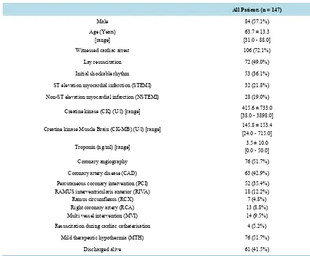
Table 1. Characteristics of those 147 patients following OHCA who could be included in our study.
All Patients (n = 147)
Male 84 (57.1%)
Age (Years) [range]
63.7 ± 13.3 [31.0 - 88.0] Witnessed cardiac arrest 106 (72.1%)
Lay resuscitation 72 (49.0%)
Initial shockable rhythm 53 (36.1%) ST elevation myocardial infarction (STEMI) 32 (21.8%) Non-ST elevation myocardial infarction (NSTEMI) 28 (19.0%)
Creatine kinase (CK) (U/l) [range] [38.0 - 3898.0] 415.6 ± 733.0
Creatine kinase Muscle Brain (CK-MB) (U/l) [range] 145.8 ± 153.4 [24.0 - 715.0]
Troponin (ng/ml) [range] 3.5 ± 10.0 [0.0 - 50.0]
Coronary angiography 76 (51.7%)
Coronary artery disease (CAD) 63 (42.9%) Percutaneous coronary intervention (PCI)
RAMUS interventricularis anterior (RIVA) Ramus circumflexus (RCX) Right coronary artery (RCA) Multi vessel intervention (MVI)
52 (35.4%) 18 (12.2%) 7 (4.8%) 13 (8.8%) 14 (9.5%) Resuscitation during cardiac catheterisation 4 (5.2%)
Mild therapeutic hypothermia (MTH) 76 (51.7%)
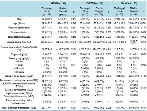
patients (50.0%) died in hospital. The only difference between these two groups was a higher Creatine kinase among patients who died (968.9 ± 1294.6 U/l vs. 280.8 ± 236.1 U/l) (p = 0.040). No differences could be seen in gender, age, rate of witnessed arrest, lay resuscitation, initial shockable rhythm, Creatine kinase Muscle Brain, Troponin at admission, cardiac catheterisation, coronary artery disease, percutaneous coronary intervention, ne-cessity of resuscitation during cardiac catheterisation and application of mild therapeutic hypothermia (Table 2).
3.3. NSTEMI
Here, 28 patients following OHCA presented with NSTEMI; 7 of them (25.0%) survived until hospital dis-charge, while 21 died in hospital (75.0%). The only difference between patients with NSTEMI who survived until hospital discharge and patients who died in hospital was a higher rate of cardiac catheterisation among those patients who could be discharged alive (71.4% vs. 19.0%) (p = 0.010). No differences could be seen in gender, age, rate of witnessed arrest, lay resuscitation, initial shockable rhythm, Creatinine kinase, Creatinine kinase Muscle Brain, Troponin at admission, coronary artery disease, percutaneous coronary intervention, ne-cessity of resuscitation during cardiac catheterisation and the application of mild therapeutic hypothermia (Table 2).
3.4. No ACS
[image:4.595.58.539.360.723.2]Overall, 87 patients following OHCA presented with neither STEMI nor NSTEMI. In this group, 38 patients (43.7%) survived until hospital discharge, while 49 patients (56.3%) died in hospital. Patients who survived until
Table 2.Differences between patients who presented with STEMI, NSTEMI or without ACS following out-of-hospital cardiac-ar- rest.
STEMI (n = 32) NSTEMI (n = 28) No ACS (n = 87)
Discharged Alive (n = 16)
Died in Hospital (n = 16)
p Discharged Alive (n = 7)
Died in Hospital (n = 21) p
Discharged Alive (n = 38)
Died in Hospital (n = 49) p Male 11 (68.8%) 9 (56.3%) 0.654 6 (85.7%) 12 (57.1%) 0.172 23 (60.5%) 23 (46.9%) 0.208 Age (Years) 59.8 ± 12.7 67.4 ± 13.6 0.108 67.3 ± 10.3 70.3 ± 13.1 0.589 63.5 ± 15.5 75.6 ± 12.1 <0.001 Witnessed cardiac arrest 14 (87.5%) 13 (81.3%) 0.831 6 (85.7%) 16 (76.2%) 0.695 29 (76.3%) 28 (57.1%) 0.175 Lay resuscitation 10 (62.5%) 8 (50.0%) 0.476 5 (71.4%) 7 (33.3%) 0.078 23 (60.5%) 19 (38.8%) 0.064 Initial shockable rhythm 11 (68.8%) 10 (62.5%) 0.690 5 (71.4%) 6 (28.6%) 0.055 15 (39.5%) 6 (12.2%) 0.002
Creatine kinase (CK) (U/l) 280.8 ± 236.1 986.9 ± 1294.6 0.040 690.1 ± 529.6 590.0 ± 971.5 0.798 171.4 ± 334.3 161.6 ± 197.5 0.871
Creatine kinase Muscle Brain (CK-MB)
(U/l) 91.6 ± 35.9 210.6 ± 200.3 0.065 170.8 ± 75.5 160.9 ± 148.6 0.879 74.8 ± 51.8 77.2 ± 35.5 0.887 Troponin (ng/ml) 1.4 ± 2.2 7.2 ± 12.9 0.087 10.0 ± 15.9 5.0 ± 11.0 0.354 0.1 ± 0.1 0.1 ± 0.1 0.068 Coronary angiography <1 hour <4 hours <24 hours >24 hours 15 (93.8%) 8 (%) 6 (%) 1 (%) 0 (0.0%) 14 (87.5%) 6 (%) 8 (%) 0 (0.0%) 0 (0.0%) 0.544 5 (71.4%) 3 (%) 1 (%) 0 (0.0%) 1 (%) 4 (19.0%) 1 (%) 1 (%) 0 (0.0%) 2 (%) 0.010 24 (63.2%) 8 (%) 4 (%) 5 (%) 7 (%) 14 (28.6%) 5 (%) 6 (%) 2 (%) 1 (%) 0.002
Coronary artery disease (CAD) 14 (87.5%) 14 (87.5%) 1.000 5 (71.4%) 4 (19.0%) 0.171 14 (36.8%) 12 (24.5%) 0.163 Percutaneous coronary intervention (PCI)
RAMUS interventricularis anterior (RIVA)
RAMUS circumflexus (RCX) Right coronary artery (RCA) Multi vessel intervention (MVI)
14 (87.5%) 6 (37.5%) 1 (6.3%) 2 (12.5%) 5 (31.3%) 14 (87.5%) 4 (25.0%) 2 (12.5%) 6 (37.5%) 2 (12.5%) 1.000 4 (57.1%) 2 (28.6%) 1 (14.3%) 1 (14.3%) 0 (0.0%) 4 (19.0%) 0 (0.0%) 2 (9.5%) 0 (0.0%) 2 (9.5%) 0.053 8 (21.1%) 3 (7.9%) 1 (2.6%) 2 (5.3%) 2 (5.3%) 8 (16.3%) 3 (6.1%) 0 (0.0%) 2 (4.1%) 3 (6.1%) 0.552
Resuscitation during cardiac
hospital discharge were significantly younger (63.5 ± 15.5 years vs. 75.6 ± 12.1 years) (p < 0.001), presented with an initial shockable rhythm more often (39.5% vs. 12.2%) and received cardiac catheterisation more often than those patients who died (63.2% vs. 28.6%) (p = 0.002). No differences could be seen in gender, rate of witnessed arrest, lay resuscitation, Creatinine kinase, Creatinine kinase Muscle Brain, Troponin at admission, coronary artery disease, percutaneous coronary intervention, necessity of resuscitation during cardiac catheteri-sation and the application of mild therapeutic hypothermia (Table 2).
4. Discussion
4.1. Patients with STEMI Following OHCA
STEMI following OHCA has been described as one of the strongest predictors for long-term survival, compara-ble to younger age or ventricular fibrillation as initial rhythm [2].
In our study that worked on this theme, there are four main findings.
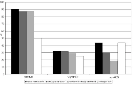
First, the prevalence of relevant coronary artery stenosis is extremely high in patients with STEMI following OHCA, but almost all culprit lesions in these patients can be successfully treated by percutaneous coronary in-tervention [6] [7] (Table 2 and Figure 1).
Second, higher Creatine kinase levels in patients who died during follow-up might underline the necessity of shortening the coronary no-flow time (Table 2). In our study, early coronary angiography within 60 minutes according to the guidelines for the treatment of patients with STEMI [4] could be achieved in almost half of all patients with STEMI (48.3%), irrespective the need for further airway management or haemodynamic stabiliza-tion in this special situastabiliza-tion following OHCA.
Third, our results underline previous findings of a better prognosis of patients with STEMI following OHCA with survival rates of 50% or better [6] [8] [9]. Treating the cause of OHCA with coronary angiography and PCI obviously reduces the incidence of adverse events and decreases mortality during hospitalisation [10].
[image:5.595.93.520.418.693.2]Fourth, enforcing early coronary angiography in patients with STEMI following OHCA is only feasible if all involved physicians and staff are prepared for further resuscitation efforts in the coronary laboratory (Table 2 and Figure 1).
4.2. Patients without STEMI following OHCA
In our study, also patients who presented with NSTEMI and no ACS following OHCA showed better survival rates if they received coronary angiography (Table 2). However, in these patients coronary angiography was at-tempted significantly later than in patients who presented with STEMI (Table 2). In our opinion, the main rea-son for this delay was a primarily focus on haemodynamic stabilisation in these patients as coronary angiogra-phy in these subgroups appeared less urgent than in patients who presented with STEMI; consequently, there were no further resuscitation attempts in the coronary laboratory in these groups.
However, in comparison with a primary focus on early PCI in patients with STEMI, a delay with primary fo-cus on haemodynamic stabilisation led to much lower rates of coronary angiography in patients with NSTEMI (32.1%) or no ACS (43.7%).
Of course, this is a single centre study and the rates of early coronary catheterisation and PCI vary enor-mously among hospitals [11]. However, our data are in line with other investigations that repetitively described rates of less than 50% for early coronary angiography if patients presented without STEMI following OHCA [12]-[14]. Following our findings, a too cautious proceeding in patients without STEMI might be one of the main reasons why there are discrepant findings and opinions about the benefit of early PCI in these patients [3] [8] [15]-[18].
However, the probability of patients who do not receive early coronary angiography on the first day after hos-pital admission receiving later coronary angiography during the same hoshos-pital stay was extremely low in our data (7.5%) (Table 2). There might be several reasons for such a low rate of later coronary angiographies during follow-up: infections, sepsis, respiratory insufficiency, kidney injury, patient’s decision, or maybe an obvious extra-cardiac cause of OHCA; however, the enormous discrepancies between our three groups underline the importance of the initial decision to undergo early coronary angiography or not.
Unfortunately, there are still hardly any objective data that can be used for the initial decision at admission. To date, no biomarker has been shown to correctly predict culprit coronary occlusion in OHCA patients [19] and also the dosage of cardiac troponin I at admission could not help in the decision of early coronary angiogram previously [20]. Also in our study, troponin and Creatine kinase at admission did not differ between patients without STEMI who survived and those who died during follow-up (Table 2). Only in patients with STEMI was higher Creatine kinase as an indirect marker of myocardial damage associated with worse survival rates (Table 2).
Likewise, also ST-segment analysis might have a good positive predictive value but a low negative predictive value in diagnosing the presence of acute or presumed recent coronary artery lesions [17]. In our study, 88.9% of the patients with NSTEMI and 42.1% of the patients with no ACS who received coronary angiography also received percutaneous coronary intervention, underlining the high prevalence of relevant coronary artery steno-sis in patients following OHCA, irrespective of the initial electrocardiographic or laboratory findings (Table 2).
Therefore, our results support previous statements that electrocardiographic findings after OHCA should not be considered as strict selection criteria for performing emergent coronary angiography in patients resuscitated from OHCA without an obvious extra-cardiac cause [8]. Even in the absence of ST-segment elevation on post- ROSC ECG, acute culprit coronary lesions may be present and considered the trigger of cardiac arrest.
4.3. Limitations
Our data are the result of a single centre study; rates of early coronary catheterisation and coronary intervention vary enormously between hospitals [11]. However, our data are in line with several previous investigations [12]-[14] and findings of better survival rates in younger patients and those with an initial shockable rhythm (Table 2) also affirm a representative patient population.
5. Conclusions
The observation of better survival rates in patients with STEMI and immediate PCI underlines the benefit of an immediate treatment of the cause of death in these patients. However, considering the high prevalence of coro-nary artery disease in patients who present without any electrocardiographic or laboratory signs of myocardial ischaemia, percutaneous coronary intervention may be a necessary causal treatment in many more cases.
References
[1] Nolan, J.P., Soar, J., Zideman, D.A., Biarent, D., Bossaert, L.L., Deakin, C., Koster, R.W., Wyllie, J. and Böttiger, B., ERC Guidelines Writing Group (2010) European Resuscitation Council Guidelines for Resuscitation 2010 Section 1. Executive Summary. Resuscitation, 81, 1219-1276. http://dx.doi.org/10.1016/j.resuscitation.2010.08.021
[2] Pleskot, M., Hazukova, R., Stritecka, H., Cermakova, E. and Pudil, R. (2009) Long-Term Prognosis after Out-of-Hospital Cardiac Arrest with/without ST Elevation Myocardial Infarction. Resuscitation, 80, 795-804.
http://dx.doi.org/10.1016/j.resuscitation.2009.04.004
[3] Anyfantakis, Z.A., Baron, G., Aubry, P., Himbert, D., Feldman, L.J., Juliard, J.M., Ricard-Hibon, A., Burnod, A., Cokkinos, D.V. and Steg, P.G. (2009) Acute Coronary Angiographic Findings in Survivors of Out-of-Hospital Cardiac Arrest. American Heart Journal, 157, 312-318. http://dx.doi.org/10.1016/j.ahj.2008.09.016
[4] Steg, P.G., James, S.K., Atar, D., Badano, L.P., Blömstrom-Lundqvist, C., Borger, M.A., Di Mario, C., Dickstein, K., Ducrocq, G., Fernandez-Aviles, F., Gershlick, A.H., Giannuzzi, P., Halvorsen, S., Huber, K., Juni, P., Kastrati, A., Knuuti, J., Lenzen, M.J., Mahaffey, K.W., Valgimigli, M., van’t Hof, A., Widimsky, P. and Zahger, D. (2012) ESC Guidelines for the Management of Acute Myocardial Infarction in Patients Presenting with ST-Segment Elevation: The Task Force on the management of ST-segment elevation acute myocardial infarction of the European Society of Cardi-ology (ESC). European Heart Journal, 33, 2569-2619.
[5] Hamm, C.W., Bassand, J.P., Agewall, S., Bax, J., Boersma, E., Bueno, H., Caso, P., Dudek, D., Gielen, S., Huber, K., Ohman, M., Petrie, M.C., Sonntag, F., Uva, M.S., Storey, R.F., Wijns, W. and Zahger, D., ESC Committee for Practice Guidelines (2011) ESC Guidelines for the Management of Acute Coronary Syndromes in Patients Presenting without Persistent ST-Segment Elevation: The Task Force for the Management of Acute Coronary Syndromes (ACS) in Pa-tients Presenting without Persistent ST-Segment Elevation of the European Society of Cardiology (ESC). European Heart Journal, 32, 2999-3054. http://dx.doi.org/10.1093/eurheartj/ehr236
[6] Zimmermann, S., Flachskampf, F.A., Alff, A., Schneider, R., Dechant, K., Klinghammer, L., Stumpf, C., Zopf, Y., Loehr, T., Brand, G., Ludwig, J., Daniel, W.G. and Achenbach, S. (2013) Out-of-Hospital Cardiac Arrest and Percuta-neous Coronary Intervention for ST-Elevation Myocardial Infarction: Long-Term Survival and Neurological Outcome. International Journal of Cardiology, 166, 236-241. http://dx.doi.org/10.1016/j.ijcard.2011.11.029
[7] Sideris, G., Voicu, S., Yannopoulos, D., Dillinger, J.G,, Adjedj, J., Deye, N., Gueye, P., Manzo-Silberman, S., Malissin, I., Logeart, D., Magkoutis, N., Capan, D.D., Makhloufi, S., Megarbane, B., Vivien, B., Cohen-Solal, A., Payen, D., Baud, F.J. and Henry, P. (2014) Favourable 5-Year Post-Discharge Survival of Comatose Patients Resuscitated from Out- of-Hospital Cardiac Arrest, Managed with Immediate Coronary Angiogram on Admission. European Heart Journal: Acute Cardiovascular Care, 3, 183-191. http://dx.doi.org/10.1177/2048872614523348
[8] Zanuttini, D., Armellini, I., Nucifora, G., Grillo, M.T., Morocutti, G., Carchietti, E., Trillò, G., Spedicato, L., Bernardi, G. and Proclemer, A. (2013) Predictive Value of Electrocardiogram in Diagnosing Acute Coronary Artery Lesions among Patients with Out-of-Hospital-Cardiac-Arrest. Resuscitation, 84, 1250-1254.
http://dx.doi.org/10.1016/j.resuscitation.2013.04.023
[9] Marcusohn, E., Roguin, A., Sebbag, A., Aronson, D., Dragu, R., Amikam, S., Boulus, M., Grenadier, E., Kerner, A., Nikolsky, E., Markiewicz, W., Hammerman, H. and Kapeliovich, M. (2007) Primary Percutaneous Coronary Interven-tion after Out-of-Hospital Cardiac Arrest: Patients and Outcomes. Israel Medical AssociaInterven-tion Journal, 9, 257-259. [10] Liu, H.W., Pan, W., Wang, L.F., Sun, Y.M., Li, Z.Q. and Wang, Z.H. (2012) Impact of Emergency Percutaneous
Coronary Intervention on Outcomes of ST-Segment Elevation Myocardial Infarction Patients Complicated by Out-of- Hospital Cardiac Arrest. Chinese Medicine Journal (English), 125, 1405-1409.
[11] Callaway, C.W., Schmicker, R.H., Brown, S.P., Albrich, J.M., Andrusiek, D.L., Aufderheide, T.P., Christenson, J., Daya, M.R., Falconer, D., Husa, R.D., Idris, A.H., Ornato, J.P., Rac, V.E., Rea, T.D., Rittenberger, J.C., Sears, G. and Stiell, I.G., ROC Investigators (2014) Early Coronary Angiography and Induced Hypothermia Are Associated with Survival and Functional Recovery after Out-of-Hospital Cardiac Arrest. Resuscitation, 85, 657-663.
http://dx.doi.org/10.1016/j.resuscitation.2013.12.028
[12] Hollenbeck, R.D., McPherson, J.A., Mooney, M.R., Unger, B.T., Patel, N.C., McMullan Jr., P.W., Hsu, C.H., Seder, D.B. and Kern, K.B. (2014) Early Cardiac Catheterisation Is Associated with Improved Survival in Comatose Survi-vors of Cardiac Arrest without STEMI. Resuscitation, 85, 88-95. http://dx.doi.org/10.1016/j.resuscitation.2013.07.027 [13] Strote, J.A., Maynard, C., Olsufka, M., Nichol, G., Copass, M.K., Cobb, L.A. and Kim, F. (2012) Comparison of Role
of Early (Less than Six Hours) to Later (More than Six Hours) or No Cardiac Catheterisation after Resuscitation from Out-of-Hospital Cardiac Arrest. The American Journal of Cardiology, 109, 451-454.
http://dx.doi.org/10.1016/j.amjcard.2011.09.036
[15] Bro-Jeppesen, J., Kjaergaard, J., Wanscher, M., Pedersen, F., Holmvang, L., Lippert, F.K., Møller, J.E., Køber, L. and Hassager, C. (2012) Emergency Coronary Angiography in Comatose Cardiac Arrest Patients: Do Real-Life Experi-ences Support the Guidelines? European Heart Journal: Acute Cardiovascular Care, 1, 291-301.
http://dx.doi.org/10.1177/2048872612465588
[16] Nanjayya, V.B. and Nayyar, V. (2012) Immediate Coronary Angiogram in Comatose Survivors of Out-of-Hospital Cardiac Arrest—An Australian Study. Resuscitation, 83, 699-704.
http://dx.doi.org/10.1016/j.resuscitation.2011.12.004
[17] Zanuttini, D., Armellini, I., Nucifora, G., Carchietti, E., Trillò, G., Spedicato, L., Bernardi, G. and Proclemer, A. (2012) Impact of Emergency Coronary Angiography on In-Hospital Outcome of Unconscious Survivors after Out-of-Hospital Cardiac Arrest. The American Journal of Cardiology, 110, 1723-1728. http://dx.doi.org/10.1016/j.amjcard.2012.08.006 [18] Radsel, P., Knafelj, R., Kocjancic, S. and Noc, M. (2011) Angiographic Characteristics of Coronary Disease and Post- Resuscitation Electrocardiograms in Patients with Aborted Cardiac Arrest outside a Hospital. The American Journal of Cardiology, 108, 634-638. http://dx.doi.org/10.1016/j.amjcard.2011.04.008
[19] Geri, G., Dumas, F. and Cariou, A. (2014) Should We Perform a Coronary Angiography in All Cardiac Arrest Survi-vors? Current Opinion in Critical Care, 20, 273-279. http://dx.doi.org/10.1097/MCC.0000000000000093