http://dx.doi.org/10.4236/oji.2015.52007
How to cite this paper: Mazzone, P., Scudiero, I., Ferravante, A., Paolucci, M., D’Andrea, L.E., Varricchio, E., Telesio, G., Pizzulo, M., Zotti, T., Reale, C., Vito, P. and Stilo, R. (2015) Functional Characterization of Porcine (Sus scrofa) BCL10. Open Journal of Immunology, 5, 64-71. http://dx.doi.org/10.4236/oji.2015.52007
Functional Characterization of Porcine
(Sus scrofa) BCL10
Pellegrino Mazzone
1*, Ivan Scudiero
1*, Angela Ferravante
1, Marina Paolucci
2,
Luca E. D’Andrea
1, Ettore Varricchio
2, Gianluca Telesio
1,3, Maddalena Pizzulo
1,
Tiziana Zotti
1,2, Carla Reale
1, Pasquale Vito
1,2, Romania Stilo
1,21Laboratory of Immunogenetics, Biogem, Via Camporeale, Ariano Irpino, Italy 2Dipartimento di Scienze e Tecnologie, Università del Sannio, Benevento, Italy
3Dipartimento di Medicina molecolare Biotecnologie mediche, Università di Napoli “Federico II”, Napoli, Italy
Email: vito@unisannio.it
Received 25 March 2015; accepted 28 May 2015; published 2 June 2015
Copyright © 2015 by authors and Scientific Research Publishing Inc.
This work is licensed under the Creative Commons Attribution International License (CC BY). http://creativecommons.org/licenses/by/4.0/
Abstract
Human BCL10 (hBCL10) protein is a signal transduction molecule originally identified because of its direct involvement in a subset of mucosa-associated lymphoid tissue (MALT) lymphomas, and later recognized as a crucial factor in regulating activation of NF-kB transcription factor following antigen receptor stimulation on lymphocytes. In this study, we characterized the NF-kB inducing activity of porcine BCL10 (pBCL10). pBCL10 oligimerizes, binds to components of the CARMA/ BCL10/MALT1 complex and forms cytoplasmic filaments. Functionally, in human cells pBCL10 is more effective in activating NF-kB compared to hBCL10, possibly due to the lack of carboxy-ter- minal inhibitory serine residues present in the human protein. Also, depletion experiments car-ried out through expression of short hairpin RNAs targeting hBCL10 indicate that pBcl10 can func-tionally replace the human protein and retains its higher NF-kB-inducing property in the absence of hBCL10. Our results contribute useful information on BCL10 protein in pigs, and may help the development of strategies based on the control of the immune response in pigs.
Keywords
BCL10, CARMA, NF-kB, CARD
1. Introduction
Human BCL10 (hBCL10) was originally identified because of its direct involvement in a subset of MALT B
*
cell lymphomas [1] [2]. As a result of the translocation t(1;14)(p22;q32), BCL10 is placed under the control of the immunoglobulin heavy chain enhancer, and is over expressed in these tumors [1] [2]. At the same time, BCL10 was identified in several other laboratories for the presence of a caspase recruitment domain (CARD) in its sequence [3]. Functionally, BCL10 regulates activation of NF-kB transcription factor, which transcribes genes that control both innate and acquired immune response and genes that play a positive effect on cell sur-vival and proliferation [4] [5]. Genetic alteration of the BCL10 locus leads to immunodeficiency in mice, due to impaired NF-kB activation following antigen receptor stimulation in both T and B cells [6]. The biological ac-tivity of BCL10 is explicated through formation of the CBM complex, a molecular complex that comprises one of three members of the family of CARMA proteins and the protease MALT1 [7]. The three CARMA proteins constitute a family of proteins conserved across many species which are characterized by the presence of differ-ent functional domains shared by all members of the family [8]. Functionally, all three CARMA proteins are able to associate BCL10 through an homophilic interaction between the corresponding CARD domains, and to cooperate with it in inducing the transcriptional activity of NF-kB [8]. Thus, correct assembly of the CBM com-plex is an essential step in the NF-kB inducing pathway mediated by BCL10. Formation of this comcom-plex in fact triggers non-conventional ubiquitination events, which eventually result in recruitment and K63-linked ubiqui-tination of the noncatalytic IKKg/NEMO subunit of the I-kB kinase complex, responsible for NF-kB transcrip-tion factor activatranscrip-tion [9]-[11].
Pork is the most highly consumed meat worldwide, and the related industry represents a crucial economical sector for many countries. Maintaining pork safety and minimizing production losses associated with diseases impacts profitability, food safety and animal health. As such, there is a major interest in characterizing aspects of the porcine immune response. A porcine homologue of hBCL10 was recently cloned [12]. pBCL10 mRNA is distributed in different tissue, and in cultured porcine cells its expression increases following treatments with li-popolysaccharide and polyriboinosinic-polyribocytidylic acid [12], two treatments that mimic bacteria and virus infections, respectively. In the present work, we have analyzed the NF-kB-inducing property of pBCL10.
2. Materials and Methods
2.1. RNA Extraction and Cloning of pBCL10 Full-Length cDNA
Spleen specimens were obtained from slaughtered Large White and Landrace hybrids F1 hybrids. Total RNA was extracted from the splenic tissue by using Trizol reagent as previously described [13], and 1 μg of total RNA was reverse-transcribed to generate a first-strand cDNA. The following primers were used to amplify pBCL10: forward 5’-ATGGAGCCCGCCGCGCC-3’ and reverse 5’-TCATTGCCGCAAAAGAGCACG-3’. PCR condi-tions were as follows: 98˚C for 30 s, 30 cycles (98˚C/15s; 63˚C/22s; 72˚C/30s). The RT-PCR product of the ex-pected size (702 bp, Genebank accession number FJ376731) was gel purified, cloned into HA- and FLAG- tagged expression vectors using standard methodologies and confirmed by sequencing.
2.2. Sequence Analysis
The BCL10 protein sequences were analyzed by using the BLAST algorithm at the NCBI web site (http://www.ncbi.nlm.nih.gov/blast).
2.3. Cell Culture and Transfection
HEK293 cells were obtained from ATCC and were maintained in Dulbecco’s modified Eagle’s medium (DMEM) supplemented with 10% FBS. The expression vectors used in transfection experiments for this study have been previously described [13]-[18]. DNA plasmids were transfected by standard calcium-phosphate me-thod. Short hairpin RNAs targeting hBCL10 were the following: shBCL10 #3 5’-CCTTAAGATCACGTA- CTGTTTCTCGAGAAACAGTACGTGATCTTAAGG-3’ and shBCL10 #5 5’-GTTGAATCTATTCGGCGA- GAACTCGAGTTCTCGCCGAATAGATTCAAC-3’ and have been already described [18]. Retroviral infec-tions were carried out as previously described [19].
2.4. Immunoblot Analysis and Coprecipitation
and a mixture of protease inhibitors). Proteins were separated by SDS-PAGE, transferred onto nitrocellulose membrane, and incubated with primary antibodies followed by horseradish peroxidase-conjugated secondary antibodies (Amersham Biosciences, Piscataway, NJ). Blots were developed using the ECL system (Amersham Biosciences). For co-immunoprecipitation experiments, cells were lysed in lysis buffer and immune complexes were bound to protein A/G, resolved by SDS-PAGE, and analyzed by immunoblot assay. Sources of antisera and monoclonal antibodies were the following: anti-FLAG, anti-β-Actin, Sigma; anti-HA, anti-MALT1, anti- CARMA3 and anti-BCL10 (H-197 SC5611, generated against an epitope corresponding to amino acids 1-197 of human BCL10), Santa Cruz Biotechnology. The calf-intestinal alkaline phosphatase was purchased from Roche.
2.5. Luciferase Assay
To assess for NF-kB activation, HEK293 were transfected with plasmidic DNAs together with pNF-κB-luc (Clontech) in 6-well plates. After transfection and treatments, luciferase activity was determined with Luciferase Assay System (Promega). A plasmids expressing β-galactosidase was added to the transfection mixture in order to normalize for the efficiency of transfection.
2.6. Immunofluorescence
1 × 10⁴ HEK293 were grown and transfected in chamber slides. Sixteen hours after transfection, cells were fixed in 4% paraformaldehyde for 15 min at room temperature and then permeabilized in PBS/0.1% Triton X-100. Cells were incubated for 30 min in 5% FCS-PBS with anti-FLAG antibody (Sigma-Aldrich) followed by several washes with 5% FCS-PBS, and then incubating for 30 min with secondary antibody in 5% FCS-PBS. All steps were done at room temperature.
3. Results and Discussion
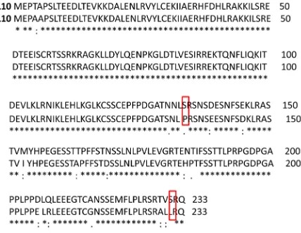
A cDNA corresponding to the porcine homologue of hBCL10 was successfully amplified by RT-PCR from por-cine spleen tissue. Sequence analysis revealed that it corresponds to the sequence already deposited in Genebank with the accession number FJ376731 [12]. Both human and porcine BCL10 proteins consist of 233 amino acidic residues, only 17 of which are dissimilar (Figure 1).
[image:3.595.199.413.488.654.2]Amino acidic differences are mainly distributed in the carboxy terminal portion of the protein, emphasizing the conservation of the card domain (aa 6 - 108), which is in fact perfectly conserved between the two species. Interestingly, some amino acidic substitutions concern serine residues present in the human protein, particularly S134 and S231, which in pBCL10 are replaced by a proline and leucine residue, respectively. This aspect is par-
Figure 1.(Alignment of human and porcine BCL10) Alignment of hBCL10 (Gene Bank NP_003912) and pBCL10 (Gene Bank FJ-376731). Identical residues are indicated by stars, conserva-tive and semi-conservaconserva-tive substitutions are indicated by double dot and single dot, respectively. Colored rectangles indicate
ticularly important, because hBCL10 phosphorylation on serine residues, including S134, were shown to nega-tively regulate hBCL10-induced NF-kB activation [20].
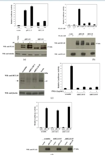
When analyzed in immunoblot assay, pBCL10 expressed in mammalian cells migrates as a 37 kDa protein (Figure 2(a)), and is recognizes by a rabbit antisera raised against hBCL10 (Figure 2(a), right panel). Similarly to hBCL10 [21] [22], pBCL10 migrates as a doublet on SDS-PAGE due to phosphorylation of the protein. In fact, the pBCL10 doublet resolves in a single band when cell lysates were treated with phosphatase prior to im-munoblot analysis (Figure 2(b)).
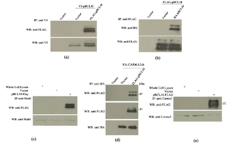
BCL10 plays a crucial role in the signal transduction pathway that leads to activation of the transcription fac-tor NF-kB [6] [7]. hBCL10-mediated activation of NF-kB requires oligomerization of hBCL10, assembly of the CBM complex and triggering of unconventional ubiquitination events, which eventually result in the recruitment of the IKK complex [7] [9]-[11]. Indeed, transfection experiments indicate that pBCL10 oligomerizes both with itself and with hBCL10 (Figures 3(a)-(b)). Furthermore, pBCL10 associates with human MALT1 (Figure 3(c)), with human CARMA2sh[14] (Figure 3(d)) and with human CARMA3 (Figure 3(e)).
Fluorescence microscopy experiments and structural studies have shown that the NF-kB-activity produced by hBCL10 is regulated through formation of cytosolic filamentous structures [23] [24]. We therefore verified whether also pBCL10 is able to form such structures. As shown in Figure 4, assembly of filamentous structures is readily visible following expression of pBCL10 in mammalian cells.
Next, we tested the NF-kB-inducing activity of pBCL10 using a luciferase-based reporter assay. The results of these experiments, shown in Figure 5(a), indicate that pBCL10 is even more effective than hBCL10 in acti-vating NF-kB in mammalian cells. In fact, while expression of hBCL10 produces a luciferase activity about 8- 10-fold higher compared to the empty vector, the luciferase activity produced by pBCL10 expression was at least 4-fold higher than that produced by hBCL10. As for hBCL10 [11] [25] [26], pBCL10-induced NF-kB ac-tivation requires ubiquitination(s) events, since NF-kB acac-tivation is completely abrogated following co-expres- sion of A20 de-ubiquitinase (Figure 5(b)).
To exclude the possibility that the higher NF-kB activation mediated by pBCL10 was due to its interaction and subsequent oligomerization of hBCL10, we abolished expression of hBCL10 in the human cell line HEK293 through retrovirus-mediated expression of short hairpin RNAs (shRNA) targeting hBCL10. As shown in Figure 5(c), introduction of hBCL10sh#3 and hBCL10sh#5 in HEK293 cells results in a great reduction of hBCL10 expression. In fact, depletion of hBCL10 in these cells abrogates their ability to activate NF-kB fol-lowing exposure to phorbol-12-myristate-13-acetate (PMA) (Figure 5(c)). However, introduction of pBCL10 in these hBCL10-depleted cells fully recovers their ability to activate NF-kB (Figure 5(d)). Thus, pBCL10 retains its higher NF-kB-inducing property even in the absence of hBCL10.
In recent years, there has been growing interest in the porcine immune system due to its potential as a model for the human immune system and because of the economic importance of pigs as livestock. Although great ad-vances have been achieved in the field of porcine immunology, there are still some important issues that require more research and development. In the work here presented, we have analyzed the NF-kB-inducing property of
Figure 3. pBCL10 olimerizes and binds to CBM proteins. (a) HEK293 cells were transiently cotransfected with tagged ver-sions of pBCL10 and hBCL10. 24 hrs later, cell lysates were prepared and immunoprecipitated with the indicated anti-tag mAb. Immuno complexes were separated by SDS-PAGE and transferred onto membranes subsequently assayed for
asso-ciated (a) pBCL10; (b) hBCL10); (c) Malt1; (d) CARMA2sh and (e) CARMA3.
Figure 4.Subcellular localization of pBCL10. HEK-293 cells were transfected with mammalian FLAG-tagged vector, empty (vector) or expressing pBCL10. 16 hrs after transfection, cells were stained with anti-FLAG mAb, followed by FITC-con-
[image:5.595.171.433.430.687.2](a) (b)
(c)
(d)
Figure 5. pBCL10 activates NF-κB (a)-(b) HEK293 cells were transiently cotransfected with expression vectors encoding for the indicated polypeptides, together with pNF-κB-luc and pRSV-βgal reporter vectors. The total amount of transfected plasmidic DNA was maintained constant by adding empty vector. 16 hrs after transfection, cell lysates were prepared and lu-ciferase activity was measured. A fraction of the cell lystes were analyzed by immunoblot to monitor protein expression, shown in the lower panels. Data shown represents relative luciferase activity normalized on β-galactosidase activity and is representative of six independent experiments done in triplicate. (c) Left panel Cell lysates from HEK293 cells infected with retroviruses encoding for shRNAs targeting hBCL10 were monitored for hBCL10 expression by immunoblot assay; Right
panel NF-κB-driven luciferase activity in HEK-293 cells silenced for hBCL10 and stimulated with PMA. (d) NF-κB-driven
luciferase activity in HEK-293 cells silenced for hBCL10 and transfected with tBCL10. Data shown represent relative luci-ferase activity normalized on β-galactosidase activity and is representative of six independent experiments done in triplicate.
[image:6.595.122.476.76.596.2]pBCL10. We found that pBCL10 owns an higher NF-kB-inducing activity compared to the human protein, pos-sibly because it lacks serine residues with inhibitory function present in hBCL10.
Given the importance of this transcription factor in regulating both normal immune response and autoimmune, immunoproliferative and tumoral disorders, and also considering the economic value of this organism, our re-sults may benefit the development of strategies based on the control of the immune response in pigs.
References
[1] Willis, T.G., Jadayel, D.M., Du, M.Q., Peng, H., Perry, A.R., et al. (1999) Bcl10 is Involved in t(1;14)(p22;q32) of MALT B Cell Lymphomas and Mutated in Multiple Tumor Types. Cell, 96, 35-45
http://dx.doi.org/10.1016/S0092-8674(00)80957-5
[2] Zhang, Q., Siebert, R., Yan, M., Hinzmann, B., Cui, X., et al. (1999) Inactivating Mutation and BCL10, a Caspase Re-cruitment Domain-Containing Gene, in MALT Lymphoma with t(1;14)(p22;q32). Nature Genetics22, 63-68.
http://dx.doi.org/10.1038/8767
[3] Vito, P. and Stilo, R. (2014) Fifteen Years of BCL10. Immunology Letters, 160, 102-103.
http://dx.doi.org/10.1016/j.imlet.2014.02.002
[4] Oeckinghaus, A., Hayden, M.S. and Ghosh, S. (2011) Crosstalk in NF-kappaB Signaling Pathways. Nature Immunol-ogy, 12, 695-708. http://dx.doi.org/10.1038/ni.2065
[5] Vallabhapurapu, S. and Karin, M. (2009) Regulation and Function of NF-kappaB Transcription Factors in the Imune System. Annual Review of Immunology, 27, 693-733.http://dx.doi.org/10.1146/annurev.immunol.021908.132641
[6] Ruland, J., Duncan, G.S., Elia, A., del Barco Barrantes, I., Nguyen, L., et al. (2001) Bcl10 is a Positive Regulator of Antigen Receptor-Induced Activation of NF-kappaB and Neural Tube Closure. Cell, 104, 33-42.
http://dx.doi.org/10.1016/S0092-8674(01)00189-1
[7] Thome, M., Charton, J.E., Pelzer, C. and Hailfinger, S. (2012) Antigen Receptor Signaling to NF-kB via CARMA1, BCL10, and MALT1.Cold Spring Harbor Perspectives in Biology, 2, Article ID: a003004.
http://dx.doi.org/10.1101/cshperspect.a003004
[8] Scudiero, I., Vito, P. and Stilo, R. (2014) The Three CARMA Sisters: So Different, So Similar. A Portrait of the Three CARMA Proteins and Their Involvement in Human Disorders. Journal of Cellular Physiology, 229, 990-997.
http://dx.doi.org/10.1002/jcp.24543
[9] Stilo, R., Liguoro, D., Di Jeso, B., et al. (2004) Physical and Functional Interaction of CARMA1 and CARMA3 with Ikappa Kinase Gamma-NF-kappaB Essential Modulator. Journal of Biological Chemistry, 279, 34323-34331.
http://dx.doi.org/10.1074/jbc.M402244200
[10] Sun, L., Deng, L., Ea, C.K., Xia, Z.P. and Chen, Z.J. (2004) The TRAF6 Ubiquitin Ligase and TAK1 Kinase Mediate IKK Activation by BCL10 and MALT1 in T lymphocytes. Molecular Cell, 14, 289-301.
http://dx.doi.org/10.1016/S1097-2765(04)00236-9
[11] Zhou, H.L., Wertz, I., O’Rourke, K., Ultsch, M., Seshagiri, S., et al. (2004) Bcl10 Activates the NF-κB Pathway
through Ubiquitination of NEMO. Nature, 427, 167-171. http://dx.doi.org/10.1038/nature02273
[12] Huang, J., Ma, G.-J., Sun, N.N., Wu, Z.F., Li X.Y. and Zhao, S.H. (2010) BCL10 as a New Candidate Gene for Im-mune Response in Pigs: Cloning, Expression and Association Analysis. International Journal of Immunogenetics, 37, 103-110. http://dx.doi.org/10.1111/j.1744-313X.2010.00898.x
[13] Mazzone, P., Scudiero, I., Coccia, E., Ferravante, A., Paolucci, M., et al. (2015) Functional Characterization of a BCL10 Isoform in the Rainbow Trout Oncorhynchus mykiss. FEBS Open Bio, 5, 175-181.
http://dx.doi.org/10.1016/j.fob.2015.01.007
[14] Scudiero, I., Zotti, T., Ferravante, A., Vessichelli, M., Vito, P., et al. (2011) Alternative Splicing of CARMA2/ CARD14 Transcripts Generates Protein Variants with Differential Effect on NF-κB Activation and Endoplasmic Reti-culum Stress-Induced Cell Death.Journal of Cellular Physiology, 226, 3121-3131.
http://dx.doi.org/10.1002/jcp.22667
[15] Zotti, T., Uva, A., Ferravante, A., Vessichelli, M., Scudiero, I., et al. (2011) TRAF7 Protein Promotes Lys-29-Linked Polyubiquitination of IκB Kinase (IKKγ)/NF-κB Essential Modulator (NEMO) and p65/RelA Protein and Represses NF-κB Activation. Journal of Biological Chemistry, 286, 22924-22933. http://dx.doi.org/10.1074/jbc.M110.215426
[16] Scudiero, I., Zotti, T., Ferravante, A., Vessichelli, M., Reale, C., et al. (2012) Tumor Necrosis Factor (TNF) Receptor- Associated Factor 7 Is Required for TNFα-Induced Jun NH2-Terminal Kinase Activation and Promotes Cell Death by Regulating Polyubiquitination and Lysosomal Degradation of c-FLIP Protein. Journal of Biological Chemistry, 287, 6053-6061. http://dx.doi.org/10.1074/jbc.M111.300137
ciated Complex Binds to and Regulates FADD Function. Biochemical and Biophysical Research Communications, 303, 1034-1041. http://dx.doi.org/10.1016/S0006-291X(03)00487-X
[18] Costanzo, A., Guet, C. and Vito, P. (1999) c-E10 Is a Caspase-Recruiting Domain-Containing Protein That Interacts with Components of Death Receptors Signaling Pathway and Activates Nuclear Factor-κB. Journal of Biological Che-
mistry, 274, 20127-20132. http://dx.doi.org/10.1074/jbc.274.29.20127
[19] Guiet, C., Silvestri, E., De Smaele, E., Franzoso, G. and Vito, P. (2002) c-FLIP Efficiently Rescues TRAF-2-/- Cells from TNF-Induced Apoptosis. Cell Death & Differentiation, 9, 138-144. http://dx.doi.org/10.1038/sj.cdd.4400947
[20] Wegener, E., Oeckinghaus, A., Papadopoulou, N., Lavitas, L., Schmidt-Supprian, M., et al. (2006) Essential Role for IκB Kinase Beta in Remodeling Carma1-Bcl10-Malt1 Complexes upon T Cell Activation. Molecular Cell, 23, 13-23.
http://dx.doi.org/10.1016/j.molcel.2006.05.027
[21] Yoneda, T., Imaizumi, K., Maeda, M., Yui, D., Manabe, T., et al. (2000) Regulatory Mechanisms of TRAF2-Mediated Signal Transduction by Bcl10, a MALT Lymphoma-Associated Protein. Journal of Biological Chemistry, 275, 11114- 11120. http://dx.doi.org/10.1074/jbc.275.15.11114
[22] Gaide, O., Martinon, F., Micheau, O., Bonnet, D., Thome, M., et al. (2001) Carma1, a CARD-Containing Binding Partner of Bcl10, Induces Bcl10 Phosphorylation and NF-κB Activation. FEBS Letters, 496, 121-127.
http://dx.doi.org/10.1016/S0014-5793(01)02414-0
[23] Qiao, Q., Yang, C.H., Zheng, C., Fontán, L., David, L., et al. (2013) Structural Architecture of the CARMA1/Bcl10/ MALT1 Signalosome: Nucleation Induced Filamentous Assembly. Molecular Cell, 51, 766-779.
http://dx.doi.org/10.1016/j.molcel.2013.08.032
[24] Guiet, C. and Vito, P. (2000) Caspase Recruitment Domain (CARD)-Dependent Cytoplasmic Filaments Mediate bcl10-Induced NF-kappaB Activation. The Journal of Cell Biology, 148, 1131-40.
http://dx.doi.org/10.1083/jcb.148.6.1131
[25] Stilo, R., Liguoro, D., Di Jeso, B., Baens, M., Kloo, B., et al. (2004) Physical and Functional Interaction of CARMA1
and CARMA3 with Iκ Kinase Gamma-NF-κB Essential Modulator. Journal of Biological Chemistry, 279, 34323-
34331. http://dx.doi.org/10.1074/jbc.M402244200
[26] Düwel, M., Welteke, V., Oeckinghaus, A., Baens, M., Kloo, B., et al. (2009) A20 Negatively Regulates T Cell Recep-tor Signaling to NF-κB by Cleaving Malt1 Ubiquitin Chains. Journal of Immunology, 182, 7718-7728.