Performance Improvement of Fuzzy C-mean Algorithm
for Tumor Extraction in MR Brain Images
Neelofar Sohi,
University College of Engineering,
Punjabi University Patiala (Pb.)
Lakhwinder Kaur
University College of Engg., Punjabi University Patiala (Pb.)
Savita Gupta
University Institute of Engg. and Tech.
Panjab University (Chd.), India.
ABSTRACT
Aim of this paper is to develop an efficient fuzzy c-mean based segmentation algorithm to extract tumor region from MR brain images. First, cluster centroids are initialized through data analysis of tumor region, which optimizes the standard fuzzy c-mean algorithm. Next, reconstruction based morphological operations are applied to enhance its performance for brain tumor extraction. The results show that simple fuzzy c-mean could not segment the region of interest properly, whereas enhanced algorithm effectively extracts the tumor region. From comparison with existing segmentation methods, enhanced fuzzy c-mean algorithm emerges as the most effective algorithm for extracting region of interest.
Keywords
segmentation, brain tumor extraction, thresholding, fuzzy c-mean, k-c-mean, morphology, markers.
1. INTRODUCTION
The image segmentation process can be considered as one of the basic, yet very important, steps in digital image processing based applications. This is being used extensively in multidisciplinary areas of medical imaging, computer vision, remote sensing, agricultural imaging, object recognition to name a few. Segmentation of medical images is a challenging task and is of great importance for medical image analysis, interpretation and understanding of images for subsequent computer aided diagnosis and treatment planning. A large number of segmentation algorithms have been developed over past few decades [1]. Medical image segmentation still continues to be a challenging problem owing to poor contrast, complex nature of medical images and dependency of segmentation method on imaging modality, image features and dimensions [2], [3]. Segmentation algorithms are based on different parameters of an image like gray-level, color, texture, depth or motion. In medical images, segmentation is mainly done based on the gray-level value of pixels, because the majority of medical images are scale representations. Image segmentation using the gray-level value of images is performed mainly using clustering, thresholding and region growing methods [1]. Medical imaging is performed using various diagnostic techniques like magnetic resonance imaging (MRI), computed tomography (CT), ultrasound, etc. Magnetic Resonance Imaging (MRI) is the most widely-used method of high quality medical imaging, especially in brain imaging where MRI's soft tissue contrast and non-invasiveness are clear advantages [3]. Segmenting MR brain image for extracting tumor or any other lesion requires knowledge of brain anatomy. Developing brain tumor extraction software relieves radiologists from the task of identifying region of interest but for reliable segmentation results it should incorporate some intervention by an expert.
Fuzzy c-means (FCM) clustering algorithm is a soft segmentation method which retains more information from input image than hard segmentation methods [4]-[6]. Various FCM based algorithms for MRI segmentation have been presented in [7]-[18]. Thresholding is one of the simplest and most widely used approaches in image segmentation. Various thresholding based algorithms for MR segmentation have been proposed in [19]-[23]. K-means clustering, an unsupervised method produces hard segmentation by restricting a pixel's membership exclusively to one class [1], [24]. K-means is suitable for MIS as the number of clusters (k) is usually known for images of particular region of human anatomy [25].K-means has been used extensively for segmentation of MR images. Various K-means based algorithms for MRI segmentation have been presented in [13], [24]-[28]. Morphology is used to transform an image into another image by eliminating undesirable features. This is done by probing the input image with other image of certain shape and size known as structuring element [29]. Various morphology based algorithms for MRI segmentation have been presented in [26], [29]-[36], Markers are generated to mark the regions of interest. Generating markers has emerged as a powerful technique for segmenting MR brain images which contain several irregularities. Examples of marker based segmentation algorithms for MR brain images include the work presented in [26], [37], [38].
The paper is organised as follows: A brief introduction to medical image segmentation is presented in section 1. Section 2 explains various existing segmentation algorithms. The enhanced fuzzy c-mean algorithm ispresented in section 3. Results and discussion are provided in section 4 and finally conclusion remarks are given in section 5.
2.
EXISTING SEGMENTATION
ALGORITHMS
2.1
Standard Fuzzy C-Mean [39]
Fuzzy c-mean (FCM) clustering algorithm was first introduced by Dunn [40] and was later extended by Bezdek [41]. FCM is a soft segmentation method which retains more information from input image than hard segmentation methods [4]-[6]. It performs segmentation through fuzzy pixel classification in the sense that pixels are allowed to belong to multiple classes with a degree of membership between 0 and 1 [7]. It imparts flexibility to segmentation process and copes with uncertainty factor by allowing fuzzy boundaries to exist between different clusters. It is based on minimization of the following objective function:
m ij C
j N
i i
m
u
J
1
x
i
c
j2where m is fuzzification parameter which determines the degree of fuzziness in the clusters, uij is the degree of membership of xi
in the cluster j, xi is the ith data point, cj is the jth cluster centre,
and ||*|| is a squared error function. The pseudo code for the fuzzy c-mean algorithm [39] is given below:
2.1.1 Cluster centroids are initialized by random numbers. 2.1.2 Distance Computation
Cluster centroids are replicated to image dimensions and then they are concatenated to each other. Image is also concatenated to itself along the third dimension.
Distance is computed as: dIc= │I-c│;
where I - input image concatenated to itself along 3rddimension
c - initial centroids replicated to image dimensions and then concatenated to each other
2.1.3 Fuzzy Membership Function
Fuzzy weight matrix is computed as inverse of distance matrix found above. Then fuzzy weights are computed along first and second dimensions. Fuzzy weights define membership value of pixels into clusters. Flexibility is achieved and outliers are dealt with, to a large extent by allowing one pixel to reside in multiple clusters.
Fuzzy weights computed as: FuzW=1/dIc;
FuzW1=1/ dIc1;
FuzW2=1/ dIc2;
where dIc1 =dIc (:,:,1)*FuzW;
dIc2 =dIc (:,:,2)*FuzW;
2.1.4 New Centroids are computed as :
Cnext1=
N
i 1
FuzW1*FuzW1*Ii/
N
i 1
FuzW1*FuzW1;
Cnext2=
N
i 1
FuzW2*FuzW2*Ii/
N
i 1
FuzW2*FuzW2;
2.1.5 Stopping Criterion :
if max { │cc1-Cnext1│ / cc1, │cc2-Cnext2) / cc2│} < ε
then stop otherwise cc1= Cnext1; cc2= Cnext2;
goto step(b)
2.2 Standard Thresholding Algorithm [42]
Thresholding is one of the simplest and most widely used approaches in image segmentation. Most of the existing thresholding methods are bi-level [43], which use two levels to categorize the image into background and object segments. However, MR images have many different parts which make these methods non-applicable. Thus, the loss of information from the image may occur and diagnosis system may mislead physicians in their clinical task. Therefore, multi-level thresholding algorithms have been developed to ensure that all important information from MR images are retained, but they become computationally expensive, because a large no. of iterations would be required for computing the optimum threshold [19]. Otsu’s global thresholding method is the most suitable image segmentation method to segment a brain tumor from a Magnetic Resonance Image [1]. It selects that gray level value as threshold for which between-class variance is maximised. In general, thresholding algorithms do not use spatial information of an image and they usually fail to segment objects with low contrast or noisy images with varyingbackground [44]. Thresholding alone is rarely used for medical image segmentation. Instead, it usually functions as a pre-processing step.The pseudocode for the thresholding based segmentation algorithm [42] is given below:
2.2.1 Initially, every gray level value in the image (I) is tested as a potential threshold to segment the image into two levels (binary image)
2.2.2 For each threshold, segment image into binary as: if ( I(x,y) > T ) then set BW(x,y) = 1
else BW(x,y)=0;
and compute the following terms: Probability of Foreground pixels, w0 is w0= No. of Foreground Pixels/ Size of image Probability of Background pixels, w1 is w1= No. of Background Pixels/ Size of image Mean of Foreground pixels, u0 is
u0= Foreground Sum/ No. of Foreground Pixels Mean of Background pixels, u1 is defined as u1=Background Sum/ No. of Background Pixels
2.2.3 Gray level value for which between-class variance [45] maximizes is chosen as threshold, T to segment image:
T=Max {wo*w1*(u0-u1)2}
2.3
Standard K-means Algorithm
K-means (KM) is one of the simplest unsupervised learning algorithms that solve the clustering problem. This clustering is convergent and its aim is to optimize the partitioning decisions based on a user-defined initial set of clustering that is updated after each iteration [47]. It produces hard segmentation by restricting a pixel’s membership exclusively to one class [1], [24]. It is a simple clustering method with low computational complexity as compared to FCM [27]. K-means is suitable for medical image segmentation as the number of clusters (k) is usually known for images of particular region of human anatomy [25]. In KM algorithm, every pixel is assigned to its closest cluster on the basis of distance between the pixel and cluster centroids. This algorithm [46] aims at minimizing an objective function:
2 j
i 1 1
||
I
||
jN
i k
j
c
P
where ║Iij-cj║2 is a chosen distance measure between a data
point Ii j
and the cluster centre cj; an indicator of the distance of
the N data points from their respective cluster centres. The generalized algorithm is composed of the following steps: 2.3.1 Place K points into the space represented by the objects that are being clustered. These points represent initial group centroids.
2.3.2 Assign each object to the group that has the closest centroid.
2.3.3 When all objects have been assigned, recalculate the positions of the K centroids.
2.3.4 Repeat Steps 2 and 3 until the centroids no longer move. This produces a separation of the objects into groups from which the metric to be minimized can be calculated.
2.4
Foreground Marker Controlled
Algorithm [26]
morphological operations are performed to achieve segmentation.
2.5
Enhanced Thresholding Algorithm [48]
Enhanced thresholding algorithm [48] is modified form of standard thresholding algorithm [42]. In this work, first instead of considering each gray value as threshold initially, threshold vector is limited to intensity values in the region of interest marked by user. This leads to selection of an appropriate threshold. This also leads to high compression by saving only region of interest. Second, to enhance the performance of thresholding for tumor area extraction, thresholding is followed by reconstruction based morphology.3. ENHANCED FUZZY C-MEAN
ALGORITHM
The proposed algorithm is an enhancement of standard Fuzzy c-mean algorithm [39]. The overall procedure of proposed algorithm can be described as follows:
3.1 In standard FCM, the cluster centers are initialized by random numbers and it requires more number of iterations for converging to a final actual cluster centre. The computation speed and memory requirement needed for executing FCM is also a big hurdle. In this algorithm, initial centroids are chosen through proper testing of values in the region of interest instead of a random initialization.
3.2 Fuzzy c-mean algorithm presented in section 2.1 is applied to the MR brain image to obtain clusters.
3.3 The image obtained from fuzzy c-means does not extract tumor region properly. Therefore morphological operations are applied as post-processing step to enhance the result. Reconstruction based morphology [26] is applied which is more effective and powerful tool than standard morphology in eliminating the undesirable features without affecting desirable ones :
3.3.1 Choose the structuring elements as per suitability for the image.
3.3.2 Opening by reconstruction
Opening by reconstruction is more effective than standard opening in removing isolated pixels or stray objects from the image without affecting overall shape of the object. This is accomplished by erosion at first followed by reconstruction. Morphological reconstruction processes one image, called the marker, based on the characteristics of another image, called the mask. Morphological reconstruction extracts the connected components of an image which are "marked" by another image.
3.3.3 Closing by reconstruction
Opening is followed by reconstruction based closing to fill the holes or gaps in the opened-up image. This is accomplished by dilation at first followed by reconstruction. Closing by reconstruction is more effective than standard closing as overall shape of the object is preserved. Complements are taken to reverse the intensities from black to white and white to black [26].
4. RESULTS AND DISCUSSION
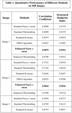
The proposed algorithm is applied to 2-D T1 and T2 post-contrast MR images of human brain containing tumor and their performance is analyzed for tumor extraction. MR brain images are collected from websites: www.radquiz.com, mri.co.nz/medimgs, newsroom.ucla.edu, www.ajnr.org. Implementation is done in Matlab 7.5 (R2007b).The performance metrics [50], [51]: coefficient of correlation (CoC) and structural similarity index (SSIM) are computed for quantitative comparison. The quantitative results are presented
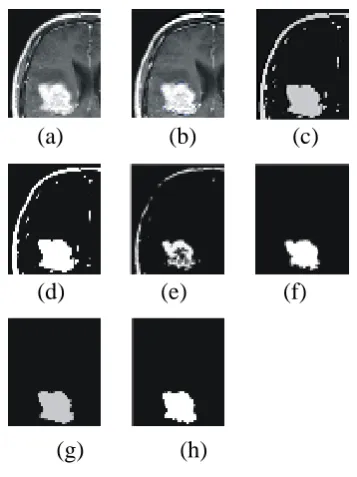
in table I. The numerical values of the metrics reveal that the proposed algorithm has higher coefficient of correlation and SSIM as compared to other existing methods and have comparable performance with enhanced thresholding method [48]. This implies that proposed algorithm extracts tumor region effectively. For visual comparison segmentation results are shown in figure 1 to 5. The segmented images reveal that standard fuzzy c-mean algorithm doesn’t extract the tumor region whereas the enhanced fuzzy c-mean algorithm is efficient enough to extract the tumor properly.
\\
(a) (b) (c)
[image:3.595.337.516.200.439.2](d) (e) (f)
Figure 2.Performance analysis of various segmentation algorithms on MR brain image (a) Original image (b) ROI
marked for segmentation (c) Standard Fuzzy c-mean (d) Standard Thresholding (e) Standard K-means (f) FMCS (g) Enhanced Fuzzy c-mean(h) Enhanced Thresholding
(g) (h)
(a)
(b) (c)
(d) (e) (f)
(g) (h)
Figure 1.Performance analysis of various segmentation algorithms on MR brain image (a) Original image (b)
ROI marked for segmentation (c) Standard Fuzzy c-mean (d) Standard Thresholding (e) Standard K-c-means
[image:3.595.309.508.493.703.2]Image Methods Correlation Coefficient
Structural Similarity
Index
Image 1
Standard Fuzzy c-mean 0.4089 0.5379
Standard Thresholding 0.4089 0.5379
Standard K-means 0.5074 0.9033
FMCS algorithm 0.9647 0.9892
Enhanced Fuzzy
c-mean 0.9879 0.9952
Enhanced Thresholding 0.9790 0.9932
Image 2
Standard Fuzzy c-mean 0.7321 0.8354
Standard Thresholding 0.7344 0.8418
Standard K-means 0.5626 0.8427
FMCS algorithm 0.9035 0.9508
Enhanced Fuzzy
c-mean 0.9844 0.9826
Enhanced Thresholding 0.9708 0.9734
Image 3
Standard Fuzzy c-mean 0.4844 0.6273
Standard Thresholding 0.4750 0.6115
[image:4.595.342.509.79.293.2]Standard K-means 0.6793 0.8871 Table 1. Quantitative Performance of Different Methods
[image:4.595.84.247.85.282.2]on MR Images
Figure 4.Performance analysis of various segmentation algorithms on MR brain image (a) Original image (b) ROI
marked for segmentation (c) Standard Fuzzy c-mean (d) Standard Thresholding (e) Standard K-means (f) FMCS (g) Enhanced Fuzzy c-mean(h) Enhanced Thresholding
(a) (b) (c)
(d) (e) (f)
(g) (h)
[image:4.595.92.259.359.572.2](g) (h)
Figure 5.Performance analysis of various segmentation algorithms on MR brain image (a) Original image (b) ROI
marked for segmentation (c) Standard Fuzzy c-mean (d) Standard Thresholding (e) Standard K-means (f) FMCS (g) Enhanced Fuzzy c-mean(h) Enhanced Thresholding
(a) (b) (c)
(d) (e) (f)
(a) (b) (c)
(d) (e) (f)
[image:4.595.299.549.365.757.2](g) (h)
Figure 3.Performance analysis of various segmentation algorithms on MR brain image (a) Original image (b) ROI
FMCS algorithm 0.9847 0.9921
Enhanced Fuzzy
c-mean 0.9853 0.9933
Enhanced Thresholding 0.9875 0.9942
Image 4
Standard Fuzzy c-mean 0.6117 0.4512
Standard Thresholding 0.6118 0.4527
Standard K-means 0.6973 0.9011
FMCS algorithm 0.9188 0.9654
Enhanced Fuzzy
c-mean 0.9857 0.9853
Enhanced Thresholding 0.9844 0.9847
Image 5
Standard Fuzzy c-mean 0.4507 0.5597
Standard Thresholding 0.4508 0.5610
Standard K-means 0.9161 0.9849
FMCS algorithm 0.4968 0.6968
Enhanced Fuzzy
c-mean 0.9096 0.9845
Enhanced Thresholding 0.9004 0.9857
5.CONCLUSIONS
From the numerical results and visual inspection, it was concluded that enhanced fuzzy c-mean algorithm is more effective and efficient than existing segmentation methods in extracting the tumor region from MR brain images. It yields comparable performance with the recently proposed enhanced thresholding algorithm. This algorithm can also be applied in other applications to segment region of interest. By saving only region of interest storage as well as bandwidth requirements can be reduced. The performance of algorithm depends on the proper choice of seed point.
6. REFERENCES
[1] Gajanayake, Randike, Yapa, Roshan Dharshana, Hewavithana and Badra. 2009. Comparison of standard image segmentation methods for segmentation of brain tumors from 2D MR images. In Proc. IEEE 4th International Conference on Industrial and Information Systems, ICIIS’09, pp. 301-305.
[2] M. Rastgarpour and J. Shanbehzadeh. 2011. Application of AI Techniques in Medical Image Segmentation and Novel Categorization of Available Methods and Tools. International MultiConference of engineers and computer scientists, IMECS’11, Vol. 1.
[3] A. Halder and D.K. Kole. 2012. Automatic Brain Tumor Detection and Isolation of Tumor Cells from MRI Images. International Journal of Computer Applications, Vol.39, No.2.
[4] J. Bezdek, L. Hall and L. Clarke. 1993. Review of MR image segmentation using pattern recognition. Journal of Medical Physics, Vol. 20, pp. 1033–1048.
[5] J. K. Udupa, L. Wei, S. Samarasekera, Y. Miki, M. A. van Buchem and R. I. Grossman. 1997. Multiple sclerosis lesion quantification using fuzzy-connectedness principles. IEEE Transactions on Medical Imaging, Vol. 16, pp. 598-609.
[6] D.L. Pham. 2003. Unsupervised Tissue Classification in Medical Images using Edge-Adaptive Clustering. In Proc. 25th Annual International Conference of the IEEE EMBS, Mexico.
[7] I. Soesanti, A. Susanto, T.S. Widodo and M. Tjokronagoro. 2011. MRI Brain Images Segmentation Based on Optimized Fuzzy Logic and Spatial Information. International Journal of Video & Image Processing and Network Security, IJVIPNS-IJENS, Vol. 11, No.4. [8] D.L. Pham and J.L. Prince. 1999. Adaptive fuzzy
segmentation of magnetic resonance images. IEEE Transactions on Medical Imaging, Vol.18, No.9, pp. 737-752.
[9] D.L. Pham and J.L. Prince. 1999. An adaptive fuzzy C-means algorithm for image segmentation in the presence of intensity inhomogeneties. Pattern Recognition Letter, Vol.20, No.1, pp. 57-68.
[10] M.N. Ahmed and S.M. Yamany. 2002. A modified fuzzy C-means algorithm for bias field estimation and segmentation of MRI data. IEEE Transactions on Medical Imaging, Vol. 21, No.3, pp. 193-199.
[11] S.Murugavalli and V. Rajamani. 2007. An Improved Implementation of Brain Tumor Detection Using Segmentation Based on Neuro Fuzzy Technique. Journal of Computer Science, Vol. 3, No. 11.
[12] M. C. Clark, L. O. Hall, D. B. Goldgof, R. Velthuizen, F. R. Murtaugh and M. S. Silbiger. Unsupervised Brain Tumor Segmentation Using Knowledge-based and Fuzzy Techniques.
[13] S. Albayrak and F. Amasyali. 2003. Fuzzy C-Means Clustering on Medical Diagnosis Systems. International 12th Turkish Symposium on Artificial Intelligence and Neural Networks, TAINN’ 03.
[14] L. Jiang and W. Yang. 2003. A Modified Fuzzy C-Means Algorithm for Segmentation of Magnetic Resonance Images. In Proc. 7th Digital Image Computing: Techniques and Applications, Sydney.
[15] C. Xu.,D.L Pham and J.L. Prince. 1997. Finding the brain cortex using fuzzy segmentation, isosurfaces and deformable surfaces. In Proc. XVth International Conference on Information Processing in Medical Imaging, IPMI, pp. 399-404.
[16] S.R. Kannan. 2005. Segmentation of MRI Using New Unsupervised Fuzzy C Mean Algorithm. ICGST-GVIP Journal, Vol. 5, No. 2.
[17] K.S. Chuang, H.L. Tzeng, S. Chen., J. Wu. and T.J. Chen. 2006. Fuzzy C-Means Clustering with Spatial Information for Image Segmentation. Computerized Medical Imaging and Graphics, Vol. 30, pp. 9-15.
[18] B. Cherradi, O. Bouattane, M. Youssfi and A. Raihani. 2011. Brain Extraction and Fuzzy Tissue Segmentation in Cerebral 2D T1-Weigthed Magnetic Resonance Images. International Journal of Computer Science Issues, Vol. 8, No.3, In Press.
Segmentation from Brain MRI Images. Journal of Bioengineering & Biomedical Science, Vol. 2, No.1. [20] S.K.S. Fan and Y. Lin. 2007. A multi-level thresholding
approach using a hybrid optimal estimation algorithm. Pattern Recognition Letter, Vol. 28, pp. 662-669.
[21] P.S. Liao, T.S. Chen and P.C. Chung. 2001. A fast algorithm for multi-level thresholding. Journal of Inf. Sci. Engg., Vol. 17, pp. 713-727.
[22] D.Y. Huang and C.H. Wang. 2009. Optimal multi-level thresholding using a two-stage Otsu optimization approach. Pattern Recognition Letter, Vol. 30, pp. 275-284.
[23] R. Al-Attas and A. El-Zaart. 2007. Thresholding of medical images using minimum cross entropy. In Proc. IFMBE, Vol. 15, pp. 296-299.
[24] H.P. Ng, S.H. Ong, K.W.C. Foong, P.S. Goh and W.L. Nowinski. 2006. Medical Image Segmentation using K-Means clustering and Improved Watershed Algorithm. IEEE.
[25] C.W. Chen, J. Luo and K.J. Parker. 1998.Image segmentation via adaptive K-mean clustering and knowledge based morphological operations with biomedical applications. IEEE Transactions on Image Processing, Vol.7, No. 12, pp 1673-1683.
[26] L. Kaur and G. Kaur. 2011. Comparison of Foreground Marker control with Watershed Segmentation Algorithms for Tumor Detection in 2D MR Images. Elsevier Journal of Digital signal processing, submitted.
[27] T. Kanungo, D.M. Mount, N.S. Netanyahu, C.D. Piatko, R. Silverman and A.Y. Wu .2002.An efficient k-means clustering algorithm: analysis and implementation. IEEE Transactions on Pattern Analysis and Machine Intelligence, Vol. 24, No. 7, pp. 881-892.
[28] V. Grau, A. U. J. Mewes, M. Alcañiz, R. Kikinis and S. K. Warfield. 2004.Improved Watershed Transform for Medical Image Segmentation Using Prior Information. IEEE Transactions on Medical Imaging, Vol. 23, No.4. [29] K. Parvati, B. S. P. Rao and M. M. Das. 2008.Image
Segmentation Using Gray-Scale Morphology and Marker-Controlled Watershed Transformation. Hindawi Publishing Corporation Discrete Dynamics in Nature and Society, Vol. 2008, Article ID 384346.
[30] Z. Yu, Y. Zhao and X.F. Wang. 2008. Research Advances and Prospects of Mathematical Morphology in Image Processing,” IEEE.
[31] L. Vincent. 1993. Morphological grayscale reconstruction in image analysis: applications and efficient algorithms. IEEE Transactions on Image Processing, Vol. 2, No. 2, pp. 176–201.
[32] C.W. Chen, J. Luo and K.J. Parker. 1998. Image segmentation via adaptive K-mean clustering and knowledge based morphological operations with biomedical applications. IEEE Transactions on Image Processing, Vol.7, No. 12, pp 1673-1683.
[33] Gui, Lisowski, Faundez, Huppi, Lazeyras and Kocher. 2011. Automatic segmentation of newborn brain MRI using mathematical morphology. In Proc. IEEE International Symposium on Biomedical Imaging: FromNanotoMacro.
[34] P.V. Ingole and K.D. Kulat. 2011. A Morphological Segmentation Based Features for Brain MRI Retrieval. In
Proc. 4th International Conference on Emerging Trends in Engineering and Technology, ICETET’11.
[35] D. Selvaraj and R. Dhanasekaran. 2010. Segmenting Internal Brain Nuclei in MRI Brain Image Using Morphological Operators. In Proc. International Conference on Computational Intelligence and Software Engineering, CiSE’10.
[36] Kharrat. 2009. Detection of brain tumor in medical images. In Proc. 3rd International Conference on Signals, Circuits and Systems, SCS’09.
[37] P. Dokladal. 2001. Segmentation of 3D head MR images using morphological reconstruction under constraints and automatic selection of markers. In Proc. International conference on Image Processing.
[38] L. Singh, R.B. Dubey, Z.A. Jaffery and Z. Zaheeruddin. 2009. Segmentation and Characterization of Brain Tumor from MR Images. In Proc. International conference on Advances in Recent Technologies in Communication and Computing, ARTCom’09.
[39]http://www.mathworks.com/matlabcentral/fileexchange/255 32-fuzzy-c-means-segmentation
[40] J.C. Dunn. 1973. A fuzzy relative of the ISODATA process and its use in detecting compact well separated clusters. Journal of Cybernetics, Vol. 3, No.3, pp. 32–57.
[41] J.C. Bezdek. 1981. Pattern Recognition with Fuzzy Objective Function Algorithms, New York: Plenum Press. [42]http://www.cnblogs.com/nktblog/archive/2012/05/08/24896
04.html
[43] M. Sezgin. 2004. Survey over image thresholding techniques and quantitative performance evaluation. Journal of Electronic Imaging. Vol. 13, pp.146-165. [44] N.R. Pal and S.K. Pal. 1993. A Review on image
segmentation techniques. Pattern Recognition Letter, Vol. 26, No.9, pp. 1277-1294.
[45] N.Otsu. 1979. A Threshold Selection Method from Gray-Level Histograms. IEEE Transactions on Systems, Man, and Cybernetics, Vol. 9, No. 1, pp. 62-66.
[46]http://www.mathworks.com/matlabcentral/fileexchange/307 40-k-means-image-segmentation
[47] A.K. Bhogal, N. Singla and M. Kaur. 2010. Color Image Segmentation using k-means clustering algorithm. International Journal on Emerging Technologies, Vol. 1, No.2, pp. 18-20.
[48] N. Sohi, L. Kaur and S. Gupta. 2012.Enhanced Thresholding algorithm to Extract Tumor region from MR brain images. Proceedings of International Conference on Electrical engineering and Computer Science.
[49] R. Kaur, L. Kaur and S. Gupta. 2011. Enhanced K-Mean Clustering Algorithm for Liver Image Segmentation to Extract Cyst Region. International Journal of Computer Applications, Special Issue on Novel Aspects of Digital Imaging Applications (DIA), Vol. 1, pp. 59–66.
[50] MATLAB statistics toolbox.