MEETING REVIEW
Blood stem cells: from beginning to end
Anna Bigas1,* and Claudia Waskow2,*ABSTRACT
In June 2016, around 200 scientists from all over the world gathered at EMBL headquarters in Heidelberg, Germany to discuss the recent advances in hematopoietic stem cells from three different angles: developmental, adulthood and aging. The meeting, aptly named ‘Hematopoietic stem cells: from the embryo to the aging organism’ also covered cutting-edge technologies applied to this subject, such as single-cell analysis, reprogramming and imaging. This Meeting review summarizes the exciting work that was presented and covers the main themes that emerged from the meeting.
KEY WORDS: Hematopoietic stem cells, Aging, Development, Single-cell genomics, Cell fate, Imaging technologies
Introduction
For two full days in early June 2016, the EMBL campus at Heidelberg, a scientific hub in the heart of Europe, hosted hematopoietic stem cell (HSC) specialists from all over the world. For this exciting conference, a new generation of HSC research scientists– Karima Kissa, Christopher Lancrin, Cristina LoCelso and Catherine Robin– brought together leading scientists with a common interest in HSC biology. The purpose of the meeting was to discuss the recent advances in understanding how HSC biology changes through time: from the early decisions that define HSC emergence in the embryo to the effects of aging on HSC maintenance and integrity (Fig. 1). The program of the meeting was carefully shaped to cover key aspects of HSC generation, homeostasis, aging and disease. A special emphasis was placed on new technologies such as single-cell genomics and advanced imaging techniques. It was the use and development of these new technologies to answer old questions that stimulated the most exciting discussions at the meeting. In this Meeting review, we summarize the main themes that emerged from the various presentations and discussions, and consider where this field is headed in the years to come.
Hematopoietic stem cells in the embryo
The meeting started with a full session on embryonic blood development in different animal models, which highlighted the advantages of using each model. Zebrafish imaging technology proved to be a key research tool in the study of hematopoietic emergence in this model system. Pioneering developers of this technology were present at the meeting (Bertrand et al., 2010; Kissa and Herbomel, 2010) and had used it previously to demonstrate that HSCs emerge directly from the aorta floor through a process called
the endothelial to hematopoietic transition (EHT). These cells then migrate to the cardinal vein before finally homing to the caudal hematopoietic tissue. Talks on this topic were focused on the use of imaging technology to understand the signals that drive HSC emergence. Karima Kissa (CNRS, Montpellier, France) showed preliminary data on how the mechanical forces shape the morphology of the aorta at the site of HSC emergence and recruitment of macrophages have a crucial role in the migration process from the aortic floor to the cardinal vein. Julien Bertrand (University of Geneva, Switzerland) presented data on the role of TFEC, an important transcription factor expressed in vascular cells of the caudal hematopoetic tissue. Here, TFEC controls expression of several cytokines, includingkitlgb. Mutants for the TFEC factor lose all their hematopoietic progenitors by 4 days post-fertilization (dpf ) and become anemic by 8-9 dpf (Mahony et al., 2016). David Traver (UCSD, San Diego, USA), who previously showed a specific requirement for non-canonical Wnt in the somites to induce Notch activation in early blood developmental stages (Clements et al., 2011), discussed a temporal requirement for canonical Wnt signaling in the vasculature, specifically mediated by Wnt9a. He demonstrated that genetic and pharmacological manipulation of Wnt signaling affects the number of HSCs that emerge in the dorsal aorta. The study of mouse embryonic hematopoiesis was the other important topic of the session. In the mouse model, HSCs are formed within hematopoietic c-Kit+ clusters that emerge in the aortic endothelium in the embryonic day (E)10-E11 aorta/gonad/ mesonephros (AGM) region. Catherine Robin (Hubrecht Institute, Utrecht, The Netherlands) introduced a key conundrum on cluster heterogeneity by showing that only 2-3 HSCs are detected in one embryonic aorta, despite the fact that at least 600-700 cells form all the clusters. Interestingly, up to 12 HSCs can be detected if maturation protocols are applied before transplantation into irradiated animals (Taoudi et al., 2008; Zhou et al., 2016) or if the cells are transplanted into the neonate fetal liver (Boisset et al., 2015), indicating that some cells are pre-HSCs. Thus, discussion in this session concentrated on the composition of the hematopoietic clusters as well as how they generate HSCs, and the important signals coming from the AGM niche that may regulate this. Robin discussed how her lab had explored the molecular signatures of the successive populations, leading to HSC production by using single-cell RNA sequencing. By taking advantage of the Gfi1 reporter (Thambyrajah et al., 2016) in conjunction with classical markers, different subpopulations of cells were analyzed and compared at E10 and E11. The niche of nascent HSCs in the AGM was widely discussed. By using a combination of genetic reporters and Cre lines, Vashe Chandrakanthan, a postdoctoral fellow in Pimanda’s lab (UNSW, Australia), dissected different elements of the aortic niche based on the expression of Nestin and PDGFRA. He reported exciting results that demonstrated the induction of LT-HSC activity from E13.5 non-hemogenic endothelial cells by re-aggregation with specific stromal populations at E11.5. Another important element of the AGM niche is the sympathetic nervous system, previously described by Katrin Ottersbach (University of Edinburgh, UK) 1
Stem Cell and Cancer Research Group, Program in Cancer Research, Institut
Hospital del Mar d’Investigacions Mèdiques (IMIM), 08003 Barcelona, Spain.
2
Regeneration in Hematopoiesis, Institute for Immunology, Technische Universitätt
Dresden, 01307 Dresden, Germany.
*Authors for correspondence ([email protected]; [email protected])
A.B., 0000-0003-4801-6899
DEVEL
O
(Fitch et al., 2012). In her talk, Ottersbach showed how embryos deficient for p57Kip2/Cdkn1c have an expanded sympathetic nervous system compartment, as detected by Gata3 expression and explained how this translates into an increase of HSC production as a direct effect via the β2-adrenergic receptor expressed on emerging HSCs.
In addition to newly identified signals, one of the old players, the Notch pathway, was also covered in this session. Anna Bigas (IMIM, Barcelona, Spain) indicated that different levels of Notch activity are used to specify different tissues. By using two reporter mouse models developed in the laboratory of Raphael Kopan (Liu et al., 2015) that trace cells with different levels of Notch-activation history, she showed how she could distinguish between two distinct Notch-dependent fates–arterial and hematopoietic–that originate from different precursor cells (Gama-Norton et al., 2015). Similarly, Michelina Iacovino (UCLA, Los Angeles, USA) presented data to suggest that cis inhibition of the Notch pathway downstream of HoxA3 could play a role in blocking blood development during embryonic stem cell (ESC) differentiation.
Finally, Thierry Jaffredo (CNRS-INSERM-UPCM, Paris, France) brought all the discussions together with his inspiring keynote lecture on how different species have evolved to generate HSCs and their associated niches. He presented his lab’s research on identification of the hemogenic endothelium using tracing techniques, ESCs and live imaging. Jaffredo also discussed recent work on the pre-somitic mesoderm as a source of endothelium, hemogenic endothelium and cells undergoing EHT (Yvernogeau et al., 2016). Some of the finer points on the formation of the dorso-ventral polarity of the aorta in the mouse, zebrafish, chick and human embryos–the size of the notochord and the influence of the Shh-VEGF axis, for example–and how it affects the polarization of HSC emergence were also discussed.
Hematopoietic stem cells in the adult The hematopoietic stem cell niche
A full day of the meeting was dedicated to talks on the localization of adult HSCs and how their differentiation is regulated. By integrating intravital microscopy, computational analysis of images, flow cytometry and mathematical modelling, Cristina Lo Celso (Imperial
College London, UK) addressed how stresses such as infection or leukemia influence normal blood cell generation in situ. Cristina showed that during infection, HSCs become either motile (although not mobilized) or very still, and that the flux through HSC and progenitor (HSPC) populations is likely to be dramatically altered (Vainieri et al., 2016). Furthermore, results presented by Delfim Duarte from her laboratory highlighted the increasing migratory behavior of leukemia cells from early bone marrow infiltration to overt chemoresistance and their relationship with bone marrow niches known to maintain HSCs (Hawkins et al., 2016). In keeping with this theme, Paul Frenette (Albert Einstein College of Medicine, New York, USA) emphasized a complex interplay between niche cells and hematopoietic cells in the bone marrow under steady state conditions in the adult. Frenette also reported exciting data on HSC localization during development, where Nestin+NG2+pericytes that are associated with portal vessels provide HSC niches that support HSC expansion in the fetal liver (Khan et al., 2016). Interestingly, HSCs appear to relocate from the fetal liver to the bone marrow upon loss of these specialized pericytes.
Xenotransplantation of human HSCs into a mouse HSC niche is a crucial tool for understanding how human HSCs interact with the niche, and new strategies of transplantation without conditioning allows the study of this interaction under steady-state conditions (Cosgun et al., 2014). To this end, Claudia Waskow (Technical University of Dresden, Germany) presented data to show that human donor HSCs can communicate with murine niche cells upon transplantation, suggesting that they shape their microenvironment according to their needs. It will be interesting to visualize HSCs in their complex environment once multicolor imaging becomes available. To this end, Timm Schroeder (ETH Zürich, Basel, Switzerland) discussed novel tools for long-term single-cell imaging and tracking (Hilsenbeck et al., 2016) and how they were used to quantify expression dynamics of transcription factors in differentiating HSCs. The results challenge the current model of myeloid lineage choice initiation by a PU.1-Gata1 stochastic switch (Hoppe et al., 2016).
Hematopoietic stem cell differentiation
A new concept of how steady-state hematopoiesis occurs in the adult mouse was presented by Katrin Busch from the Rodewald
Specification Amplification
Leukemia
HSCs in development
HSCs in the adult
Homeostasis
Aging Differentiation
Mouse embryo
Yolk sac
Zebrafish embryo
[image:2.612.44.568.56.293.2]Adult mouse
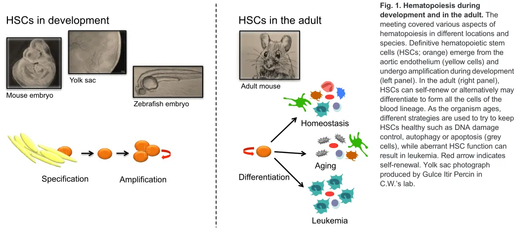
Fig. 1. Hematopoiesis during development and in the adult.The meeting covered various aspects of hematopoiesis in different locations and species. Definitive hematopoietic stem cells (HSCs; orange) emerge from the aortic endothelium (yellow cells) and undergo amplification during development (left panel). In the adult (right panel), HSCs can self-renew or alternatively may differentiate to form all the cells of the blood lineage. As the organism ages, different strategies are used to try to keep HSCs healthy such as DNA damage control, autophagy or apoptosis (grey cells), while aberrant HSC function can result in leukemia. Red arrow indicates self-renewal. Yolk sac photograph produced by Gulce Itir Percin in C.W.’s lab.
DEVEL
O
laboratory (DKFZ, Heidelberg, Germany). She presented a self-generated elegantin situfate mapping mouse tool that allows tracing of differentiating progeny that arise in the bone marrow from the most primitive HSCsin situ(Busch et al., 2015). Most immature HSCs were only rarely involved in the daily generation of new blood cells, implying that steady-state hematopoiesis is largely independent of the activity of HSCs over long periods of time. In contrast, despite the low contribution of individual HSCs, many HSCs participate in hematopoiesis under steady state–just at very low frequencies. This is in stark contrast to hematopoiesis in post-transplantation settings that relies on the activity of just a few clones.
How is HSC function regulated? Martijn Nolte (Sanquin, The Netherlands) provided insight into the regulation of HSC self-renewal by T cells, particularly by memory CD8+T cells and also virus-specific memory CD8+ T cells, by a yet to be determined soluble factor. To uncover new regulators of HSC function, Nina Cabezas-Wallscheid, a postdoctoral fellow in Andreas Trumpp’s laboratory (DKFZ and HI-STEM, Heidelberg, Germany), showed by bulk and single-cell RNA-seq analyses that the transition from dormancy towards cell cycle entry is not achieved by a simple binary on/off switch, but rather that it follows a continuous upregulation of all major biosynthetic processes: transcription, splicing, translation, metabolism and so on. During dormancy, cells tune down these processes to a very low base level. The team also reported a novel transgenic reporter mouse that specifically labels dormant HSCs and thus avoids time consuming label retention assays to identify, isolate or characterize these extremely rare cells. One of the key regulators controlling entry and exit from dormancy in HSCs and in pluripotent cells of the pre-implantation embryo is the Myc oncogene (Laurenti et al., 2008; Scognamiglio et al., 2016). While the JAK-STAT pathway controls Myc expression in pluripotent cells, data were provided suggesting that retinoic acid signaling is upstream of Myc in HSCs.
To better understand HSC commitment and differentiation and how these processes are regulated, Simon Haas from Marieke Essers’lab (DKFZ and HI-STEM, Heidelberg, Germany) together with the Steinmetz (EMBL, Heidelberg/Stanford University) and Trumpp groups developed a novel multiplexed approach that integrates flow-cytometric, transcriptomic and functional lineage-fate data at the single-cell level. The data were used to characterize developmental transitions during HSC commitment and differentiation and point to a novel model for a continuous differentiation flow conceptualized by a ‘developmental continuum’. The data sets were used to identify the molecular and cell biological processes, transcription factors and signaling pathways associated with lineage commitment and maturation. Adding an interesting metabolic angle to the story, Satish Khurana (The Indian Institute of Science Education and Research, Thiruvananthapuram, India) used a comparative gene expression approach between fetal liver (FL)- and adult bone marrow-derived HSCs, to show that highly proliferative FL HSCs met their increased energy demand by upscaling the number and activity of mitochondria (Manesia et al., 2015). Importantly, niche-HSC interactions were retrieved by that screen and outside-in integrin signaling via integrin-αv (Itgav) identified as a key pathway regulating the proliferation of HSCs. Consistent with these data, loss of Itgav led to increased HSC proliferation and loss of stemness in vivo. Continuing with the metabolic theme, Toshio Suda (National University of Singapore, Singapore) gave an excellent keynote lecture about HSC metabolism, focusing on the difference between steady state and stress hematopoiesis with regard to HIF-1α
function and p38 signaling. He described how the folliculin (Flcn)-Tfe3 axis is also involved in suppression of oxidative metabolism, and how Flcn-deleted mice show multi-abnormal phenotypes, which include bone marrow failure, abnormal function of macrophages (hemophagocytosis) and osteoclasts (resulting in osteoporosis) (Baba et al., 2016). These phenotypes might be explained by a common cause, which is the result of a hyper-oxidative metabolism and nucleic acid disturbance within the niche. He discussed how metabolic state is not a result of cell status, but that it can cause changes in cell differentiation and activity.
Hematopoietic stem cell aging
HSC aging was specifically covered in the meeting and introduced by Emmanuelle Passague from the University of California, USA, who discussed her work showing that autophagy protects HSCs from aging-associated functional decline (Warr et al., 2013). Passague showed how HSCs from aged mice display striking heterogeneity with respect to their autophagy levels, with high autophagy levels maintaining a low metabolic state and protecting the cell’s intrinsic robust long-term engraftment activity. Hartmut Geiger (Cincinnati Children’s Hospital Medical Center, Cincinnati, USA and University of Ulm, Germany) showed that inhibition of the small Rho GTPase Cdc42 rejuvenates HSC function. In fact, a shift from canonical to non-canonical Wnt signaling as a result of increased expression of Wnt5a in aged HSCs switches the aging process‘on’via activation of Cdc42. Consistent with this, Wnt5a haploinsufficiency and knockdown approaches both result in rejuvenation of HSCs (Florian et al., 2013). Subsequently, Novella Guidi, a graduate student from Geiger’s laboratory, presented exciting data on the essential role of stroma-derived osteopontin in HSC aging in vivo. A rejuvenating effect was observed following brief exposure to thrombin-cleaved osteopontin, suggesting that HSC aging might in fact be reversible. Karl Lenhard Rudolph (Fritz Lipmann Institute for Aging, Jena, Germany) turned our focus towards DNA damage control. Rudolph presented an update on his previous work showing that DNA damage limits the self-renewal of HSCs by inducing BATF-dependent lymphoid differentiation (Wang et al., 2012). His current work is now aimed at analyzing the consequences of BATF deletion on hematopoiesis and leukemia formation in aging mice. Moving from aging to disease, Richard Groen (VU University Medical Center, Amsterdam, The Netherlands) presented recent advances on his lab’s efforts to build a bone marrow-like HSC niche using ossicles that were previously shown to be optimized tools for the engraftment of leukemic cells (Sontakke et al., 2016). Groen showed how adding human blood vessels to the bone marrow-like ossicle system resulted in improved human HSC, acute myeloid leukemia and multiple myeloma engraftment and differentiation upon xenotransplantation intoRag2−/−Ll2rg(γc)−/− recipient mice.
Emerging technologies for hematopoietic stem cell research Emerging technologies are providing new avenues to explore old questions in HSC biology. In particular, single-cell RNA-seq has become a powerful tool to decipher the trajectory of cells within a given differentiation pathway. To this end, Dana Pe’er (Columbia University, New York, USA) described the use of single-cell datasets to map developmental trajectories and how these go awry in disease. By taking a multi-dimensional approach, combining single-cell data from mass cytometry and RNA-seq, she showed how it was possible to order cells according to their developmental progression and label each cell as pre-bifurcation or as one of two post-
DEVEL
O
bifurcation cell fates (Setty et al., 2016). She highlighted the importance of distinguishing between the noise/artifact and biological signal in these high-throughput technologies, stressing that methods that do not account for this can be misleading. Once cells were aligned on a trajectory, she was able to follow the order of events during development and identify transitional populations, as well as the populations that go awry in disease.
Single-cell RNA-seq is an equally powerful tool in the study of developmental HSC biology, as it can be used to decipher the trajectory of embryonic cells. Bertie Gottgens (University of Cambridge, UK) reported a regulatory network model for hematopoiesis based on extensive experimental evidence and validated by single-cell expression profiling (Schütte et al., 2016). He also demonstrated how ectopic activation of cardiac genes in Tal1+yolk sac endothelium is unlikely to be the immediate result of a binary fate switch (Scialdone et al., 2016). Taking a more global look at the regulatory dynamics of hematopoietic specification, Constanze Bonifer (University of Birmingham, UK) presented the work of a UK-wide consortium aimed at obtaining a comprehensive genome-wide map of transcription factor occupancy, chromatin accessibility, histone modifications and gene expression from a complete developmental pathway (Goode et al., 2016). To this end, the groups performedin vitrodifferentiation of mouse ESCs and purified the cells from six different stages of hematopoietic specification and differentiation. They were able to identify the dynamic gene regulatory networks that regulate the transition between these the different cell types: from mesodermal precursors to macrophages. One of the important pathways identified in the hemogenic endothelium was Hippo signaling, which may be an important regulator in the early stages of hematopoietic differentiation. A relatively new and innovative approach to understanding gene expression at the four-dimensional level–that is, through both space and time – is ‘Tomo-seq’, a genome-wide RNA tomography approach introduced by Laurent Yvernogeau, a postdoctoral fellow in Catherine Robin’s Lab (Junker et al., 2014). He collected and sequenced sequential sections of whole aortas (cut along the anterior-to-posterior axis) and thick transversal embryo slices (cut along the ventral-to-dorsal axis) from various embryo species isolated at different developmental stages: before, during and after cluster/HSC emergence. This approach will provide a comprehensive evolutionary map of gene expression in the developing AGM region. Lineage reprogramming may still be considered a relatively recent technology in the HSC field, and this meeting heard progress from two labs on this topic. Shahin Rafii (Cornell University Ithaca, USA) introduced an innovative reprogramming approach whereby endothelial cells (ECs) could be converted into bona-fide transplantable HSCs (Sandler et al., 2014). By employing Runx1 reporter mice, his group could distinguish and track the emergence of reprogrammed ECs into authentic HSCs, which were capable of serial and long-term multilineage repopulation, including the production of polarized T-cells. Data from reprogramming fibroblasts was presented by Carlos-Filipe Pereira (University of Coimbra, Portugal), who then used this information to isolate early precursor HSC cells from the mid-gestation placenta. Pereira characterized these cells by single-cell RNA-seq and further showed that they expressed Prom1, Sca1 and CD34, and localized to the placental vascular labyrinth at the maternal-to-fetal interface (Pereira et al., 2016).
Concluding remarks
The study of hematopoiesis has a long history and, although the field has come far, many of the key questions remain. One of the
most exciting aspects of this EMBL meeting was the realization that we may now be close to definitively answering some of these questions, thanks to recent advances in single-cell data collection and analysis, as well as new approaches to imaging. The majority of meeting participants agreed that these technologies will soon allow a clearer picture of hematopoiesis and leukemogenesis to emerge. There is no doubt that this is a unique and exciting time to be studying hematopoietic development, and we look forward to discussing future breakthroughs at the next EMBL meeting on hematopoiesis in 2018.
Acknowledgements
We would like to thank the meeting organizers and supporters. We apologize to all the speakers and references that are not mentioned directly owing to space limitations.
Competing interests
The authors declare no competing or financial interests.
Funding
A.B. is funded by Ministerio de Economıa y Competitividad [SAF2013-40922-R];́
Red Temática de Investigación Cooperativa en Cáncer [RD12/0036/0054]; and Agència de Gestiód’Ajuts Universitaris i de Recerca (AGAUR) [2014SGR-124]. C.W. is funded by the Deutsche Forschungsgemeinschaft (DFG) [WA2837, FOR2033-A03, SFB655-B9, TRR127-A5]; and the Else Kröner-Fresenius-Stiftung.
References
Bertrand, J. Y., Chi, N. C., Santoso, B., Teng, S., Stainier, D. Y. R. and Traver, D.
(2010). Haematopoietic stem cells derive directly from aortic endothelium during development.Nature464, 108-111.
Boisset, J.-C., Clapes, T., Klaus, A., Papazian, N., Onderwater, J.,
Mommaas-Kienhuis, M., Cupedo, T. and Robin, C.(2015). Progressive maturation toward
hematopoietic stem cells in the mouse embryo aorta.Blood125, 465-469.
Busch, K., Klapproth, K., Barile, M., Flossdorf, M., Holland-Letz, T., Schlenner,
S. M., Reth, M., Höfer, T. and Rodewald, H.-R.(2015). Fundamental properties
of unperturbed haematopoiesis from stem cells in vivo.Nature518, 542-546.
Clements, W. K., Kim, A. D., Ong, K. G., Moore, J. C., Lawson, N. D. and Traver, D.(2011). A somitic Wnt16/Notch pathway specifies haematopoietic stem cells.
Nature474, 220-224.
Cosgun, K. N., Rahmig, S., Mende, N., Reinke, S., Hauber, I., Schäfer, C.,
Petzold, A., Weisbach, H., Heidkamp, G., Purbojo, A. et al. (2014). Kit
Regulates HSC Engraftment across the Human-Mouse Species Barrier.Cell Stem Cell15, 227-238.
Fitch, S. R., Kimber, G. M., Wilson, N. K., Parker, A., Mirshekar-Syahkal, B.,
Göttgens, B., Medvinsky, A., Dzierzak, E. and Ottersbach, K.(2012). Signaling
from the sympathetic nervous system regulates hematopoietic stem cell emergence during embryogenesis.Cell Stem Cell11, 554-566.
Florian, M. C., Nattamai, K. J., Dörr, K., Marka, G., Überle, B., Vas, V., Eckl, C.,
Andrä, I., Schiemann, M., Oostendorp, R. A. J. et al.(2013). A canonical to
non-canonical Wnt signalling switch in haematopoietic stem-cell ageing.Nature503, 392-396.
Gama-Norton, L., Ferrando, E., Ruiz-Herguido, C., Liu, Z., Guiu, J., Islam,
A. B. M. M. K., Lee, S.-U., Yan, M., Guidos, C. J., López-Bigas, N. et al.(2015).
Notch signal strength controls cell fate in the haemogenic endothelium.Nat.
Commun.6, 8510.
Goode, D. K., Obier, N., Vijayabaskar, M. S., Lie-A-Ling, M., Lilly, A. J., Hannah,
R., Lichtinger, M., Batta, K., Florkowska, M., Patel, R. et al.(2016). Dynamic
gene regulatory networks drive hematopoietic specification and differentiation.
Dev. Cell36, 572-587.
Hawkins, E. D., Duarte, D., Khorshed, R., Akinduro, O., Passaro, D., Nowicka,
M., Scott, M., Rothery, S., Foster, K., Ruivo, N. et al.(2016). T cell acute
leukaemia exhibits dynamic interactions with bone marrow microenvironments.
Nature(in press) doi:10.1038/nature19801.
Hilsenbeck, O., Schwarzfischer, M., Skylaki, S., Schauberger, B., Hoppe, P. S., Loeffler, D., Kokkaliaris, K. D., Hastreiter, S., Skylaki, E., Filipczyk, A. et al.
(2016). Software tools for single-cell tracking and quantification of cellular and molecular properties.Nat Biotech.34, 703-706.
Hoppe, P. S., Schwarzfischer, M., Loeffler, D., Kokkaliaris, K. D., Hilsenbeck, O., Moritz, N., Endele, M., Filipczyk, A., Gambardella, A., Ahmed, N. et al.
(2016). Early myeloid lineage choice is not initiated by random PU.1 to GATA1 protein ratios.Nature535, 299-302.
Junker, J. P., Noël, E. S., Guryev, V., Peterson, K. A., Shah, G., Huisken, J.,
McMahon, A. P., Berezikov, E., Bakkers, J. and van Oudenaarden, A.(2014).
Genome-wide RNA tomography in the Zebrafish embryo.Cell159, 662-675.
DEVEL
O
Khan, J. A., Mendelson, A., Kunisaki, Y., Birbrair, A., Kou, Y., Arnal-Estapé, A.,
Pinho, S., Ciero, P., Nakahara, F., Maayan, A. et al. (2016). Fetal liver
hematopoietic stem cell niches associate with portal vessels. Science 351, 176-180.
Kissa, K. and Herbomel, P. (2010). Blood stem cells emerge from aortic
endothelium by a novel type of cell transition.Nature464, 112-115.
Laurenti, E., Varnum-Finney, B., Wilson, A., Ferrero, I., Blanco-Bose, W. E., Ehninger, A., Knoepfler, P. S., Cheng, P.-F., MacDonald, H. R., Eisenman,
R. N. et al.(2008). Hematopoietic stem cell function and survival depend on c-Myc
and N-Myc activity.Cell Stem Cell3, 611-624.
Liu, Z., Brunskill, E., Boyle, S., Chen, S., Turkoz, M., Guo, Y., Grant, R. and
Kopan, R.(2015). Second-generation Notch1 activity-trap mouse line (N1IP::
CreHI) provides a more comprehensive map of cells experiencing Notch1 activity.
Development142, 1193-1202.
Mahony, C. B., Fish, R. J., Pasche, C. and Bertrand, J. Y.(2016). tfec controls the
hematopoietic stem cell vascular niche during zebrafish embryogenesis.Blood 128, 1336-1345.
Manesia, J. K., Xu, Z., Broekaert, D., Boon, R., van Vliet, A., Eelen, G.,
Vanwelden, T., Stegen, S., Van Gastel, N., Pascual-Montano, A. et al.(2015).
Highly proliferative primitive fetal liver hematopoietic stem cells are fueled by oxidative metabolic pathways.Stem Cell Res.15, 715-721.
Pereira, C.-F., Chang, B., Gomes, A., Bernitz, J., Papatsenko, D., Niu, X., Swiers,
G., Azzoni, E., de Bruijn, M. F. T. R., Schaniel, C. et al.(2016). Hematopoietic
reprogramming in vitro informs in vivo identification of hemogenic precursors to definitive hematopoietic stem cells.Dev. Cell36, 525-539.
Sandler, V. M., Lis, R., Liu, Y., Kedem, A., James, D., Elemento, O., Butler, J. M.,
Scandura, J. M. and Rafii, S.(2014). Reprogramming human endothelial cells to
haematopoietic cells requires vascular induction.Nature511, 312-318.
Schütte, J., Wang, H., Antoniou, S., Jarratt, A., Wilson, N. K., Riepsaame, J.,
Calero-Nieto, F. J., Moignard, V., Basilico, S., Kinston, S. J. et al.(2016). An
experimentally validated network of nine haematopoietic transcription factors reveals mechanisms of cell state stability.eLife5, e11469.
Scialdone, A., Tanaka, Y., Jawaid, W., Moignard, V., Wilson, N. K., Macaulay,
I. C., Marioni, J. C. and Göttgens, B. (2016). Resolving early mesoderm
diversification through single-cell expression profiling.Nature535, 289-293.
Scognamiglio, R., Cabezas-Wallscheid, N., Thier, M. C., Altamura, S., Reyes,
A., Prendergast, Á. M., Baumgärtner, D., Carnevalli, L. S., Atzberger, A.,
Haas, S. et al.(2016). Myc depletion induces a pluripotent dormant state
mimicking diapause.Cell164, 668-680.
Setty, M., Tadmor, M. D., Reich-Zeliger, S., Angel, O., Salame, T. M., Kathail, P.,
Choi, K., Bendall, S., Friedman, N. and Pe’er, D.(2016). Wishbone identifies
bifurcating developmental trajectories from single-cell data.Nat Biotech. 34, 637-645.
Sontakke, P., Carretta, M., Jaques, J., Brouwers-Vos, A. Z., Lubbers-Aalders, L., Yuan, H., de Bruijn, J. D., Martens, A. C., Vellenga, E., Groen, R. W. et al.
(2016). Modeling BCR-ABL and MLL-AF9 leukemia in a human bone marrow-like scaffold-based xenograft model.Leukemia(in press). doi:10.1038/leu.2016.108.
Taoudi, S., Gonneau, C., Moore, K., Sheridan, J. M., Blackburn, C. C., Taylor, E.
and Medvinsky, A.(2008). Extensive hematopoietic stem cell generation in the
AGM region via maturation of VE-cadherin+CD45+ pre-definitive HSCs.Cell Stem Cell3, 99-108.
Thambyrajah, R., Mazan, M., Patel, R., Moignard, V., Stefanska, M.,
Marinopoulou, E., Li, Y., Lancrin, C., Clapes, T., Möröy, T. et al.(2016).
GFI1 proteins orchestrate the emergence of haematopoietic stem cells through recruitment of LSD1.Nat. Cell Biol.18, 21-32.
Vainieri, M. L., Blagborough, A. M., MacLean, A. L., Haltalli, M. L., Ruivo, N.,
Fletcher, H. A., Stumpf, M. P., Sinden, R. E. and Celso, C. L.(2016). Systematic
tracking of altered haematopoiesis during sporozoite-mediated malaria development reveals multiple response points.Open Biol.6, 160038.
Wang, J., Sun, Q., Morita, Y., Jiang, H., Groß, A., Lechel, A., Hildner, K.,
Guachalla, L. M., Gompf, A., Hartmann, D. et al.(2012). A differentiation
checkpoint limits hematopoietic stem cell self-renewal in response to DNA damage.Cell148, 1001-1014.
Warr, M. R., Binnewies, M., Flach, J., Reynaud, D., Garg, T., Malhotra, R.,
Debnath, J. and Passegué, E.(2013). FOXO3A directs a protective autophagy
program in haematopoietic stem cells.Nature494, 323-327.
Yvernogeau, L., Gautier, R., Khoury, H., Menegatti, S., Schmidt, M., Gilles, J.-F.
and Jaffredo, T.(2016). Anin vitromodel of hemogenic endothelium commitment
and hematopoietic production.Development143, 1302-1312.
Zhou, F., Li, X., Wang, W., Zhu, P., Zhou, J., He, W., Ding, M., Xiong, F., Zheng, X.,
Li, Z. et al.(2016). Tracing haematopoietic stem cell formation at single-cell
resolution.Nature533, 487-492.