The anaphylatoxin C3a downregulates the Th2
response to epicutaneously introduced antigen
Seiji Kawamoto, … , Craig Gerard, Raif S. Geha
J Clin Invest. 2004;
114(3)
:399-407.
https://doi.org/10.1172/JCI19082
.
Mechanical injury to the skin results in activation of the complement component C3 and
release of the anaphylatoxin C3a. C3a binds to a seven-transmembrane G protein–coupled
receptor, C3aR. We used C3aR
–/–mice to examine the role of C3a in a mouse model of
allergic inflammation induced by epicutaneous sensitization with OVA. C3aR
–/–mice
exhibited an exaggerated Th2 response to epicutaneous but not to intraperitoneal
sensitization with OVA, as evidenced by significantly elevated levels of serum OVA-specific
IgG1 and significantly increased secretion of the Th2 cytokines IL-4, IL-5, and IL-10 by
antigen-stimulated splenocytes. Presentation of OVA peptide by C3aR
–/–APCs caused
significantly more IL-4 and IL-5 secretion by T cells from OVA–T cell receptor (OVA-TCR)
transgenic mice compared with presentation by WT APCs. C3a inhibited the ability of
splenocytes, but not of highly purified T cells, to secrete Th2 cytokines in response to TCR
ligation. This inhibition was mediated by IL-12 secreted by APCs in response to C3a. These
results suggest that C3a-C3aR interactions inhibit the ability of APCs to drive Th2 cell
differentiation in response to epicutaneously introduced antigen and may have important
implications for allergic skin diseases.
Article
Immunology
Find the latest version:
http://jci.me/19082/pdf
The anaphylatoxin C3a downregulates
the Th2 response to epicutaneously
introduced antigen
Seiji Kawamoto,1,2 Ali Yalcindag,1 Dhafer Laouini,1 Scott Brodeur,1 Paul Bryce,1 Bao Lu,3
Alison A. Humbles,3 Hans Oettgen,1 Craig Gerard,3 and Raif S. Geha1
1Division of Immunology, Children's Hospital and the Department of Pediatrics, Harvard Medical School, Boston, Massachusetts, USA. 2Graduate School of Advanced Sciences of Matter, Hiroshima University, Higashi-Hiroshima, Japan. 3Division of Pulmonary Medicine and
Ina Sue Perlmutter Laboratory, Children’s Hospital and the Department of Pediatrics, Harvard Medical School, Boston, Massachusetts, USA.
Mechanical injury to the skin results in activation of the complement component C3 and release of the
anaphylatoxin C3a. C3a binds to a seven-transmembrane G protein–coupled receptor, C3aR. We used C3aR
–/–mice to examine the role of C3a in a mouse model of allergic inflammation induced by epicutaneous
sensitiza-tion with OVA. C3aR
–/–mice exhibited an exaggerated Th2 response to epicutaneous but not to intraperitoneal
sensitization with OVA, as evidenced by significantly elevated levels of serum OVA-specific IgG1 and
signifi-cantly increased secretion of the Th2 cytokines IL-4, IL-5, and IL-10 by antigen-stimulated
splenocytes.
Pre-sentation of OVA peptide by C3aR
–/–APCs caused significantly more IL-4 and IL-5 secretion by T cells from
OVA–T cell receptor (OVA-TCR) transgenic mice compared with presentation by WT APCs. C3a inhibited the
ability of splenocytes, but not of highly purified T cells, to secrete Th2 cytokines in response to TCR ligation.
This inhibition was mediated by IL-12 secreted by APCs in response to C3a. These results suggest that
C3a-C3aR interactions inhibit the ability of APCs to drive Th2 cell differentiation in response to epicutaneously
introduced antigen and may have important implications for allergic skin diseases.
Introduction
The complement cascade is an important system that consists of proenzymes that become activated sequentially to perform diverse biologic functions (1). The third component of the complement system, C3, is the pivotal molecule in this cascade, and its activa-tion leads to the release of several peptides from the parent mol-ecule. In a first step, C3 is cleaved to C3a and C3b. Subsequently, C3b is cleaved to iC3b and C3dg. Each of these peptides acts as a ligand for specific receptor(s) to mediate classical C3-dependent functions such as opsonization, leukocyte chemotaxis and smooth muscle cell contraction (2). In the past few years, C3 has been also been found to play a role in the adaptive immune response. C3 has been shown to be necessary for an optimal antibody response to T-dependent antigens (3). Most C3 present in the serum is syn-thesized by the liver (4), but local synthesis by hematopoietically derived cells plays a more important role in immunomodulatory functions, since C3–/– mice reconstituted with C3+/+ bone
mar-row are rescued from their impaired antibody response to T cell– dependent antigens (5). Extrahepatic synthesis of C3 occurs in macrophages, endothelial cells, and kidney tubular cells (2). Skin keratinocytes are also a rich source of C3, and C3 can be activated in the skin by mechanical and UV radiation injury (6–8).
Murine C3a is a 78-AA peptide derived from the N-terminal end of C3 upon its activation by the classical, alternative, and lectin path-ways (9, 10). Well-described functions of C3a include chemotaxis for mast cells (11) and eosinophils (12), and contraction of smooth
muscle cells (13). C3a performs these functions by engaging its receptor, C3aR, on target cells. C3aR is a seven transmembrane G protein–coupled protein with structural resemblance to chemokine receptors (10). Expression of C3aR by human DCs (14), T cells (15), and B cells (3) suggests that the C3a-C3aR interactions might play a role in immune modulation. In fact, C3a has been shown to enhance IL-6 release by human PBMCs stimulated with LPS or IL-1β (16), and to inhibit IgG and cytokine (IL-6, TNF-α) synthe-sis by human B cells stimulated with Staphylococcus aureus and IL-2 (17). C3aR–/– mice have increased IL-1β in the plasma and exhibit
lethality in response to injection of LPS (18), suggesting that C3aR can act in vivo as an anti-inflammatory receptor by attenuating LPS-induced proinflammatory cytokine production. Following intraperitoneal (i.p.) sensitization, C3aR–/– mice and guinea pigs
with a natural C3aR defect exhibit diminished airway reactivity in response to airway challenge with antigen (19, 20). This is consis-tent with a role for C3a in airway smooth muscle contraction.
Following antigen stimulation, Th cells can develop into Th1 cells that secrete IFN-γ or Th2 cells that secrete IL-4, IL-5, and IL-13 (21). Cytokine microenvironment (22), antigen dose (23), affinity of anti-gens (24), MHC haplotypes and costimulatory factors (25) have all been implicated in Th1/Th2 polarization. DCs also play an impor-tant role in Th cell polarization. Based on their ability to favor Th1 or Th2 differentiation, mature DCs have been called DC1 or DC2 respectively (26). Production of IL-12 by DC1s favors the develop-ment of Th1 cells (27). Bacterial and viral products, and IFN-γ, which are potent stimuli for IL-12 secretion, result in DC1 and subsequent Th1 development, while PGE2, cholera toxin, and extracellular ATP are reported to favor DC2 differentiation (28–31).
We have developed a mouse model of atopic dermatitis (AD) using repeated epicutaneous (EC) sensitization with OVA to tape-stripped skin (28, 29). This model displays many of the features of human
Nonstandard abbreviations used: atopic dermatitis (AD); epicutaneous (EC); intraperitoneal (i.p.); T cell receptor (TCR).
Conflict of interest: The authors have declared that no conflict of interest exists.
research article
AD and results in a vigorous local and systemic Th2 response. This is evidenced by elevated total and antigen-specific IgE and a vigor-ous Th2 cytokine response of splenocytes to stimulation with the immunizing antigen. Mechanical injury to the skin by scratching is an important feature of AD. C3a has been shown to accumulate in blisters induced by mechanical injury to the skin (8). This prompted us to assess the role of C3a in the immune response induced by EC sensitization.
Results
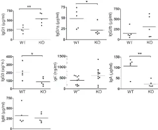
C3aR–/– mice have elevated serum IgG1 and decreased serum IgG2a
lev-els.Analysis of thymocytes, bone marrow, and spleens revealed no detectable differences in cell numbers or distribution of lympho-cyte subsets (CD3+, CD4+, B220+, IgM+, CD40+) between C3aR–/–
mice and WT littermates (data not shown). However, C3aR–/– mice
had significantly higher levels of IgG1 than WT littermates (Figure 1). In contrast, they had significantly lower IgG2a, IgG3, and IgA levels. Since the Th2 cytokines IL-4 and IL-13 play a critical role in isotype switching to IgG1, while the Th1 cytokine IFN-γ plays a critical role in isotype switching to IgG2a, these results suggested that C3a may play an important role in Th cell polarization.
C3aR–/– mice mount an exaggerated Th2 response to EC sensitization with
OVA antigen.Mechanical injury to the skin results in C3 activation and local generation of C3a (8). Since EC sensitization involves tape-stripping of the skin, which results in mechanical injury, we examined the effect of C3aR deficiency on the immune response to epicutaneously introduced antigen. As we previously reported, WT BALB/C mice mounted OVA-specific IgG1, IgE, and IgG2a antibod-ies following EC sensitization (Figure 2), while no OVA-specific anti-bodies were detected in the sera of saline-sensitized mice. C3aR–/–
mice mounted significantly higher levels of OVA-specific IgG1 anti-bodies following EC sensitization than WT littermates. There were no significant differences in the levels of OVA-specific serum IgE or IgG2a levels between C3aR–/– and WT controls.
We next examined cytokine production in response to in vitro stimulation of splenocytes with OVA antigen. OVA stimulation of splenocytes from epicutaneously sensitized BALB/C mice induced the secretion of IL-4, IL-5, and IL-10 (Figure 3). Splenocytes from OVA-sensitized C3aR–/– mice secreted
signifi-cantly higher levels of IL-4, IL-5, and IL-10 in response to OVA. There was no induction of IFN-γ secretion in vitro by splenocytes of epi-cutaneously sensitized WT or C3aR–/– mice.
The increased secretion of Th2 cytokines was specific to the immunizing antigen, because the levels of IL-4, IL-5, and IL-10, as well as of IFN-γ, secreted by splenocytes following stimulation with anti-CD3 was comparable in C3aR–/– mice and WT controls (data not
shown). Taken together, these results suggest that interaction of C3a with its receptor C3aR inhibits Th2 cell differentiation in response to epicutaneously introduced antigen.
Dermal infiltration with eosinophils and expression of Th2 cytokines are an important feature of our model. We also examined Th1 and Th2 cytokine expression in the skin. There was no differ-ence in the numbers of eosinophils in saline-treated skin of C3aR–/–
and WT mice. OVA sensitization elicited a significantly stronger eosinophil influx in OVA-sensitized skin of C3aR–/– compared with
WT controls (Figure 4A). There was negligible IL-4 mRNA expres-sion in saline-sensitized skin of both C3aR–/– mice and WT controls.
OVA-sensitized skin sites of C3aR–/– mice exhibited a trend towards
higher IL-4 mRNA expression compared with WT controls, but the difference was not statistically significant (Figure 4B). There was no significant increase in skin IFN-γ mRNA levels between saline- and OVA-sensitized sites in either WT or C3aR–/– mice, and the IFN-γ
mRNA levels were similar in both strains.
C3aR–/– mice do not exhibit a deviated Th response to i.p. immunization
with OVA antigen.We next examined whether the effect of C3aR defi-ciency on the Th response was selective to the EC route of immu-nization. To this end, we examined the response of C3aR–/– mice
to i.p. immunization with OVA in alum, a mode of immunization that also elicits an immune response with a strong Th2 component. There were no significant differences in the serum OVA-specific IgG1, IgE or IgG2a antibody levels of intraperitoneally immunized C3aR–/– mice and WT controls (Figure 5A). In addition, splenocytes
from intraperitoneally immunized C3aR–/– mice and WT controls
produced similar levels of IL-4, IL-5, IL-10, and IFN-γ when stimu-lated by OVA in vitro (Figure 5B).
We considered the possibility that the antigen dose used for immunization and in vitro antigen stimulation might have resulted in maximal Th2 cytokine production by intraperitoneally immunized mice. This could have prevented the detection of an enhanced Th2 response in intraperitoneally immunized C3aR–/–
[image:3.585.222.541.471.734.2]mice. For this reason, we used an i.p. immunization protocol that consisted of two rather than three injections of antigen (on days 0 and 14) and stimulated the splenocytes in vitro with a range
Figure 1
of OVA concentrations (5 to 50 μg/ml). Figure 5C shows that even under conditions in which Th2 cytokine secretion by WT splenocytes was suboptimal, there was no difference in cytokine secretion by splenocytes of C3aR–/–. These results suggest that the
effect of C3aR deficiency on the Th2 response to antigen is selec-tive to the EC route of immunization.
APCs from C3aR–/– mice enhance the secretion of Th2 cytokines by OVA
transgenic T cells.We directly examined the effect of C3aR deficiency on the ability of APCs to drive Th cell differentiation. Irradiated splenocytes from C3aR–/– mice and WT littermates on a BALB/
C background were used to present OVA323–339 peptide to highly
purified T cells obtained from DO11.10 T cell receptor (TCR) transgenic mice on the same genetic background. Presentation of OVA peptide to the T cells by C3aR–/– APCs resulted in significantly
higher IL-4 and IL-5 secretion compared with presentation by WT APCs (Figure 6). C3aR–/– APCs and WT APCs elicited equivalent
amounts of IFN-γ secretion by OVA-TCR transgenic T cells. These results suggest that the effect of C3a-C3aR interactions on Th cell polarization is exerted, at least in part, at the level of the APCs.
C3a inhibits Th2 responses in vitro.Taken together, the above results suggest that C3a-C3aR interactions inhibit Th2 responses, with no detectable effect on Th1 responses. We directly examined the effect of C3a on the development of Th cells in vitro following stimulation of splenocytes with anti-CD3. Since C3 may be secreted by APCs, we used splenocytes from C3–/– mice to obviate the potential
con-founding effect of endogenously generated C3a. C3a by itself did not induce cytokine secretion (data not shown). Addition of C3a to anti-CD3–stimulated cells significantly inhibited the secretion of IL-4 and IL-13 in a dose-dependent manner (Figure 7A). In contrast, it had no effect on the secretion of IFN-γ. These results are consis-tent with the idea that C3a-C3aR interactions normally inhibit Th2 cytokine secretion but do not affect Th1 cytokine secretion.
Since both APCs and T cells may express C3aR (14, 15, 30, 31), we examined the effect of C3a-C3aR interactions on the secretion of Th2 cytokines by highly purified T cells in response to anti-CD3 and anti-CD28. C3a had no effect on Th2 cytokine produc-tion by these cells (Figure 7B). The inhibitory effect of C3a on
Th2 cytokine secretion by T cells stimulated with anti-CD3 in the presence of APCs may have been exerted at the induction phase or on cells after they differentiated into Th2 cells. To distinguish between these two possibilities, we examined the effect of C3a on Th2 cytokine secretion by T cells that had been previously skewed to Th2 by stimulating TCR-OVA transgenic T cells with OVA in the presence of IL-4 and anti–IL-12. Figure 7C shows that addition of 10 nM C3a, a concentration that virtually completely inhibited IL-4 and IL-13 synthesis by naive T cells, had no effect on the secre-tion of these cytokines by differentiated Th2 cells. Furthermore, C3a had no effect on IFN-γ secretion by TCR-OVA transgenic T cells skewed to Th1 by previous stimulation with OVA in the pres-ence of IL-12 and anti–IL-4.
C3a inhibition of the IL-4 production is mediated by IL-12.Taken togeth-er, the above results suggest that C3a-C3aR interactions modulate Th2 cytokine secretion by targeting the APCs and not the T cell. To examine whether this inhibition is mediated by cell-to-cell con-tact or by secreted factors, we prepared splenic DCs from BALB/C mice by positive magnetic selection with anti-CD11c. The resulting population was more than 80% CD11c+. DCs were preincubated
[image:4.585.299.539.457.700.2]for 24 hours with human C3a (10 nM). This concentration of C3a inhibited IL-4 secretion by TCR-OVA DO11.10 transgenic T cells stimulated with OVA peptide in the presence of DCs by 60–75% and did not affect IFN-γ secretion by these cells. Supernatants from C3a-stimulated DCs and from control DCs incubated with medium were collected, and the cells were washed. Control supernatants col-lected were reconstituted with C3a, and all supernatants were pas-saged through a filter with a cutoff of 10 kDa. ELISA assay revealed depletion of more than 90% of the C3a. Figure 8A shows that DCs pretreated with C3a did not differ from control DCs in their abil-ity to support IL-4 and IFN-γ secretion by DO11.10 T cells. In con-trast, supernatants from DCs pretreated with C3a, but not control
Figure 2
Serum levels of IgG1, IgE, and IgG2a anti-OVA antibodies in epicu-taneously immunized C3aR–/– and WT littermates. Horizontal lines represent means. **P < 0.01. SAL, saline-sensitized group; OVA, OVA-sensitized group.
Figure 3
research article
supernatants, inhibited IL-4, but not IFN-γ secretion, by DO11.10 T cells stimulated with OVA peptide presented by fresh DCs (Figure 8B). The inhibition of IL-4 by the supernatants was dose dependent (Figure 8C). These results suggest that C3a induces DCs to secrete a soluble factor that selectively inhibits Th2 cytokine production.
IL-12 is a Th2-inhibitory cytokine secreted by DCs (32). We therefore examined whether IL-12 could be the IL-4–inhibitory factor secreted by C3a-stimulated DCs. Figure 9A shows that stim-ulation with C3a triggered IL-12 p70 secretion by splenic CD11c+
DCs from BALB/c mice. To test whether IL-12 is involved in the IL-4–inhibitory activity of supernatants from C3a-treated DCs, the supernatants were first depleted of C3a by ultrafiltration, then immunodepleted of IL-12 by anti-p40 and anti-p35 immunoaf-finity chromatography, and tested as above. Depletion with either anti-p40 or with anti-p35 mAb, but not with IgG isotype controls, abrogated their ability to inhibit IL-4 secretion by T cells (Figure 9B). To further confirm the involvement of IL-12 in our system, we tested whether supernatants from C3a-stimulated DCs from p40–/– and p35–/– mice were capable of inhibiting IL-4 secretion
by TCR-OVA transgenic T cells. Supernatants of C3a-treated DCs from both p40–/– and p35–/– mice failed to suppress IL-4 secretion
by DO11.10 T cells (Figure 9C). Taken together, these results sug-gest that IL-12 secreted by DCs in response to stimulation with C3a plays an essential role in the inhibition of IL-4 production.
Discussion
The present study shows that absence of C3aR results in an exag-gerated Th2 response to epicutaneously introduced antigen. This effect is exerted, at least in part, at the level of APCs.
The immune system of C3aR–/– mice has not previously been
well characterized. Our results show that C3aR–/– mice have
nor-mal numbers and subset distribution of T and B lymphocytes in their lymphoid organs, suggesting that C3a-C3aR interactions are not important for T and B cell development. However, we found significantly increased levels of serum IgG1 and decreased serum IgG2a, IgG3, and IgA in C3aR–/– mice compared with WT
littermate controls. The Th2 cytokine IL-4 is essential for normal IgG1 isotype switching, while the Th1 cytokine IFN-γ is important for IgG2a switching (33). The elevated IgG1 and decreased IgG2a levels in C3aR–/– mice suggest that the immune response of these
mice to normally encountered environmental antigens is skewed towards Th2. The fact that baseline IL-4 and IFN-γ production were low and not significantly different in OVA-immunized WT and C3aR–/– mice may simply reflect the fact the frequency of
[image:5.585.43.402.82.209.2]cytokine-producing OVA-specific T cells may not be sufficient to result in a difference in cytokine production by total splenocytes. In vitro stimulation with antigen that results in expansion of these cells is required to give rise to detectable differences. IL-4 inhib-its IgA synthesis by B cells from both normal and atopic subjects
Figure 4
(A) Number of infiltrating eosinophils and (B) cytokine expression in sen-sitized skin of C3aR–/– mice and WT controls. Levels were normalized to β2-microglobulin. Pooled results of experiments using 6 OVA-sensi-tized mice and six saline-sensiOVA-sensi-tized controls. Horizontal lines represent mean ± SEM. *P < 0.05, **P < 0.01. hpf, high-power field.
Figure 5
[image:5.585.186.538.508.741.2](34, 35). Increased IL-4 production may underlie the decreased IgA in C3aR–/– mice. Studies in TACI–/– mice and APRIL–/– mice
(36, 37) suggest that BAFF/APRIL, which are expressed on DCs, are critical for normal serum IgA and for IgA isotype switching in vivo in response to mucosal immunization. Since C3a modulates IL-12 production by DCs, it will be important to test whether it also modulates their BAFF/APRIL expression. The fact that serum IgE was not elevated in C3aR–/– mice may reflect the tight control of IgE
synthesis by Th1 cytokines such as IFN-γ, the production of which is not decreased in C3aR–/– mice.
Keratinocytes are a rich source of C3 (38–40), and trauma to the skin has been shown to result in the generation of C3a (8). Epicu-taneously immunized C3aR–/– mice had a significantly increased
OVA-specific IgG1 antibody response. More importantly, their splenocytes secreted significantly increased amounts of the Th2 cytokines IL-4, IL-5, and IL-10 in response to in vitro stimulation with OVA, although cells other than Th2 cells, for example regula-tory T cells, may have contributed to increased IL-10 production. There was significantly increased infiltration with eosinophils and a trend towards increased expression of IL-4 mRNA in epicuta-neously immunized skin sites of C3aR–/– mice. The fact that the
IL-4 mRNA increase was not significant, in the face of a significant increase in Th2 cytokine secretion by splenocytes, suggests that local factors limit the infiltration of Th2 cells and/or the expres-sion of Th2 cytokines in the skin.
EC immunization results in a weak Th1 response with low anti-gen-specific IgG2a levels (28), and no detectable induction of IFN-γ secretion in antigen-stimulated splenocytes (41). The IgG2a anti-OVA response of C3aR–/– mice to
EC immunization was comparable to that of WT BALB/C controls. Fur-thermore, splenocytes from epicuta-neously immunized C3aR–/– mice and
WT controls failed to secrete IFN-γ in response to in vitro stimulation with OVA. The reason why serum IgE and
IgE anti-OVA responses were not elevated in the face of increased Th2 cytokine production may relate to the tight control of IgE synthesis, for example by Th1 cytokines such as IFN-γ, which is not decreased in C3aR–/– mice.
Although splenocytes from epicutaneously immunized C3aR–/–
mice were skewed towards Th2 when stimulated with OVA, they secreted normal levels of IL-4, IL-5, and IL-10, as well as of IFN-γ, following stimulation with anti-CD3. This observation suggested that in vitro stimulation with anti-CD3 does not replicate the con-ditions of T cell priming after in vivo EC immunization. Further-more, the enhanced Th2 response of C3aR–/– mice was selective to
[image:6.585.45.340.82.165.2]EC exposure to antigen. The Th immune response of these mice to i.p. immunization with OVA was comparable to that of WT con-trols, as evidenced by a normal isotype profile of antigen-specific antibody and normal secretion of Th2 and Th1 cytokines by their antigen-stimulated splenocytes. These results suggest that C3a-C3aR interaction differentially affects the Th response to EC and i.p. immunization. C3a is generated upon activation of peritoneal cavity macrophages by LPS, dextran sulfate, or C3 (30). In addition, mesothelial cells synthesize C3 and thus may be a potential source of C3a (42). The lack of a demonstrable role for C3aR in Th polar-ization following i.p. immunpolar-ization may have several explanations. First, i.p. immunization with alum as adjuvant may not provide a sufficient stimulus for C3 activation and C3a generation. Sec-ond, C3a generated in the peritoneal space may be rapidly diluted by peritoneal fluid, while C3a generated in the skin may remain trapped in that tissue and reach concentrations that exceed the threshold needed to exert an effect on immune cells. Furthermore,
Figure 6
[image:6.585.199.542.508.742.2]Cytokine production by splenic T cells from TCR-OVA transgenic mice stimulated in vitro with APCs from C3aR–/– mice and WT controls in the presence or absence of OVA323–339 peptide. Bars and error bars rep-resent mean ± SEM of three experiments. **P < 0.01.
Figure 7
research article
peritoneal macrophages have been reported to secrete a monocar-boxypeptidase that inactivates C3a (43). Finally, we cannot rule out a differential effect of C3a on skin versus peritoneal APCs.
The normal systemic Th cell response of C3aR–/– mice to i.p.
immunization is consistent with our previous observation that intraperitoneally immunized C3aR–/– mice on a BALB/C
back-ground develop normal BAL eosinophilia and a normal BAL cytokine response following antigen inhalation challenge (19). It was recently reported that C3aR–/– mice on a C57BL6 background
immunized intraperitoneally with a mixture of OVA and Aspergillus fumigatus extract have decreased OVA-specific IgE response and a diminished airway inflammatory response, with decreased num-bers of cells that secrete Th2 cytokines in the lungs (44). Although cytokine production by antigen-stimulated spleen cells was not measured, the results suggested that C3aR deficiency inhibits Th2 cell development. The different results obtained in this study and in ours may relate to differences in antigen preparation, genetic background, and route of immunization. The gut and airways are usually major portals of normal antigen encounter. Given the fact that C3aR–/– mice had increased levels of serum IgG1 and IgE and
decreased serum IgG2a, it will be important to examine in future studies the role of C3a-C3aR interactions on the Th response to antigens absorbed through these portals.
We directly demonstrated that APCs that lack C3aR skew cytokine secretion by T cells towards Th2. In the presence of the cognate peptide, C3aR–/– APCs induced significantly more IL-4 and IL-5
secretion by OVA-TCR transgenic T cells than WT APCs. There
was no detectable difference in the ability of C3aR–/– and WT APCs
to support IFN-γ secretionby the transgenic T cells. These results strongly suggest that C3a interaction with its receptor on APCs nor-mally regulates their ability to induce Th2 cell differentiation.
Addition of C3a inhibited in a dose-dependent manner the capac-ity of splenocytes from C3–/– mice to secrete the Th2 cytokines IL-4
and IL-13 in response to anti-CD3 stimulation but had no effect on the secretion of the Th1 cytokine IFN-γ. This directly demon-strates that C3a inhibits Th2 cytokine production. C3a
inhibit-Figure 8
[image:7.585.41.312.83.255.2](A) Effect of pretreatment of BALB/C splenic DCs with C3a on their ability to support cytokine production by DO11.10 TCR-OVA transgenic T cells in response to OVA peptide. (B) Effect of supernatants of splenic DCs treated with C3a, then depleted of C3a, on cytokine production by DO11.10 TCR-OVA transgenic T cells in response to TCR-OVA peptide presented by fresh DCs. Columns and error bars represent mean ± SEM of three experiments. *P < 0.05. (C) Dose-response analysis of the effect of supernatants from C3a-stimulated DCs on IL-4 secretion. Sup, supernatant.
Figure 9
[image:7.585.300.539.403.739.2]ed Th2 cytokine production at concentrations well below those reported to accumulate in skin blister fluids following mechanical injury (8). The inhibitory effect of C3a on Th2 cytokine secretion was likely exerted at the induction phase because C3a had no effect on cytokine secretion by differentiated Th2 cells. C3a had also no effect on Th2 cytokine production by purified T cells stimulated with anti-CD3 and anti-CD28. This suggested that the inhibitory action of C3a was exerted primarily at the level of the APCs rather than on T cells.
Experiments using C3a-treated DCs and their supernatants sug-gest that the inhibitory effect of C3a on Th2 cytokine secretion is mediated by a soluble factor secreted by C3a-stimulated DCs rather than by cell-to-cell contact between C3a-treated DCs and T cells. We have identified this factor as IL-12. C3a induced IL-12 release from purified DCs. More importantly, immunodepletion of supernatants with anti-p40 and anti-p35 mAbs abrogated their IL-4–inhibitory activity. Finally, DCs from p35- and p40-deficient mice did not release IL-4–inhibitory activity in response to C3a. Because p40 is a shared subunit of IL-12 and IL-23 (7), we can-not rule out a contribution of IL-23 in concert with IL-12 to the IL-4–inhibitory activity in supernatants of DCs stimulated with C3a. A recent report shows that human DCs fail to secrete IL-12 after C3a stimulation (45) In contrast to our freshly isolated DCs, these human DCs were derived from PBMCs stimulated with IL-4+GM-CSF. Their failure to secrete IL-12 in response to C3a may be due to species differences, a different level of C3aR expres-sion, or prior treatment with IL-4+GM-CSF.
While EC application of antigen normally induces a strong Th2 response in WT mice, the fact that C3aR–/– mice had an even more
exaggerated Th2 response after EC sensitization suggests that there are factors released during skin injury that favor a Th2 response and that other factors, such as C3a, counterbalance this effect. Fol-lowing EC immunization, antigen-laden skin DCs take up antigen, express receptors for CCR7, downregulate E-cadherin, and secrete metalloproteinases (43). This allows them to migrate to regional lymph nodes in response to the chemokine, secondary lymphoid-tissue chemokine (46), where they encounter antigen-specific T cells. Local factors released in the skin in response to injury include iC3b (6), PGE2 (47), and IL-10 (48). All three factors are known to downregulate IL-12 production by DCs and to induce DCs to drive the differentiation of Th2 cells. Our results suggest that, by promoting IL-12 production, C3a interaction with its receptor on skin DCs counterbalances these Th2-inductive signals and limits the ability of the DCs to drive Th2 cell differentiation.
C3a-C3aR interactions play a critical role in the effector phase of asthma as evidenced by the diminished airway hyperresponsiveness to OVA inhalation of C3aR–/– mice immunized intraperitoneally
with OVA (19). Given the strong evidence that EC exposure to antigens plays an important role in AD (47), our present observa-tions suggest that C3a-C3aR interacobserva-tions play a role in AD. Fur-thermore, they raise the possibility that mutations in this ligand receptor pair may exist in some patients with AD. Furthermore, our findings have important implications for devising therapies that interfere with the development of a Th2 response following EC antigen exposure.
Methods
Mice and sensitization.C3aR–/– mice were generated as described and have
been bred for nine generations on a BALB/C background (19). Age- and weight-matched littermates were used as WT controls. The animals were
kept in a specific pathogen-free environment. All procedures performed on the animals were in accordance with the Animal Care and Use Committee of the Children’s Hospital, Boston, USA.
EC sensitization of 4- to 6-week-old female mice was performed as described previously (28). Briefly, the skin of anesthetized mice was shaved and tape-stripped six times by transparent IV dressing (Tegaderm; Owens & Minor Inc., Franklin, Massachusetts, USA). One hundred micrograms of chicken egg OVA (OVA Grade V; Sigma-Aldrich, St Louis, Missouri, USA) in 100 μl of normal saline, or placebo (100 μl of normal saline), was placed on a patch of sterile gauze (1 × 1 cm), which was secured to the skin with a transparent bio-occlusive dressing. Each mouse had a total of three 1-week exposures to the patch separated from each other by 2-week intervals. For i.p. immunization, mice were injected with 50 μg OVA in alum on days 0, 14, and 21 and were killed on day 28.
Flow cytometry analysis.Single cell suspensions were stained with FITC- or phycoerythrin- conjugated (PE-conjugated) antibodies in PBS con-taining 5% rat serum (Sigma-Aldrich), 0.05% sodium azide, and Fc-block (BD — Pharmingen, San Diego, California, USA), washed, fixed in 2% paraformaldehyde, and analyzed on a FACSCalibur cytometer (BD, Moun-tain View, California, USA). FITC- or PE-conjugated mAbs used in these studies were: CD3 (145-2C11), CD4 (L3T4), CD8α (53-6.7), B220 (RA3-6B2), anti-CD40 (3/23), and anti-IgM (R6-60.2) (all from BD — Pharmingen.
Serum immunoglobulins and antibody determinations.The BD — Pharmingen protocol for sandwich ELISA was used to quantify serum immunoglobu-lins. Specific IgG1, IgG2a, and IgE anti-OVA antibodies were determined by ELISA following the procedures we previously described (28).
In vitro cytokine synthesis.Single-cell suspensions of spleen cells were pre-pared in complete RPMI 1640 (JRH Biosciences Inc., Lenexa, Kansas, USA) supplemented by 10% FCS, 1 mM sodium pyruvate, 2mM L-glutamine, 0.05mM 2-ME, 100 U/ml penicillin, and 100 μg/ml streptomycin. Cells were cultured in the above medium at 2 × 10⁶/ml in 24-well plates in the
presence of OVA (50 μg/ml) or in plates coated with anti-CD3ε mAb. (1
μg/ml; BD — Pharmingen). Supernatants were collected after 96 hours of culture, centrifuged, and frozen until use. IL-4, IL-5, IL-10, and IFN-γ
in supernatants were determined by ELISA following the manufacturer’s instructions (BD — Pharmingen).
Eosinophil infiltration and expression of cytokines in the skin. Skin biopsies obtained at the end of the third sensitization were fixed in 10% buffered formalin and embedded in paraffin. Multiple 4-μm sections were stained with H&E. Eosinophils were counted blinded in 15–20 high-power fields at a magnification of ×1000. Biopsies were also immediately frozen in dry ice. RNA isolation, cDNA synthesis, and RT-PCR amplification and quantifi-cation of IL-4 and IFN-γ mRNA were as described previously (29). Results were expressed as a ratio of cytokine cDNA to β2-microglobulin cDNA.
Cytokine secretion by T cells from OVA-specific TCR transgenic mice.T cells from spleens of TCR transgenic mice BALB/C-TgN(DO11.10)10Loh (Jackson Laboratories, Bar Harbor, Maine, USA) were purified over mouse T cell enrichment columns (R&D Systems Inc., Minneapolis, Minnesota, USA) and consisted of 90–95% CD3+ cells. APCs consisted of irradiated (2000 rad)
splenocytes. T cells were cultured in 24-well plates (106 cells/well) in the
presence of APCs (1.0 × 10⁶ irradiated cells/well) obtained from either WT
BALB/C mice or C3aR–/– mice and were stimulated with OVA323–339 peptide
(0.5 μM)(Sigma-Aldrich, The Woodlands, Texas, USA). After 72 hours, supernatants were collected and assayed for cytokines by ELISA.
Effect of C3a on cytokine production.Splenocytes from C3–/– mice (3), a kind
gift from M. Carroll (The CBR Institute for Biomedical Research Inc., Harvard Medical School, Boston, Massachusetts, USA), were cultured in 96-well plates in the presence of plate-bound anti-mouse CD3ε mAb (1
μg/ml). Highly purified T cells were obtained from spleens of C3–/– mice
plate-research article
bound anti-CD3ε (10 μg/ml) and anti-CD28 mAb (10 μg/ml). Recom-binant human C3a (Calbiochem-Novabiochem International Inc., San Diego, California, USA) was added to the wells at the beginning of the culture. Supernatants were collected after 72 hours of culture, centrifuged and frozen at –20°C until cytokines were quantified by ELISA following the manufacturer’s instructions (BD — Pharmingen).
Differentiation of T cells from OVA-specific TCR transgenic mice into Th2 and Th1 cells. Splenocytes (2 × 10⁵ cells) from OVA-specific TCR transgenic mice
were stimulated with OVA323–339 peptide (0.5 μM) for 72 hours under Th2
(10 ng/ml IL-4 and 10 μg/ml anti–IL-12; BD — Pharmingen) or Th1 (10 ng/ml IL-12 and 10 μg/ml anti–IL-4) skewing conditions. After washing with medium, cells were restimulated with immobilized anti-CD3ε (1 μg/ ml) for 24 hours in the presence of C3a (10 nM). Cytokines secreted into the supernatants were analyzed by ELISA as described above.
Effect of C3a on splenic DCs and preparation and assay of DCs supernatants. CD11c+ DCs were purified from BALB/c splenocytes by MACS (Miltenyi
Biotec GmbH, Bergisch Gladbach, Germany) with a purity of approxi-mately 80%. DCs (2 × 10⁶ cells) were pretreated with human C3a (10 nM)
for 24 hours in the presence of OVA peptide (0.25 μM) and GM-CSF (10 ng/ml). The supernatants were collected, and the cells were washed twice and cocultured (2 × 10⁴ DCs) with purified OVA-specific TCR transgenic
T cells (4 × 10⁵ cells) for 72 hours. Supernatants of DCs cultured with
medium and spiked with C3a (10 nM) as well as supernatants from C3a-treated DCs were depleted of C3a by ultrafiltration using NANOSEP 10K OMEGA filter units (Pall Corp., Ann Arbor, Michigan, USA). The efficiency of C3a depletion by this protocol was more than 90% as assessed by ELISA using a kit from BD — Pharmingen with a lower sensitivity limit of 1 pM. OVA-TCR transgenic T cells (4 × 10⁵ cells) were then cultured in undiluted
supernatants with freshly isolated BALB/C splenic DCs (2 × 10⁴ cells) and
OVA peptide (0.25 μM). After 72 hours culture, secreted cytokines were analyzed by ELISA.
Analysis and immunodepletion of IL-12 in supernatants of C3a-treated DCs. BALB/c splenic CD11c+ DCs (2 × 10⁶ cells) were stimulated with human
C3a (10 nM) for 24 hours in the presence of GM-CSF (10 ng/ml). IL-12 secreted into the supernatants was analyzed by ELISA using kits from BD
— Pharmingen. To immunodeplete IL-12, C3a-depleted DC supernatant (500 μl) was mixed with 25 μl slurry of Protein G Sepharose (Amersham Biosciences AB, Uppsala, Sweden) bound with 5 μg of rat anti–mouse IL-12 p40 (IgG1 isotype, BD — Pharmingen), or anti–IL-12 p35 (IgG2a; eBio-science, San Diego, California, USA) monoclonal antibody. After incuba-tion for 2 hours at 4°C with rotaincuba-tion, the C3a-treated DC supernatant was recovered by centrifugation and used to culture DO11.10 T cells as described above. For the control experiment, the DC supernatant was treat-ed with protein G column immobiliztreat-ed with isotype control IgG1 or IgG2a antibody (BD — Pharmingen).
C3a-stimulated DC supernatant from IL-12–deficient mice. Supernatants of C3a-stimulated splenic CD11c+ DCs from IL-12 p40- or p35-deficient mice
(C57BL/6 background; Jackson Laboratory) were prepared as described above and assessed for their effect on IL-4 secretion by DO11.10 T cells. WT C57BL/6 mice were purchased from Taconic (Germantown, New York, USA).
Statistical analysis.Student’s t test was used to compare the differences between groups. A P value less than 0.05 was considered statistically significant.
Acknowledgments
We would like to express our gratitude to Harri Alenius for his technical assistance, Haifa Jabara for her critical review of the manuscript, and Richard Blumberg for useful discussions. This work was supported by USPHS Grant AR 47417. S. Kawamoto was supported by a postdoctoral fellowship for research abroad from the Japan Society for the Promotion of Science.
Received for publication May 30, 2003, and accepted in revised form June 1, 2004.
Address correspondence to: Raif S. Geha, Division of Immunol-ogy, Children’s Hospital, One Blackfan Circle, Karp 10th Floor, Boston, Massachusetts 02115, USA. Phone: (617) 919-2482; Fax: (617) 730-0528; E-mail: raif.geha@tch.harvard.edu.
Seiji Kawamoto and Ali Yalcindag contributed equally to this work.
1. Ember, J.A., and Hugli, T.E. 1997. Complement factors and their receptors. Immunopharmacology.
38:3–15.
2. Carroll, M.C. 1998. The role of complement and complement receptors in induction and regulation of immunity. Annu. Rev. Immunol.16:545–568. 3. Fischer, M.B., et al. 1996. Regulation of the B cell
response to T-dependent antigens by classical path-way complement. J. Immunol.157:549–556. 4. Alper, C.A., Johnson, A.M., Birtch, A.G., and Moore,
F.D. 1969. Human C’3: evidence for the liver as the primary site of synthesis. Science.163:286–288. 5. Fischer, M.B., Ma, M., Hsu, N.C., and Carroll, M.C.
1998. Local synthesis of C3 within the splenic lym-phoid compartment can reconstitute the impaired immune response in C3-deficient mice. J. Immunol.
160:2619–2625.
6. Yoshida, Y., et al. 1998. Monocyte induction of IL-10 and down-regulation of IL-12 by iC3b depos-ited in ultraviolet-exposed human skin. J. Immunol.
161:5873–5879.
7. Hammerberg, C., Katiyar, S.K., Carroll, M.C., and Cooper, K.D. 1998. Activated complement com-ponent 3 (C3) is required for ultraviolet induction of immunosuppression and antigenic tolerance.
J. Exp. Med.187:1133–1138.
8. Ohkohchi, K., Takematsu, H., and Tagami, H. 1986. Determination of anaphylatoxin concentrations in suction blisters in patients with psoriasis. J. Invest. Dermatol.87:65–67.
9. Hugli, T.E. 1990. Structure and function of C3a
anaphylatoxin. Curr. Top. Microbiol. Immunol.
153:181–208.
10. Hollmann, T.J., Haviland, D.L., Kildsgaard, J., Watts, K., and Wetsel, R.A. 1998. Cloning, expres-sion, sequence determination, and chromo-some localization of the mouse complement C3a anaphylatoxin receptor gene. Mol. Immunol.
35:137–148.
11. Legler, D.F., et al. 1996. Expression of high- and low-affinity receptors for C3a on the human mast cell line, HMC-1. Eur. J. Immunol.26:753–758. 12. Daffern, P.J., Pfeifer, P.H., Ember, J.A., and Hugli,
T.E. 1995. C3a is a chemotaxin for human eosinophils but not for neutrophils. I. C3a stimu-lation of neutrophils is secondary to eosinophil activation. J. Exp. Med.181:2119–2127.
13. Stimler, N.P., Bloor, C.M., and Hugli, T.E. 1983. C3a-induced contraction of guinea pig lung paren-chyma: role of cyclooxygenase metabolites. Immu-nopharmacology.5:251–257.
14. Kirchhoff, K., et al. 2001. Detection of anaphylatoxin receptors on CD83+ dendritic cells derived from human skin. Immunology.103:210–217.
15. Werfel, T., et al. 2000. Activated human T lympho-cytes express a functional C3a receptor. J. Immunol.
165:6599–6605.
16. Fischer, W.H., Jagels, M.A., and Hugli, T.E. 1999. Regulation of IL-6 synthesis in human peripheral blood mononuclear cells by C3a and C3a(desArg).
J. Immunol.162:453–459.
17. Fischer, W.H., and Hugli, T.E. 1997. Regulation
of B cell functions by C3a and C3a(desArg): sup-pression of TNF-alpha, IL-6, and the polyclonal immune response. J. Immunol.159:4279–4286. 18. Kildsgaard, J., et al. 2000. Cutting edge: targeted
disruption of the C3a receptor gene demonstrates a novel protective anti-inflammatory role for C3a in endotoxin-shock. J. Immunol.165:5406–5409. 19. Humbles, A.A., et al. 2000. A role for the C3a
anaphylatoxin receptor in the effector phase of asthma. Nature.406:998–1001.
20. Bautsch, W., et al. 2000. Cutting edge: guinea pigs with a natural C3a-receptor defect exhibit decreased bronchoconstriction in allergic airway disease: evidence for an involvement of the C3a anaphylatoxin in the pathogenesis of asthma.
J. Immunol.165:5401–5405.
21. Liew, F.Y. 2002. T(H)1 and T(H)2 cells: a historical perspective. Nat. Rev. Immunol.2:55–60.
22. O’Garra, A. 1998. Cytokines induce the develop-ment of functionally heterogeneous T helper cell subsets. Immunity.8:275–283.
23. Hosken, N.A., Shibuya, K., Heath, A.W., Murphy, K.M., and O’Garra, A. 1995. The effect of antigen dose on CD4+ T helper cell phenotype develop-ment in a T cell receptor-alpha beta-transgenic model. J. Exp. Med.182:1579–1584.
costimulatory molecules activate differentially the Th1/Th2 developmental pathways: application to autoimmune disease therapy. Cell.80:707–718. 26. Kalinski, P., Hilkens, C.M., Wierenga, E.A., and
Kapsenberg, M.L. 1999. T-cell priming by type-1 and type-2 polarized dendritic cells: the concept of a third signal. Immunol. Today.20:561–567. 27. Macatonia, S.E., et al. 1995. Dendritic cells produce
IL-12 and direct the development of Th1 cells from naive CD4+ T cells. J. Immunol.154:5071–5079. 28. Spergel, J.M., et al. 1998. Epicutaneous
sensiti-zation with protein antigen induces localized allergic dermatitis and hyperresponsiveness to methacholine after single exposure to aerosolized antigen in mice. J. Clin. Invest.101:1614–1622. 29. Spergel, J.M., Mizoguchi, E., Oettgen, H., Bhan,
A.K., and Geha, R.S. 1999. Roles of TH1 and TH2 cytokines in a murine model of allergic dermatitis.
J. Clin. Invest.103:1103–1111.
30. Ferluga, J., Schorlemmer, H.U., Baptista, L.C., and Allison, A.C. 1978. Production of the complement cleavage product, C3a, by activated macrophages and its tumorolytic effects. Clin. Exp. Immunol.
31:512–517.
31. Zwirner, J., Werfel, T., Wilken, H.C., Theile, E., and Gotze, O. 1998. Anaphylatoxin C3a but not C3a(desArg) is a chemotaxin for the mouse macro-phage cell line J774. Eur. J. Immunol.28:1570–1577. 32. Trinchieri, G. 2003. Interleukin-12 and the
regula-tion of innate resistance and adaptive immunity.
Nat. Rev. Immunol.3:133–146.
33. Snapper, C.M., and Paul, W.E. 1987. Interferon-gamma and B cell stimulatory factor-1 reciprocally
regulate Ig isotype production. Science.236:944–947. 34. van Vlasselaer, P., Gascan, H., de Waal Malefyt, R.,
and de Vries, J.E. 1992. IL-2 and a contact-medi-ated signal provided by TCR alpha beta + or TCR gamma delta + CD4+ T cells induce polyclonal Ig production by committed human B cells. Enhance-ment by IL-5, specific inhibition of IgA synthesis by IL-4. J. Immunol.148:1674–1684.
35. Nies, J.H., et al. 2002. IL-4 supplemented B-cell cultures of allergic children show reduced IgA and IgG production in response to additional stimu-lation with IL-10. J. Investig. Allergol. Clin. Immunol.
12:99–106.
36. von Bulow, G.U., van Deursen, J.M., and Bram, R.J. 2001. Regulation of the T-independent humoral response by TACI. Immunity.14:573–582. 37. Castigli, E., et al. 2004. Impaired IgA class
switch-ing in APRIL-deficient mice. Proc. Natl. Acad. Sci. U. S. A.101:3903–3908.
38. Basset-Seguin, N., Caughman, S.W., and Yancey, K.B. 1990. A-431 cells and human keratinocytes synthesize and secrete the third component of complement. J. Invest. Dermatol.95:621–625. 39. Terui, T., et al. 1997. C3 production of cultured
human epidermal keratinocytes is enhanced by IFNgamma and TNFalpha through different path-ways. J. Invest. Dermatol.108:62–67.
40. Pasch, M.C., Van Den Bosch, N.H., Daha, M.R., Bos, J.D., and Asghar, S.S. 2000. Synthesis of comple-ment components C3 and factor B in human kera-tinocytes is differentially regulated by cytokines.
J. Invest. Dermatol.114:78–82.
41. Ma, W., et al. 2002. CCR3 is essential for skin
eosinophilia and airway hyperresponsiveness in a murine model of allergic skin inflammation. J. Clin. Invest.109:621–628. doi:10.1172/JCI200214097. 42. Barbano, G., et al. 1999. Peritoneal mesothelial cells
produce complement factors and express CD59 that inhibits C5b-9-mediated cell lysis. Adv. Perit. Dial.15:253–257.
43. Kreuzpaintner, G., Damerau, B., Zimmermann, B., Plummer, T.H., Jr., and Brade, V. 1986. Inactivation of C3a by a monocarboxypeptidase present in cul-ture supernatants of stimulated guinea pig perito-neal macrophages. J. Immunol.136:3384–3389. 44. Drouin, S.M., Corry, D.B., Hollman, T.J.,
Kilds-gaard, J., and Wetsel, R.A. 2002. Absence of the complement anaphylatoxin C3a receptor sup-presses Th2 effector functions in a murine model of pulmonary allergy. J. Immunol.169:5926–5933. 45. Gutzmer, R., et al. 2004. Human monocyte-derived
dendritic cells are chemoattracted to C3a after up-regulation of the C3a receptor with interferons.
Immunology.111:435–443.
46. Saeki, H., Moore, A.M., Brown, M.J., and Hwang, S.T. 1999. Cutting edge: secondary lymphoid-tis-sue chemokine (SLC) and CC chemokine receptor 7 (CCR7) participate in the emigration pathway of mature dendritic cells from the skin to regional lymph nodes. J. Immunol.162:2472–2475. 47. Sugiyama, S., et al. 1978. Prostaglandins released into
human burn blister fluid. Biomedicine.29:51–53. 48. Sato, Y., Ohshima, T., and Kondo, T. 1999.