Reactive oxygen species produced by
macrophage-derived foam cells regulate the
activity of vascular matrix metalloproteinases in
vitro. Implications for atherosclerotic plaque
stability.
S Rajagopalan, … , D G Harrison, Z S Galis
J Clin Invest.
1996;
98(11)
:2572-2579.
https://doi.org/10.1172/JCI119076
.
Vulnerable areas of atherosclerotic plaques often contain lipid-laden macrophages and
display matrix metalloproteinase activity. We hypothesized that reactive oxygen species
released by macrophage-derived foam cells could trigger activation of latent proforms of
metalloproteinases in the vascular interstitium. We showed that in vivo generated
macrophage foam cells produce superoxide, nitric oxide, and hydrogen peroxide after
isolation from hypercholesterolemic rabbits. Effects of these reactive oxygens and that of
peroxynitrite, likely to result from simultaneous production of nitric oxide and superoxide,
were tested in vitro using metalloproteinases secreted by cultured human vascular smooth
muscle cells. Enzymes in culture media or affinity-purified (pro-MMP-2 and MMP-9) were
examined by SDS-PAGE zymography, Western blotting, and enzymatic assays. Under the
conditions used, incubation with xanthine/xanthine oxidase increased the amount of active
gelatinases, while nitric oxide donors had no noticeable effect. Incubation with peroxynitrite
resulted in nitration of MMP-2 and endowed it with collagenolytic activity. Hydrogen
peroxide treatment showed a catalase-reversible biphasic effect (gelatinase activation at
concentrations of 4 microM, inhibition at > or = 10-50 microM). Thus, reactive oxygen
species can modulate matrix degradation in areas of high oxidant stress and could therefore
contribute to instability of atherosclerotic plaques.
Research Article
Find the latest version:
J. Clin. Invest.
© The American Society for Clinical Investigation, Inc. 0021-9738/96/12/2572/08 $2.00
Volume 98, Number 11, December 1996, 2572–2579
Reactive Oxygen Species Produced by Macrophage-derived Foam Cells Regulate
the Activity of Vascular Matrix Metalloproteinases In Vitro
Implications for Atherosclerotic Plaque Stability
Sanjay Rajagopalan, Xiao Ping Meng,* Santhini Ramasamy, David G. Harrison, and Zorina S. Galis
Department of Medicine, Division of Cardiology, Emory University School of Medicine, Atlanta, Georgia 30322; and the *Second Clinic Hospital and the Norman Bethune University of Medical Sciences, Changchun, China 1320021
Abstract
Vulnerable areas of atherosclerotic plaques often contain lipid-laden macrophages and display matrix metallopro-teinase activity. We hypothesized that reactive oxygen spe-cies released by macrophage-derived foam cells could trig-ger activation of latent proforms of metalloproteinases in the vascular interstitium. We showed that in vivo generated macrophage foam cells produce superoxide, nitric oxide, and hydrogen peroxide after isolation from hypercholester-olemic rabbits. Effects of these reactive oxygens and that of peroxynitrite, likely to result from simultaneous production of nitric oxide and superoxide, were tested in vitro using metalloproteinases secreted by cultured human vascular smooth muscle cells. Enzymes in culture media or affinity-purified (pro-MMP-2 and MMP-9) were examined by SDS-PAGE zymography, Western blotting, and enzymatic as-says. Under the conditions used, incubation with xanthine/ xanthine oxidase increased the amount of active gelatinases, while nitric oxide donors had no noticeable effect. Incuba-tion with peroxynitrite resulted in nitraIncuba-tion of MMP-2 and endowed it with collagenolytic activity. Hydrogen peroxide treatment showed a catalase-reversible biphasic effect (ge-latinase activation at concentrations of 4 mM, inhibition at
$ 10–50 mM). Thus, reactive oxygen species can modulate matrix degradation in areas of high oxidant stress and could therefore contribute to instability of atherosclerotic plaques. (J. Clin. Invest. 1996. 98:2572–2579.) Key words: atheroma•
extracellular matrix degradation
Introduction
Dysregulated metabolism of extracellular matrix, principally due to focal overexpression of matrix metalloproteinases
(MMPs),1 may contribute to weakening of the atherosclerotic
plaque (1–3). This results in plaque rupture (4), which under-lies as many as 90% of acute myocardial infarctions (5). The regions of the plaques prone to rupture are the “shoulder ar-eas,” which often contain macrophages (6, 7). Colocalization of macrophage foam cells and active forms of MMPs within these vulnerable regions is likely relevant for disruption of ath-erosclerotic lesions (1, 8). While there is a strong connection between plaque vulnerability and the presence of macro-phages (6, 7, 9, 10), the mechanisms whereby macromacro-phages in-fluence MMP activity remain poorly defined.
MMP regulation occurs both at the level of gene transcrip-tion (11) and activatranscrip-tion of proenzymes (12). Several biological mediators have been noted to induce the expression of MMPs, thereby disturbing the tenuous balance between them and their endogenous inhibitors—the tissue inhibitors of MMPs or TIMPs. MMPs are secreted in a latent, zymogen form in which the prodomain is thought to fold over and shield the catalytic site. This conformation of zymogens is maintained due to thiol interactions between cysteine residues in the prodomain and the zinc atom present in the catalytic site of all MMPs. In vitro, MMP activation can occur when the prodomain is cleaved by other proteases (13–15) or when the zinc-cysteine bond is in-terrupted (16). Such an interruption leads to autoactivation (17). In vivo, the presence of proteolytic activators has not been established definitively in situations where MMPs are clearly active. This raises the possibility that activation by other proteases may not be necessary and that alternate path-ways of MMP activation may occur in vivo.
Reactive oxygen species are known to react with thiol groups, such as those involved in preserving MMP latency, so they could modulate the activity of MMPs. The sources of re-active oxygen species in the vasculature are diverse and in-clude vascular smooth muscle cells (SMC) (18), endothelial cells (19, 20) and, importantly, macrophages (21). During the past several years, it has become obvious that an increase in the steady state levels of reactive oxygen species occurs in a number of pathological processes that affect the blood vessels, such as atherosclerosis (22), certain forms of hypertension (23), and diabetes mellitus (24, 25). We hypothesized that in areas of atherosclerotic plaques rich in macrophages activation of extracellular MMPs could occur through interaction with reactive oxygen species. Therefore, we initially defined the na-ture of reactive oxygen species released by lipid-laden mac-rophages isolated from tissues of hypercholesterolemic experi-The results described in this study have been published in abstract
form (1996. Circulation. 94:A0099).
Address correspondence to Zorina S. Galis, Ph.D., Emory Uni-versity School of Medicine, Division of Cardiology, 1639 Pierce Drive, WMB319, Atlanta, GA 30322. Phone: 404-727-9204; FAX: 404-727-3330; E-mail: zgalis@emory.edu
Received for publication 6 August 1996 and accepted in revised form 1 October 1996.
mental animals. These experiments provided a basis for a rational choice of reactive oxygen species whose effects upon MMP activity were to be tested. Then, we used exogenously generated reactive oxygen species and performed in vitro ex-periments with latent MMPs produced by vascular SMC.
Methods
Isolation of lipid-laden macrophages from experimental aortic atheroma and carrageenan-induced granuloma
New Zealand White rabbits were fed a 0.5% cholesterol and 4.5% co-conut oil diet for 9 wk. To induce granulomas, sterile 1% carrageenan in saline was injected subcutaneously. The injection of carrageenan in hypercholesterolemic rabbits has been shown to induce the formation of macrophages which accumulate intracellular lipid and are indistin-guishable from atheromatous foam cells when evaluated by scanning electron microscopy, oil red O, and nonspecific esterase staining (26). Furthermore, the lipid metabolism and content of macrophage foam cells in granuloma or in aorta of hypercholesterolemic rabbits were found previously to be similar (27). Rabbit aortic atheroma was pro-duced by balloon injury as described previously (28). At the end of the feeding period, the animals were killed using 100 mg/ml pentobar-bital.
Lipid-laden, macrophage-derived foam cells were isolated from aortic atheromas and subcutaneous granulomas. Briefly, the aorta and the granulomas were separately harvested, minced, and then in-cubated under sterile conditions with HBSS containing collagenase (type I; Worthington Biochemical Corp., Freehold, NJ), elastase, and soybean trypsin inhibitor (Sigma Chemical Co., St. Louis, MO) (28). The resultant turbid fluid was filtered through a sterile nylon mesh filter and cells were collected in sterile tubes. Foam cells were then isolated by metrizamide density centrifugation as described previ-ously (28, 29). Aliquots of cells were resuspended in Opti-MEM (Gibco Laboratories, Grand Island, NY) and used for cell counting and measurement of reactive oxygen species. The presence of intrac-ellular lipid was confirmed by intense Nile red (Molecular Probes, Inc., Eugene, OR) staining of the cytoplasm.
To study non–lipid-laden macrophages, we obtained alveolar macrophages by bronchoalveolar lavage in the same rabbits. After anesthesia, but before death, the trachea was cannulated with sterile plastic tubing (4 mm) and gently lavaged twice with 25 cm3 of sterile
normal saline. The lavages were then centrifuged and pelleted out at 500 g for 5 min. The alveolar macrophages were resuspended in Opti-MEM and then subjected to the same handling procedures as the foam cells (i.e., enzymatic digestion, centrifugation) to check for the possible effect of the isolation protocol on reactive oxygen species production.
The protocol for animal use has been approved by the Emory University Committee on Institutional Animal Care and Use.
SMC culture
SMC were grown from explants of human saphenous veins obtained at bypass surgery. Cells from passages 3 to 4 were grown to conflu-ency in DME (Cellgro/Fisher, Herndon, VA) containing an antibiotic– antimycotic mixture (penicillin 100 U/ml; streptomycin 100 mg/ml; amphotericin B: 0.25 mg/ml; Cellgro/Fisher) and supplemented with 10% FCS. Cells were then washed twice with HBSS and transferred into serum-free medium (DME/F12; 1:1), supplemented with 1 mM in-sulin and 5 mg/ml transferrin, for 24 h at the end of which SMC-condi-tioned culture media containing pro-MMP-2 were harvested. In some experiments, SMC were incubated for 24 h with PMA (100 ng/ml, Sigma Chemical Co.) for induction of pro-MMP-9.
Affinity purification of MMPs
MMP-2 and -9, also called gelatinases due to their activity toward gel-atin, can be isolated based on their affinity for this substrate. Briefly, 1 ml of SMC-conditioned culture medium from unstimulated cells or
cells stimulated with PMA (to induce secretion of MMP-9) was added to 100 ml gelatin-agarose beads (Sigma Chemical Co.) and the mix-ture was incubated for 1 h at 48C. The mixture was then centrifuged briefly, washed, and the gelatinases were eluted from the agarose by adding 100 ml cold 10% DMSO. The mixture was incubated for 5 min and pulse-centrifuged, after which the supernatant containing the eluted gelatinases was loaded onto polysulfone ultrafuge filters (30,000 NMWC; Micron Separations Inc., Westboro, MA) and centri-fuged again for 5 min at 3,500 rpm to remove DMSO. The filtered su-pernatant (containing the MMPs) was subsequently used to study ac-tivity of MMP-2 and -9.
Cell-free incubation experiments
In cell-free experiments, we tested the effect of reactive oxygen spe-cies on MMPs present either in the conditioned culture medium har-vested from SMC or affinity-purified MMPs. Incubations were car-ried out in a total volume of 500 ml for times ranging from 30 min to 24 h. To generate O.22 and H2O2, we used mixtures of xanthine and
xanthine oxidase (X/XO). In most experiments, culture media or purified gelatinases were incubated with 100 mM xanthine and 5 mU/ml xanthine oxidase (Sigma Chemical Co.). Under these conditions, 16.52 mM O.22 was generated, as measured by lucigenin
chemilumi-nescence. In other experiments, similar samples were incubated with H2O2 (Sigma Chemical Co.), at final concentrations varying from 4 to
50 mM. To examine the effect of nitric oxide, we used the ?NO donors,
S-nitroso-N-acetyl-D, L penicillamine (SNAP) or SPER/NO, (Z)-1-(N-[3-aminopropyl]-N -[4-(3-amino-propylammonio)butyl]-amino)-dia-zen-1-ium-1,2-diolate] (Spermine NONO-ate) (Research Biochemicals Inc., Natick, MA) at 50–500 mM. Peroxynitrite anion (ONOO2,
pur-chased as a 0.17 M stock in 0.3 M NaOH; Alexis Corp., San Diego, CA) was also added directly to culture medium (final concentration of 10–500 mM). The effect of adding the highest volume of 0.3 M NaOH (vehicle corresponding to the highest concentration of ONOO2)
to the samples was also examined. Since H2O2 and ONOO2 were not
continuously generated and have a short life time, we added repeti-tive equal doses of these reacrepeti-tive oxygen species in some experi-ments. At the end of each incubation, samples were directly loaded on to gels for SDS-PAGE zymography or Western blotting.
SDS-PAGE zymography
Proteins with gelatinolytic activity were identified by electrophoresis in the presence of SDS in 10% discontinuous polyacrylamide gels containing 1 mg/ml gelatin. In this method, after electrophoretic mi-gration, proteins with gelatinolytic activity can be detected due to their capacity to digest the gelatin substrate incorporated into SDS-PAGE gels. Culture media were loaded on gels directly or after affin-ity purification. The proteins in the gels were renatured by exchang-ing SDS with Triton X-100 (two 15-min incubations with 2.5% Triton X-100). The gels were subsequently incubated overnight at 378C in 50 mM Tris-HCl, pH 7.4, containing 10 mM CaCl2 and 0.05% Brij 35. At
the end of the incubation, gels were stained with Colloidal Brilliant Blue G (Sigma Chemical Co.). Proteins having gelatinolytic activity were then visualized as areas of lytic activity on an otherwise blue gel. Migration of proteins was compared with that of prestained low mo-lecular weight range markers (Bio-Rad, Hercules, CA). Identical gels were incubated in parallel in the presence of 0.01 M EDTA. Disap-pearance of lytic bands in these gels confirmed the metal dependence of gelatinolytic activity characteristic of MMPs. Photographs of the gel were scanned by an imaging densitometer and quantified using the NIH Image 1.55 software program.
Enzymatic assays
The type IV collagenase activity of SMC-conditioned culture medium after incubation with different reactive oxygen species was detected using 3H-collagen type IV (Du Pont-NEN, Wilmington, DE).
Ali-quots (50 and 100 ml) of either treated or untreated SMC culture me-dium were incubated with 2 mg 3H-collagen IV (specific activity 0.14
(reac-tive oxygen species or reac(reac-tive oxygen species–generating systems) to test their direct effect upon degradation of collagen IV. All samples were incubated at 378C for 18 h, then reactions were stopped by addi-tion of reducing SDS-PAGE sample buffer and samples were boiled for 10 min and loaded onto 10% SDS-polyacrylamide gels. Degrada-tion of radiolabeled collagen was assessed by fluorography of gels dried after impregnation with EN3HANCE (DuPont-NEN).
Western blotting
SMC culture media were separated on 10% SDS-PAGE mini gels and transferred onto nitrocellulose membranes (Bio-Rad), using a semidry blotting system (Bio-Rad). Blocking of nonspecific binding was achieved with incubation of the membrane in 5% milk in 50 mM Na phosphate buffer, pH 7.2, 150 mM Na chloride (PBS) containing 0.1% Tween 20. Rabbit polyclonal antibodies to MMP-2 were ob-tained from Dr. William Stetler-Stevensen (National Institutes of Health, Bethesda, MD). Monoclonal antibodies against nitro-tyro-sine were kindly provided by Dr. Joseph S. Beckman (University of Alabama at Birmingham). Antigen detection was performed with a chemiluminescent detection system as per the manufacturer’s instruc-tions (ECLTM, Amersham International, Buckinghamshire, UK).
Reactive oxygen species produced by macrophages
O2?2 production. O.22 production by macrophages was measured by
lucigenin chemiluminescence. Lucigenin is a sensitive and specific
measure of O.22 release (30). Details of this assay have been
pub-lished previously (20, 22). Scintillation vials containing 250 mM lu-cigenin solution in 50 mM phosphate buffer (pH 7.4) were placed in a scintillation counter in the out-of-coincidence mode and allowed to dark adapt for 4 min to obtain background counts. After counting the number of cells, 106 macrophages were suspended in 50 mM
phos-phate buffer and added to the vial containing lucigenin. Counts were obtained at 1-min intervals for 12 min. Steady state counts corrected for the background values were then expressed as counts/106 cells/
min. To study the effect of exogenous stimuli, macrophages were in-cubated with PMA (final concentration 100 mM) for 5 min before measurement of O.22.
Measurement of NO synthase activity by conversion of L-[14
C]argi-nine to L-[14C]citrulline. Freshly isolated macrophages were homog-enized in the presence of protease inhibitors (1 mM pepstatin A, 2 mM leupeptin, 1 mM bestatin, and 1 mM PMSF). NO synthase activity was assayed in the particulate fraction of the homogenate. Each sam-ple, normalized by protein (100 mg), was incubated in the presence of cofactors [final concentration: 100 nM calmodulin, 2.5 mM CaCl2, 1
mM NADPH, 3 mM tetrahydrobiopterin] and the substrate 100 mM/ liter L-arginine combined with L-[2,3-3H]arginine (0.2 mCi; specific
activity, 55 Ci/mmol) for 15 min at 378C. The mixture also contained 1 mM L-citrulline to minimize any conversion of the formed
L-[2,3-3H]citrulline back to L-[2,3-3H]arginine (31). After the incubation
pe-riod, the reaction was quenched by addition of 1 ml of stop buffer (20 mM Hepes, pH 5.5; 2 mM EDTA, and 2 mM EGTA). The reaction mix was applied to a 1-ml column that had been preequilibrated with the stop buffer. Radioactivity associated with L-[2,3-3H]citrulline
was eluted twice from a Dowex AG 50WX-8 column (Na1 form,
Bio-Rad) and measured by liquid scintillation counting.
Detection of H2O2 production by 2,7-dichlorofluorescein (DCF)
fluorescence. This test is based on the H2O2-mediated conversion of
2,7-dichlorofluorescein diacetate (DCFA; Molecular Probes, Inc.) into fluorescent DCF, with increased fluorescence emission reflecting enhanced H2O2 production (32). Briefly foam cells in tissue culture
dishes were loaded with 10 mM DCFA by incubation for 30 minutes. Before analysis, cells were washed two times with PBS and then im-aged by confocal scanning laser microscopy using 488 nm excitation and 510 nm emission filters.
Results
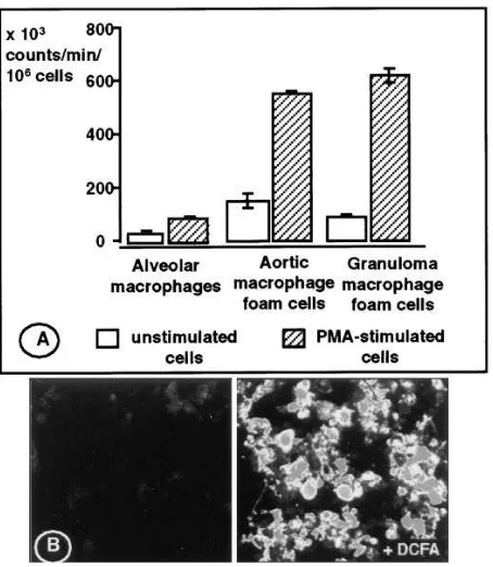
Reactive oxygen species production in lipid-laden macrophages Superoxide production. We found that lipid-laden macro-phages isolated from either atherosclerotic aortas or granulo-mas of hypercholesterolemic rabbits generated O.22 (Fig. 1 A).
Counts reflecting O.22 production were higher in lipid-laden
macrophages than those generated by non–lipid-laden alveo-lar macrophages from the same animals (e.g., Fig. 1 A, steady state chemiluminescence levels of 153.7613.0 and 81.165.2 from foam cells vs. 31.765.6 3 103 counts/min/106 alveolar
macrophages, respectively). Interestingly, aortic foam cells, in the absence of exogenous stimulation, produced even more O.22 than alveolar macrophages stimulated with PMA
(153.7613.0 vs. 86.064.3 3 103 counts/min/106 cells,
respec-tively). The level of O.22 production was increased severalfold
by PMA stimulation in both aortic (from 153.7613.0 3 103
counts/min/106 cells to 561.569.0 3 103 counts/min/106 cells)
and in granuloma-derived foam cells (from 81.165.2 to 616.4625.0 3 103 counts/min/106 cells), suggesting a high
oxi-dative potential.
NOS activity. NOS activity in foam cells from several granulomas was highly variable. In some, as little as 0.2660.03 pmol/mg protein/min of 3H-citrulline was detected, while in
[image:4.612.59.286.331.592.2]others as much as 4.7 pmol/mg protein/min of 3H-citrulline was Figure 1. (A) Lucigenin-enhanced chemiluminescence, reflecting
production of superoxide by alveolar macrophages, aortic foam cells, and granuloma foam cells isolated from cholesterol-fed rabbits. Un-stimulated cells as well as cells Un-stimulated with PMA were assayed. The figure shows the average of triplicate measurements of steady state levels of chemiluminescence 15 min after adding cells to the lu-cigenin-containing buffer. In this assay, 10 pmol of generated from known amounts of xanthine and xanthine oxidase yielded z 4,700 counts. (B) Detection of hydroperoxides as shown by the fluores-cence of DCF in lipid-laden macrophages. The left panel shows the autofluorescence of cells incubated in the absence of DCFA and the right panel shows cells 30 min after addition of DCFA. Cells were im-aged with a confocal microscope using a magnification of 40 and an FITC filter.
formed. In each preparation NOS activity was not affected by addition of EGTA (average 3H-citrulline in the presence of
EGTA was 0.8260.30 pmol/mg protein/min), suggesting that the NO synthase responsible for this activity was the inducible isoform. Due to limited cell yield for aortic foam cells, we mea-sured formation of 3H-citrulline only in a pair of samples
ob-tained from one experiment, in which cells from four rabbits were pooled. This was 1.32 pmol/mg protein/min. Alveolar macrophages isolated from the same rabbits had no detectable .NO synthase activity.
H2O2 detection. Lipid-laden macrophages cultured for 24 h,
then incubated with DCFA (10 mM), showed intense fluores-cent staining by confocal microscopy, indicative of peroxide generation (Fig. 1 B).
Effect of reactive oxygen species on vascular MMP activity in vitro
X/XO system activates latent MMPs. Incubation of human vascular SMC culture media containing latent MMP-2 (pro-MMP-2) with X/XO (100 mM:5 U/liter) resulted in activation of pro-MMP-2. Activation also occurred when culture media were incubated with X/XO after harvesting from tissue culture dishes. To rule out the contribution of other components in the culture media that could serve as intermediaries in this activa-tion sequence, latent MMP-2 and MMP-9 (induced by PMA treatment of vascular SMC) were purified using gelatin-aga-rose. The purified gelatinases were then exposed to the X/XO system, which led again to activation of pro-MMP-2 as well as pro-MMP-9 (Fig. 2). Fig. 2 presents the typical appearance of gelatinolytic bands with lower molecular weights after incuba-tion with X/XO. XO by itself had no effect upon latent MMP-2 (data not shown), but was responsible for production of lytic bands that migrate close to the bottom of 10% gels (Fig. 2) and are not inhibited by incubation with EDTA. Xanthine neither affected pro-MMP-2 nor produced any lytic pattern in gelatin gels, suggesting that pro-MMP-2 activation was due to a prod-uct of the X/XO system. In additional experiments, we tested the effect of adding SOD to the incubates (not shown), but ac-tivation of pro-MMP-2 by X/XO was not inhibited, probably due to the simultaneous generation of other reactive oxygen species (H2O2 and/or hydroxyl, ?OH).
Effect of H2O2 on MMP-2 activity. H2O2 is likely
pro-duced by lipid-laden macrophages in vivo. In vitro, H2O2 is a
main product of the X/XO system, which we used to generate reactive oxygen species in vitro, and thus could have
[image:5.612.55.290.57.246.2]contrib-Figure 2. SDS-PAGE zymography showing the effect of reactive ox-ygen species generated by X/XO (100 mM:5 mU/ml) on activity of la-tent gelatinases, which were affinity-purified from culture medium harvested from human vascular SMC. White areas of lysis produced in the gel, which contained 1 mg/ml gelatin, shows presence of acti-vated, as well as latent, forms of gelatinases. The left panel illustrates activation of latent MMP-2 (pro-MMP-2), purified from the condi-tioned medium of unstimulated human SMC, after incubation with X/XO. The right panel shows activation of pro-MMP-2 and latent MMP-9 (pro-MMP-9) isolated from medium of SMC stimulated with PMA (which induces MMP-9 expression). Treatment with X/XO re-sulted in the conversion of latent forms of both gelatinases to the ac-tive, lower molecular weight forms (MMP-2 and MMP-9).
Figure 3. (A) SDS-PAGE zy-mography of media conditioned by human SMC. Identical ali-quots were exposed to various concentrations of H2O2 for 4 h.
The upper arrow points to the latent form of MMP-2 (pro-MMP-2). Notice that incubation with 4 mM H2O2 activated
pro-MMP-2 to the lower molecular weight active form MMP-2 (lower arrow) and even to smaller forms with gelatinolytic activity. Higher doses of H2O2
led to inactivation (disappear-ance of gelatinolytic activity). The effect of the highest con-centration of H2O2 (50 mM) was
prevented by addition of cata-lase (250 U/ml). (B) Effect of H2O2 upon the gelatinolytic activity associated with the active form of MMP-2, as measured from SDS-PAGE
zy-mography gels. Areas of lysis have been measured by laser scanning densitometry and plotted as the percentage of gelatin lysis produced by con-trol. Data shown have been collected from four independent experiments. Compared with control samples (no H2O2), gelatinolytic activity was
[image:5.612.59.548.509.694.2]uted to the activation of MMP-2 that we observed using X/XO. The direct effect of H2O2 on pro-MMP-2 activity was tested by
incubating aliquots of cell culture media with various concen-trations of H2O2. At low concentrations, incubation with H2O2
increased gelatinolysis associated with the activated form of MMP-2 and also induced generation of lower molecular weight gelatinolytic bands (Fig. 3 A). Higher doses of H2O2
(10–50 mM) resulted in concentration-dependent inactivation of gelatinolytic activity (Fig. 3, A and B). The effects of H2O2
could be blocked completely by addition of catalase (Fig. 3 A). We confirmed that H2O2 initiates molecular processing of
la-tent MMP-2 by identifying the lower molecular species with anti-MMP-2 antibodies (shown in Fig. 6). Compared with con-trol culture media, aliquots incubated with H2O2 displayed
in-creased immunoreactivity of lower molecular weight species and additional immunoreactive bands.
Effect of .NO and peroxynitrite (ONOO2) on MMP-2
activ-ity. Generation of .NO by SNAP and Spermine NONO-ate (50–500 mM) in the culture media had no discernible effect upon activation of MMP-2 zymogen (data not shown). How-ever, since the simultaneous production of .NO and O.22 in
vivo may lead to the generation of ONOO2 (33), we also tested the effect of this reactive oxygen species upon MMP-2 produced by cultured vascular SMC. We incubated cells in cul-ture with ONOO2 and analyzed culture media by SDS-PAGE zymography. We found that after incubation with ONOO2 the intensity of the MMP-2–associated lytic band, migrating at z 60 kD, was increased and was accompanied by the appear-ance of several lower molecular weight gelatinolytic bands (Fig. 4). Initially, we suspected that in addition to activation of pro-MMP-2, incubation of SMC with ONOO2 may induce the production and release of new proteins with gelatinolytic activ-ity. However, the same effect was achieved by incubating the
culture media after harvesting from unstimulated SMC (cell-free experiments) with ONOO2 (Fig. 4). This showed that the generation of several gelatinolytic bands running at appar-ent molecular masses , 60 kD did not require the presence of cells and was likely due to sequential extracellular process-ing of MMP-2. Additional confirmation that incubation with ONOO2 results in activation of latent MMP-2 was obtained through a collagenolytic assay using radiolabeled type IV col-lagen, a typical MMP-2 substrate. Latent MMP-2 incubated with ONOO2 displayed enzymatic activity against collagen IV (Fig. 5). Culture media incubated with pH-inactivated ONOO2, containing the stable decomposition products NO
22
and NO32, had no collagenolytic activity. The activation of
[image:6.612.60.283.61.240.2]pro-MMP-2 by ONOO2 was accompanied by tyrosine nitration of MMP-2 (Fig. 6), as shown by consecutive immunodetection of MMP-2 followed by detection of nitro-tyrosine residues in samples of SMC culture media. Incubation of culture media
Figure 4. Effect of peroxynitrite on gelatinolytic activity in culture media of human SMC. In left lanes, we applied culture media from SMC incubated with increasing concentrations of ONOO2 for 24 h.
One cell culture dish was incubated with the highest concentration of pH-inactivated ONOO2 (Inact.). The right lanes show activity in
me-dia harvested from unstimulated SMC, then incubated with similar concentrations of ONOO2. The same gelatinolytic pattern was
[image:6.612.319.550.74.251.2]ob-tained in both cases (6SMC). Molecular weight of prestained mark-ers (MWM), loaded in between samples obtained from the two exper-iments, is indicated at right in kiloDaltons (kD).
Figure 5. Fluorographic image of 3H-type IV
collagen incubated with SMC-conditioned medium treated with ONOO2. Typical
col-lagen bands are indi-cated with arrows. Treatment with ONOO2 imparts
col-lagenolytic activity to SMC-derived culture media, as suggested by the diminishing radio-activity associated with
a 20 chains and pro-teolytic processing of other higher molecular weight collagen chains.
Figure 6. Immunoblotting of SMC culture media incubated with per-oxynitrite (ONOO2) or H
2O2. The top panels show detection of
la-tent MMP-2 (top arrow) and activated MMP-2 (bottom arrow). The bottom left panel shows detection of nitrotyrosine residues using spe-cific antibodies. The membrane containing ONOO2-treated culture
media, presented in the left two panels, was first blotted with anti– MMP-2 antibodies (top left panel), then stripped, and reprobed with antinitrotyrosine antibodies (bottom left panel). Incubation with ONOO2 led to a decrease in immunoreactivity to MMP-2 antibodies
and to an increased immunoreactivity to antinitrotyrosine antibodies. In contrast incubation of culture media with H2O2 actually increased
[image:6.612.317.545.487.600.2]with 100–250 mM of ONOO2 abolished the immunoreactivity of the anti–MMP-2 antibody for bands associated with acti-vated forms of MMP-2, while increasing the immunoreactivity of antinitrotyrosine antibodies for the corresponding bands. This effect suggested that, during activation, the MMP-2 epitopes recognized by the MMP-2 antibody were modified by nitration. Further supporting the hypothesis of a specific modi-fication of MMP-2 by ONOO2 treatment was the finding that the same anti–MMP-2 antibody recognized an MMP-2 form activated by H2O2 (Fig. 6).
Discussion
This study was undertaken to examine the possibility that reac-tive oxygen species produced by lipid-laden macrophages modulate vascular MMP activity. We found that macrophage foam cells, isolated from atheroma, steadily produce O.22,
H2O2, and .NO without additional exogenous stimulation. We
also showed that these reactive oxygens can modulate MMP-2 and -9 activity, either directly or via a derivative radical, ONOO2. This may be an important mechanism for modulat-ing MMP activity in atherosclerotic plaques. Similar radicals may also be responsible for MMP activation in inflammation or other conditions associated with oxidative stress.
Traditionally, the theory regarding MMP activation in vivo postulates the attack of susceptible regions in the propeptide MMP domain by soluble proteases (13), or by a membrane-bond protease in the case of MMP-2 (34, 35). Studies from Van Wart’s group (16) led to the formulation of the “cysteine-switch” hypothesis to explain the seemingly disparate means by which activation could be achieved in vitro by a wide vari-ety of agents that share one common characteristic: thiol reac-tivity. In this model, the zinc atom at the active site is coordi-nately bound to an unpaired cysteine thiol group located approximately at the 80th residue of the propeptide domain.
Disruption of this interaction is believed to represent the criti-cal step in initiating the process of MMP autoactivation. Reac-tive oxygen species undergo facile reactions with thiol groups and may serve as a common mechanism of activation for sev-eral different MMPs. Macrophage-derived reactive oxygen species may thus provide a link between the presence of acti-vated forms of MMPs and macrophage-rich areas in athero-sclerotic plaques.
Rosenfeld et al. showed previously that macrophage-derived foam cells isolated from hypercholesterolemic rabbit aortas are capable of oxidatively modifying lipoproteins (29), but the nature of reactive oxygen species released by these cells has not been defined. To our knowledge, all other previous studies have examined reactive oxygen species production by mono-cytes differentiated in vitro and subsequently activated with exogenous stimuli. Properties of cells differentiated and loaded with lipid in vitro may differ significantly from foam cells gen-erated in vivo. In this study, we examined two types of mac-rophages that accumulate lipid in vivo, in tissues of hypercho-lesterolemic animals. By producing lipid-laden macrophages in granulomas we have generated large quantities of foam cells for study, in contrast to aortic foam cells isolated from ather-oma, where the yield of isolated cells is low. Further, rapid iso-lation (, 4 h from time of killing of the animal) precludes the influence of extraneous factors that may influence radical pro-duction.
Macrophage foam cells, whether derived from aortic
ather-oma or granulather-oma of hypercholesterolemic animals, produced substantial amounts of superoxide compared with non–lipid-laden macrophages obtained from the same animal. Lipid-non–lipid-laden macrophages expressed varying amounts of NO synthase activity. In contrast, NO synthase activity was undetectable in alveolar macrophages. These findings provided a basis for ex-periments testing the effects of O.22, H2O2, .NO, and ONOO2
on activity of vascular MMPs. To directly examine the effects of these, we either generated or exogenously added the vari-ous reactive oxygen species of interest. MMPs investigated were produced by cultured human SMC. These cells can pro-duce a spectrum of MMPs (36) and likely contribute substan-tially to the MMPs produced in human vessels.
We concentrated on vascular gelatinases for two reasons. First, MMP-2 is ubiquitous in normal and atherosclerotic hu-man vessels (1) and is produced in vitro by vascular huhu-man en-dothelium (37) and SMC (36). Generation of activated forms of MMP-2 is associated with SMC migration and proliferation in vitro and in vivo in animal models of restenosis (38–41). In-duction of MMP-9 also occurs under these conditions (38–41). Pro-MMP-9, the main MMP product of monocyte-macrophages (42), is expressed in human atheroma (1) and can be detected in specimens collected from patients with unstable angina (43). Second, the biochemical characteristics of these two gelati-nases, which have prodomains rich in cysteine residues, would presumably increase their susceptibility to the action of reac-tive oxygen species.
The X/XO reaction, commonly used to generate O.22, also
generates other reactive oxygen species: X/XO can reduce di-oxygen univalently to O.22 or divalently to H2O2. The balance
between these two pathways depends on the state of reduction of the enzyme, favoring divalent reduction when the enzyme is fully reduced. Conditions such as substrate concentration, pO2,
and pH affect the reduction state of the enzyme. Thus, it is possible that, in our experiments with the X/XO system, gelati-nases were subjected to the action of reactive oxygen species other than O.22. Furthermore, addition of SOD did not block
activation of MMP-2 in the presence of X/XO, which could mean that a radical other than O.22 may be involved. H2O2
generated by X/XO or through dismutation of O.22 could have
reacted with pro-MMP-2. We tested this hypothesis and found that direct addition of H2O2 caused modulation of MMP-2
ac-tivity. Activation of MMP-2 at low doses of H2O2 (4 mM) and
inactivation at higher doses (10–50 mM) suggest that there may be a biphasic response to oxidant stress. High concentrations of H2O2 may inhibit MMP-2 enzymatic activity through
mobi-lization of the zinc atom at the catalytic site (44). Also, higher H2O2 concentrations may modify the zymogen more
exten-sively, leading to protein degradation, and could act as a regu-latory mechanism for MMP activity. Similarly, the highest con-centration of ONOO2 that we used (500 mM) seemed to inactivate pro-MMP-2. We cannot exclude the possibility that, in our in vitro conditions, the OH2 radical was also formed via the H2O2-dependent Fenton reaction which could be
sup-ported by free metals in the culture medium.
its decomposition products NO32 and NO22, had no effect on
the activation of MMP-2. It has been estimated that the aver-age rate of ONOO2 formation may reach 1 mM/min in the rat lung epithelial fluid upon stimulation of alveolar macrophages (52). The concentrations used in our study are thus within the range found in pathophysiologic states. Immunoblotting with specific antibodies showed that activation of MMP-2 was asso-ciated with almost exclusive tyrosine nitration of proteins mi-grating at z 70 kD (likely MMP-2) and concomitant loss of re-activity to anti–MMP-2 antibodies. It is interesting to speculate that nitration of two tyrosine residues, present within a five amino acid stretch in the hinge region between the propeptide and active domains of pro-MMP-2, could assist in unfolding of the zymogen. Using the same antinitrotyrosine antibody to stain atherosclerotic lesions, Beckman et al. (48) demonstrated an intense immunopositive reaction. Therefore it is possible that nitration of MMPs could contribute to this immunostain-ing pattern in atheroma. Importantly, the reaction with ONOO2 generated around activated macrophages provides a possible mechanism by which MMP activity previously detected in vul-nerable areas of the atherosclerotic plaque (1, 8) is unleashed. .NO donors used in our experiments had no effect on MMP-2 activation. A recently published study suggested that .NO acti-vates pro-MMP-2 (53). However, it is possible that ONOO2, rather than .NO, was directly responsible for activation of MMP-2 in that study, in which cells were simultaneously incu-bated in culture with cytokines, lipopolysaccharide, and .NO donors. Under these conditions both .NO and O.22 may have
been generated, leading to formation of ONOO2 (33). Macrophage-derived foam cells express MMP mRNA (54), produce MMP proteins (1, 28), and colocalize with MMP activ-ity in human and experimental atherosclerotic plaques (1, 8). Shah et al. (3) showed that MMPs secreted by monocyte-derived macrophages may be responsible for breakdown of tissue collagen by incubating these cells, or their conditioned culture medium, with explanted atherosclerotic lesions in vitro. Since the presence of cells enhanced collagenolysis, it is possible that generation of active MMPs depended on the ac-tion of cell-released reactive oxygen species. These reactive species have a short life time and their effects would otherwise rapidly diminish in conditioned culture media.
Based on the findings of this study and of previously pub-lished reports, it appears that macrophages may be able to par-ticipate in MMP matrix degradation at several levels. These include: (a) inducing MMP expression in other cells (via secre-tion of cytokines); (b) producing MMPs; and (c) activating la-tent forms of secreted MMPs (via production of reactive oxy-gen species). It is of note that antiprotease inhibitors (serpins) are susceptible to degradation by MMPs (55, 56), possibly leading to an increase in proteolytic activity in the vicinity of activated macrophages. Similarly, activated neutrophils, which generate hypochlorous acid through the action of myeloperox-idase, seem capable of autoactivating their latent collagenase (57). Since myeloperoxidase is also present in atherosclerotic vessels (58), hypochlorous acid may contribute to activation of MMPs and inactivation of serpins.
The results of this investigation provide support for a mechanism by which macrophage-derived foam cells could ac-tivate MMPs in the atherosclerotic plaque. Activation of latent MMPs by reactive oxygen species may also be relevant in other pathological conditions associated with high oxidative stress. Our observations may explain some of the benefits of
antioxidant therapy (59, 60) and may help direct future inter-ventions to improve plaque stability.
Acknowledgments
The authors wish to acknowledge Dr. Joe Beckman and Dr. William Stetler-Stevensen for their generous gift of antibodies, and Dr. Sam-path Parthasarathy for interesting discussions.
This work was supported through a development fund from Em-ory University School of Medicine and funds from the Whittaker Foundation to Zorina Galis, and from National Institutes of Health grants HL-48667, DK-45215, and HL-39006, and a merit grant from the Veterans Administration to David Harrison.
References
1. Galis, Z.S., G.K. Sukhova, M.W. Lark, and P. Libby. 1994. Increased ex-pression of matrix metalloproteinases and matrix degrading activity in vulnera-ble regions of human atherosclerotic plaques. J. Clin. Invest. 94:2493–2503.
2. Nikkari, S.T., K.D. O’Brien, M. Ferguson, T. Hatsukami, H.G. Welgus, C.E. Alpers, and A.W. Clowes. 1995. Interstitial collagenase (MMP-1) expres-sion in human carotid atherosclerosis. Circulation. 92:1393–1398.
3. Shah, P.K., E. Falk, J.J. Badimon, A. Fernandez-Ortiz, A. Mailhac, G. Villareal-Levy, J.T. Fallon, J. Regnstrom, and V. Fuster. 1995. Human mono-cyte-derived macrophages induce collagen breakdown in fibrous caps of ath-erosclerotic plaques. Potential role of matrix-degrading metalloproteinases and implications for plaque rupture. Circulation. 92:1565–1569.
4. Constantinides, P. 1989. Plaque hemorrhages, their genesis and their role in supra-plaque thrombosis and atherogenesis. In Pathobiology of the Human Atherosclerotic Plaque. S. Glagov, W.P.I. Newman, and S.A. Schaffer. Springer-Verlag, New York. 393–412.
5. Falk, E., P.K. Shah, and V. Fuster. 1995. Coronary plaque disruption. Circulation. 92:657–671.
6. Richardson, P.D., M.J. Davies, and G.V. Born. 1989. Influence of plaque configuration and stress distribution on fissuring of coronary atherosclerotic plaques. Lancet. 2:941–944.
7. van der Wal, A.C., A.E. Becker, C.M. van der Loos, and P.K. Das. 1994. Site of intimal rupture or erosion of thrombosed coronary atherosclerotic plaques is characterized by an inflammatory process irrespective of the domi-nant plaque morphology. Circulation. 89:36–44.
8. Galis, Z.S., G.K. Sukhova, and P. Libby. 1995. Microscopic localization of active proteases by in situ zymography: detection of matrix metalloprotein-ase activity in vascular tissue. FASEB (Fed. Am. Soc. Exp. Biol.) J. 9:974–980.
9. Lendon, C.L., M.J. Davies, G.V. Born, and P.D. Richardson. 1991. Ath-erosclerotic plaque caps are locally weakened when macrophage density is in-creased. Atherosclerosis. 87:87–90.
10. Moreno, P.R., E. Falk, I.F. Palacios, J.B. Newell, V. Fuster, and J.T. Fal-lon. 1994. Macrophage infiltration in acute coronary syndromes. Implications for plaque rupture. Circulation. 90:775–778.
11. Matrisian, L.M. 1994. Matrix metalloproteinase gene expression. Ann. NY Acad. Sci. 732:42–50.
12. Murphy, G., F. Willenbrock, T. Crabbe, M. O’Shea, R. Ward, S. Atkin-son, J. O’Connell, and A. Docherty. 1994. Regulation of matrix metalloprotein-ase activity. Ann. NY Acad. Sci. 732:31–41.
13. Okada, Y., T. Morodomi, J.J. Enghild, K. Suzuki, A. Yasui, I. Naka-nishi, G. Salvesen, and H. Nagase. 1990. Matrix metalloproteinase 2 from hu-man rheumatoid synovial fibroblasts. Purification and activation of the precur-sor and enzymic properties. Eur. J. Biochem. 194:721–730.
14. Nagase, H., J.J. Enghild, K. Suzuki, and G. Salvesen. 1990. Stepwise ac-tivation mechanisms of the precursor of matrix metalloproteinase 3 (stromel-ysin) by proteinases and (4-aminophenyl)mercuric acetate. Biochemistry. 29: 5783–5789.
15. Galis, Z.S., R. Kranzhöfer, and P. Libby. 1995. Thrombin promotes acti-vation of matrix metalloproteinase-2 (MMP-2) produced by cultured smooth muscle cells. FASEB (Fed. Am. Soc. Exp. Biol.) J. 9:A413.
16. Van Wart, H.E., and H. Birkedal-Hansen. 1990. The cysteine switch: a principle of regulation of metalloproteinase activity with potential applicability to the entire matrix metalloproteinase gene family. Proc. Natl. Acad. Sci. USA. 87:5578–5582.
17. Suzuki, K., J.J. Enghild, T. Morodomi, G. Salvesen, and H. Nagase. 1990. Mechanisms of activation of tissue procollagenase by matrix metallopro-teinase 3 (stromelysin). Biochemistry. 29:10261–10270.
18. Griendling, K., J.D. Ollerenshaw, C.A. Minieri, and R.W. Alexander. 1994. Angiotensin II stimulates NADH and NADPH activity in cultured vascu-lar smooth muscle cells. Circ. Res. 74:1141–1148.
20. Mohazzab, K.M., P.M. Kaminski, and M.S. Wolin. 1994. NADH oxi-doreductase is a major source of superoxide anion in bovine coronary artery en-dothelium. Am. J. Physiol. 266:H2568–H2572.
21. Segal, A.W., and A. Abo. 1993. The biochemical basis of the NADPH oxidase of phagocytes. Trends Biochem. Sci. 18:43–47.
22. Ohara, Y., T.E. Peterson, and D.G. Harrison. 1993. Hypercholester-olemia increases endothelial superoxide anion production. J. Clin. Invest. 91: 2546–2551.
23. Rajagopalan, S., S. Kurz, T. Münzel, M. Tarpey, B. Freeman, K. Griend-ling, and D. Harrison. 1996. Angiotensin II–mediated hypertension in the rat increases vascular superoxide production via membrane NADH/NADPH oxi-dase activation. Contribution to alterations of vasomotor tone. J. Clin. Invest. 97:1916–1923.
24. Tesfamariam, B. 1994. Free radicals in diabetic endothelial cell dysfunc-tion. Free Radical Biol. Med. 16:383–391.
25. Giugliano, D., A. Ceriello, and G. Paolisso. 1995. Diabetes mellitus, hy-pertension, and cardiovascular disease: which role for oxidative stress? Metab. Clin. Exp. 44:363–368.
26. Schwartz, C.J., J.J. Ghidoni, J.L. Kelley, E.A. Sprague, A.J. Valente, and C.A. Suenram. 1985. Evolution of foam cells in subcutaneous rabbit carra-geenan granulomas. I. Light-microscopic and ultrastructural study. Am. J. Pathol. 118:134–150.
27. Bell, F.P., and R.G. Schaub. 1989. Comparison of lipid accumulation and metabolism in carrageenan-induced granulomas to aorta and blood mono-cytes of normal and cholesterol-fed rabbits. Exp. Mol. Pathol. 50:327–336.
28. Galis, Z.S., G.K. Sukhova, R. Kranzhöfer, S. Clark, and P. Libby. 1995. Macrophage foam cells from experimental atheroma constitutively produce matrix-degrading proteinases. Proc. Natl. Acad. Sci. USA. 92:402–406.
29. Rosenfeld, M.E., J.C. Khoo, E. Miller, S. Parthasarathy, W. Palinski, and J.L. Witztum. 1991. Macrophage-derived foam cells freshly isolated from rabbit atherosclerotic lesions degrade modified lipoproteins, promote oxidation of low-density lipoproteins, and contain oxidation-specific lipid-protein ad-ducts. J. Clin. Invest. 87:90–99.
30. Gyllenhammar, H. 1987. Lucigenin chemiluminescence in the assess-ment of neutrophil superoxide production. J. Immunol. Methods. 97:209–213.
31. Hecker, M., W.C. Sessa, H.J. Harris, E.E. Ånggard, and J.R. Vane. 1990. The metabolism of L-arginine and its significance for the biosynthesis of endothelium-derived relaxing factor: cultured endothelial cells recycle L-citrul-line to L-arginine. Proc. Natl. Acad. Sci. USA. 87:8612–8616.
32. Royall, J.A., and H. Ischiropoulos. 1993. Evaluation of 29,79 -dichloroflu-orescin and dihydrorhodamine 123 as fluorescent probes for intracellular H2O2 in cultured endothelial cells. Arch. Biochem. Biophys. 302:348–355.
33. Huie, R.E., and S. Padmaja. 1993. The reaction of NO with superoxide. Free Radical Res. Commun. 18:195–199.
34. Strongin, A.Y., I. Collier, G. Bannikov, B.L. Marmer, G.A. Grant, and G.I. Goldberg. 1995. Mechanism of cell surface activation of 72-kDa type IV collagenase. Isolation of the activated form of the membrane metalloprotease. J. Biol. Chem. 270:5331–5338.
35. Cao, J., H. Sato, T. Takino, and M. Seiki. 1995. The C-terminal region of membrane type matrix metalloproteinase is a functional transmembrane do-main required for pro-gelatinase A activation. J. Biol. Chem. 270:801–805.
36. Galis, Z.S., M. Muszynski, G.K. Sukhova, E. Simon-Morrissey, E.N. Unemori, M.W. Lark, E. Amento, and P. Libby. 1994. Cytokine-stimulated hu-man vascular smooth muscle cells synthesize a complement of enzymes re-quired for extracellular matrix digestion. Circ. Res. 75:181–189.
37. Hanemaaijer, R., P. Koolwijk, L. le Clercq, W.J. de Vree, and V.W. van Hinsbergh. 1993. Regulation of matrix metalloproteinase expression in human vein and microvascular endothelial cells. Effects of tumour necrosis factor al-pha, interleukin 1 and phorbol ester. Biochem. J. 296:803–809.
38. Southgate, K.M., M. Davies, R.F. Booth, and A.C. Newby. 1992. In-volvement of extracellular-matrix-degrading metalloproteinases in rabbit aortic smooth-muscle cell proliferation. Biochem. J. 288:93–99.
39. Pauly, R.R., A. Passaniti, C. Bilato, R. Monticone, L. Cheng, N. Papa-dopoulos, Y.A. Gluzband, L. Smith, C. Weinstein, E.G. Lakatta, and M.T. Crow. 1994. Migration of cultured vascular smooth muscle cells through a
base-ment membrane barrier requires type IV collagenase activity and is inhibited by cellular differentiation. Circ. Res. 75:41–54.
40. Bendeck, M.P., N. Zempo, A.W. Clowes, R.E. Galardy, and M.A. Reidy. 1994. Smooth muscle cell migration and matrix metalloproteinase expression after arterial injury in the rat. Circ. Res. 75:539–545.
41. Zempo, N., R.D. Kenagy, Y.P.T. Au, M. Bendeck, M.M. Clowes, M.A. Reidy, and A.W. Clowes. 1994. Matrix metalloproteinases of vascular wall cells are increased in balloon-injured rat carotid artery. J. Vasc. Surg. 20:209–217.
42. Welgus, H.G., R.M. Senior, W.C. Parks, A.J. Kahn, T.J. Ley, S.D. Sha-piro, and E.J. Campbell. 1992. Neutral proteinase expression by human mono-nuclear phagocytes: a prominent role of cellular differentiation. Matrix Suppl. 1:363–367.
43. Brown, D.L., M.S. Hibbs, M. Kearney, C. Loushin, and J.M. Isner. 1995. Identification of 92-kD gelatinase in human coronary atherosclerotic lesions. Association of active enzyme synthesis with unstable angina. Circulation. 91: 2125–2131.
44. Fliss, H., and M. Menard. 1992. Oxidant-induced mobilization of zinc from metallothionein. Arch. Biochem. Biophys. 293:195–199.
45. Radi, R., J.S. Beckman, K.M. Bush, and B.A. Freeman. 1991. Peroxyni-trite-induced membrane lipid peroxidation: the cytotoxic potential of superox-ide and nitric oxsuperox-ide. Arch. Biochem. Biophys. 288:481–487.
46. Radi, R., J.S. Beckman, K.M. Bush, and B.A. Freeman. 1991. Peroxyni-trite oxidation of sulfhydryls. The cytotoxic potential of superoxide and nitric oxide. J. Biol. Chem. 266:4244–4250.
47. Beckman, J.S., T.W. Beckman, J. Chen, P.A. Marshall, and B.A. Free-man. 1990. Apparent hydroxyl radical production by peroxynitrite: implications for endothelial injury from nitric oxide and superoxide. Proc. Natl. Acad. Sci. USA. 87:1620–1624.
48. Beckman, J.S., Y.Z. Ye, P.G. Anderson, J. Chen, M.A. Accavitti, M.M. Tarpey, and C.R. White. 1994. Extensive nitration of protein tyrosines in hu-man atherosclerosis detected by immunohistochemistry. Biol. Chem. Hoppe Seyler. 375:81–88.
49. Haddad, I.Y., G. Pataki, P. Hu, C. Galliani, J.S. Beckman, and S. Mata-lon. 1994. Quantitation of nitrotyrosine levels in lung sections of patients and animals with acute lung injury. J. Clin. Invest. 94:2407–2413.
50. Ischiropoulos, H., and A.B. al-Mehdi. 1995. Peroxynitrite-mediated oxi-dative protein modifications. FEBS Lett. 364:279–282.
51. van der Vliet, A., J.P. Eiserich, C.A. O’Neill, B. Halliwell, and C.E. Cross. 1995. Tyrosine modification by reactive nitrogen species: a closer look. Arch. Biochem. Biophys. 319:341–349.
52. Ischiropoulos, H., L. Zhu, and J.S. Beckman. 1992. Peroxynitrite forma-tion from macrophage-derived nitric oxide. Arch. Biochem. Biophys. 298:446–451.
53. Trachtman, H., S. Futterweit, P. Garg, K. Reddy, and P.C. Singhal. 1996. Nitric oxide stimulates the activity of a 72-kDa neutral matrix metalloprotein-ase in cultured rat mesangial cells. Biochem. Biophys. Res. Commun. 218:704–708.
54. Henney, A.M., P.R. Wakeley, M.J. Davies, K. Foster, R. Hembry, G. Murphy, and S. Humphries. 1991. Localization of stromelysin gene expression in atherosclerotic plaques by in situ hybridization. Proc. Natl. Acad. Sci. USA. 88:8154–8158.
55. Desrochers, P.E., J.J. Jeffrey, and S.J. Weiss. 1991. Interstitial collage-nase (matrix metalloproteicollage-nase-1) expresses serpicollage-nase activity. J. Clin. Invest. 87:2258–2265.
56. Wallaert, B., C. Aerts, B. Gressier, P. Gosset, and C. Voisin. 1993. Oxi-dative inactivation of alpha 1-proteinase inhibitor by alveolar epithelial type II cells. J. Appl. Physiol. 75:2376–2382.
57. Weiss, S.J., G. Peppin, X. Ortiz, C. Ragsdale, and S.T. Test. 1985. Oxi-dative autoactivation of latent collagenase by human neutrophils. Science (Wash. DC). 227:747–749.
58. Daugherty, A., J.L. Dunn, D.L. Rateri, and J.W. Heinecke. 1994. My-eloperoxidase, a catalyst for lipoprotein oxidation, is expressed in human ath-erosclerotic lesions. J. Clin. Invest. 94:437–444.
59. Gaziano, J.M. 1994. Antioxidant vitamins and coronary artery disease risk. Am. J. Med. 97:18S–21S; Discussion 22S–28S.