PKC
ll
in liver mediates insulin-induced
SREBP-1c expression and determines both hepatic lipid
content and overall insulin sensitivity
Michihiro Matsumoto, … , Masato Kasuga, Tetsuo Noda
J Clin Invest.
2003;
112(6)
:935-944.
https://doi.org/10.1172/JCI18816
.
PKC
l
is implicated as a downstream effector of PI3K in insulin action. We show here that
mice that lack PKC
l
specifically in the liver (L-
l
KO mice), produced with the use of the
Cre-loxP system, exhibit increased insulin sensitivity as well as a decreased triglyceride content
and reduced expression of the sterol regulatory element–binding protein-1c (SREBP-1c)
gene in the liver. Induction of the hepatic expression of
Srebp1c
and of its target genes
involved in fatty acid/triglyceride synthesis by fasting and refeeding or by hepatic expression
of an active form of PI3K was inhibited in L-
l
KO mice compared with that in control animals.
Expression of
Srebp1c
induced by insulin or by active PI3K in primary cultured rat
hepatocytes was inhibited by a dominant-negative form of PKC
l
and was mimicked by
overexpression of WT PKC
l
. Restoration of PKC
l
expression in the liver of L-
l
KO mice
with the use of adenovirus-mediated gene transfer corrected the metabolic abnormalities of
these animals. Hepatic PKC
l
is thus a determinant of hepatic lipid content and whole-body
insulin sensitivity.
Article
Endocrinology
Find the latest version:
Introduction
The liver is essential for both carbohydrate and lipid homeostasis. Individuals with type 2 diabetes often exhibit impairment of insulin action in the liver (1), and liver-specific inhibition of insulin signaling in mice results in glucose intolerance and dyslipidemia (2), indi-cating the physiological importance of hepatic insulin action in energy homeostasis. Among the signaling mol-ecules activated by insulin, PI3K plays a key role in the metabolic actions of this hormone (3, 4). Prevention of the insulin-induced activation of PI3K in the liver of mice thus results in glucose intolerance and dyslipidemia (5).
However, the signaling molecules that function down-stream of PI3K to mediate the metabolic actions of insulin in living animals have remained unclear.
Two atypical PKC (aPKC) isozymes, PKCλand PKCζ
(6), have been identified in mammals. We have previous-ly shown that PKCλacts downstream of PI3K in PDGF or insulin signaling in cultured cells (7, 8). Furthermore, evidence suggests that 3′-phosphoinositide–dependent kinase-1 (PDK-1), thought to be a key mediator of PI3K signaling, contributes to the activation of AGC protein kinases (which include Akt, p70 S6 kinase, p90rsk, and
aPKC) by phosphorylating these enzymes on residues located in their activation loops (9, 10). On the basis of these various observations, we hypothesized that PKCλ
participates in insulin action in vivo. To verify this hypothesis, using homologous recombination, we gen-erated mice that harbor a PKCλgene containing loxP sites. Disruption of the corresponding aPKC genes in
Caenorhabditis elegansand Drosophila melanogasterresulted in embryonic death (11, 12). As expected, PKCλ-deficient mice generated from the floxed PKCλtransgenic animals also died during embryonic development (K. Akimoto et al., unpublished observations).
Given that the liver is one of the most important tar-get organs of insulin, we next generated mice in which
PKC
λ
in liver mediates insulin-induced SREBP-1c
expression and determines both hepatic lipid
content and overall insulin sensitivity
Michihiro Matsumoto,
1Wataru Ogawa,
1Kazunori Akimoto,
2Hiroshi Inoue,
1Kazuaki Miyake,
1Kensuke Furukawa,
1Yoshitake Hayashi,
3Haruhisa Iguchi,
4Yasushi Matsuki,
4Ryuji Hiramatsu,
4Hitoshi Shimano,
5Nobuhiro Yamada,
5Shigeo Ohno,
2Masato Kasuga,
1and Tetsuo Noda
61Department of Clinical Molecular Medicine, Division of Diabetes and Digestive and Kidney Diseases, Kobe University
Graduate School of Medicine, Kobe, Japan
2Department of Molecular Biology, Yokohama City University Graduate School of Medical Science, Yokohama, Japan 3Division of Molecular Medicine and Medical Genetics, International Center for Medical Research, Kobe University
Graduate School of Medicine, Kobe, Japan
4Genomics Science Laboratories, Sumitomo Pharmaceuticals Co. Ltd., Takarazuka, Japan
5Department of Internal Medicine, Division of Endocrinology/Metabolism, Institute of Clinical Medicine,
University of Tsukuba, Tsukuba, Japan
6Department of Experimental Pathology, Cancer Institute, Tokyo, Japan
PKCλis implicated as a downstream effector of PI3K in insulin action. We show here that mice that lack PKCλspecifically in the liver (L-λKO mice), produced with the use of the Cre-loxP system, exhib-it increased insulin sensexhib-itivexhib-ity as well as a decreased triglyceride content and reduced expression of the sterol regulatory element–binding protein-1c (SREBP-1c) gene in the liver. Induction of the hepat-ic expression of Srebp1cand of its target genes involved in fatty acid/triglyceride synthesis by fasting and refeeding or by hepatic expression of an active form of PI3K was inhibited in L-λKO mice com-pared with that in control animals. Expression of Srebp1cinduced by insulin or by active PI3K in pri-mary cultured rat hepatocytes was inhibited by a dominant-negative form of PKCλand was mim-icked by overexpression of WT PKCλ. Restoration of PKCλexpression in the liver of L-λKO mice with the use of adenovirus-mediated gene transfer corrected the metabolic abnormalities of these animals. Hepatic PKCλis thus a determinant of hepatic lipid content and whole-body insulin sensitivity.
J. Clin. Invest.112:935–944 (2003). doi:10.1172/JCI200318816.
Received for publication May 2, 2003, and accepted in revised form July 15, 2003.
Address correspondence to: Masato Kasuga, Department of Clinical Molecular Medicine, Division of Diabetes and Digestive and Kidney Diseases, Kobe University Graduate School of Medicine, 7-5-1 Kusunoki-cho, Chuo-ku, Kobe 650-0017, Japan. Phone: 81-78-382-5861; Fax: 81-78-382-2080;
E-mail: kasuga@med.kobe-u.ac.jp.
Conflict of interest:The authors have declared that no conflict of interets exists.
the floxed PKCλgene was specifically deleted in the liver as a result of Cre recombinase expression in this organ. Characterization of these animals has now revealed that PKCλmediates the regulatory effect of insulin on hepat-ic triglyceride content by contributing to the expression of the gene for sterol regulatory element–binding pro-tein-1c (SREBP-1c), and that the lack of PKCλin the liver results in increased insulin sensitivity.
Methods
Animals. Mice harboring a floxed PKCλgene in which exon 5 was flanked by loxP sequences (PKCλlox/+mice) were generated by homologous recombination (K. Aki-moto et al., unpublished observations). Mice that express Cre recombinase under the control of the albumin gene promoter (Alb-Cremice) (13) were pro-vided by D. LeRoith (Diabetes Branch, National Insti-tute of Diabetes and Digestive and Kidney Diseases, NIH, Bethesda, Maryland, USA). We used only male mice for the present studies. For the fasting-refeeding experiments, mice in the fasted group were deprived of food for 24 hours; mice in the refed group were deprived of food for 24 hours and then allowed access to food for 12 hours before analysis. The liver X recep-tor agonist T0901317 (kindly provided by K. Muraka-mi, Kyorin Pharmaceutical Co., Tokyo, Japan) was administered daily by oral gavage at a dose of 10 mg/kg body mass for 4 days. For oral glucose intake experi-ments, mice deprived of food for 16 hours were loaded orally with glucose (2 g/kg body mass). For in vivo ade-novirus-mediated gene transfer experiments, mice were injected with the indicated adenovirus vector (1 ×10⁸
PFU) via the tail vein 72 hours before experiments.
Analysis of metabolic parameters. Blood glucose and plasma insulin concentrations were determined as described (5). For glucose tolerance and insulin toler-ance tests, mice deprived of food for 16 hours were injected intraperitoneally with glucose (2 g/kg body mass); mice in the randomly fed state were injected intraperitoneally with human regular insulin (0.75 U/kg). Serum leptin and adiponectin concentrations were measured with a mouse leptin ELISA kit (Mori-naga Institute of Biological Science, Yokohama, Japan) and a mouse/rat adiponectin ELISA kit (Otsuka Phar-maceutical Co. Ltd., Tokyo, Japan), respectively. Serum cholesterol, triglyceride, and FFA concentrations were determined with a cholesterol C-II kit, a triglyceride G kit, and a NEFA C kit, respectively (Wako Pure Chemi-cal Industries Ltd., Osaka, Japan). For assay of the cho-lesterol and triglyceride contents of liver or hind limb skeletal muscle, lipids were extracted from the tissue as described (14) and the concentrations of the analytes in the extract were determined with a cholesterol C-II kit or a triglyceride G kit.
Northern blot, immunoblot, kinase activity, real-time quan-titative RT-PCR analyses, and primary culture of hepatocytes. Total RNA (∼15 µg) was subjected to Northern blot analysis essentially as described (15); autoradiograms were visualized and signal intensity was quantitated
with a BAS2000 image analyzer (Fujifilm Co., Tokyo, Japan). The probes for the genes encoding PPAR-α, acyl-CoA oxidase-1, and uncoupling protein-2 mRNA’s (mouse full-length cDNAs) were synthesized by PCR; the other probes were as described (5, 16, 17). Primary cultures of rat hepatocytes were prepared and subject-ed to adenovirus infection as describsubject-ed (15).
For assay of the expression of the genes encoding SREBP-1 and fatty acid synthase, cells infected or not with adenoviruses were incubated for 6 hours and 20 hours, respectively, with 100 nM insulin or with 10 µM T0901317; for assay of the expression of the genes encoding phosphoenolpyruvate carboxykinase-1 (PKC-1) and glucose-6-phosphatase catalytic subunit (G6PC), cells were incubated for 6 hours with 500 nM dexamethasone and 0.1 mM pCPT-cAMP in the absence or presence of 100 nM insulin. Adenovirus vectors encoding a dominant-negative mutant of PKCλ(AxCAλKD), WT PKCλ(AxCAλWT) (8), or a Myc epitope–tagged active form of PI3K (the 110-kDa catalytic subunit fused with a myristoylation signal sequence at its NH2-terminus; AxCAMyr-p110) (15,
18) were described previously, and that encoding β-gal (AxCALacZ) was kindly provided by I. Saito (Universi-ty of Tokyo, Tokyo, Japan). The antibodies specific for mouse SREBP-1c were as described (14).
For assay of the abundance of PKCλand PKCζ, total tis-sue homogenates were subjected to immunoprecipitation with antibodies to PKCλ(αλ190) or to PKCζ(αζ170) (8), and the resulting precipitates were subjected to immunoblot analysis with antibodies to PKCλ/ι (Trans-duction Laboratories, Lexington, Kentucky, USA) or to PKCζ(Life Technologies Inc., Rockville, Maryland, USA), respectively. Antibodies that recognize both PKCλand PKCζ(C-20, a rabbit polyclonal antibody, and C-20-G, a goat polyclonal antibody) were obtained from Santa Cruz Biotechnology (Santa Cruz, California, USA). For immunodepletion of aPKC, liver homogenates were subjected to three sequential immunoprecipitations for 120 minutes with αλ190 or with αζ170. The super-natants of the immunoprecipitations were then sub-jected to immunoprecipitation with C-20-G and the precipitates were subjected to immunoblot analysis with C-20. The kinase activity of aPKC was assayed in the immunoprecipitates with αλ190 or with αζ170 as described previously (8). For real-time quantitative reverse transcription and PCR analysis, cDNA synthe-sized from total RNA was evaluated in a sequence detec-tor (model 7900; Applied Biosystems, Foster City, Cali-fornia, USA) with specific primers and SYBR Green PCR Master Mix (Applied Biosystems). The relative abun-dance of mRNA’s was calculated with 36B4mRNA as the invariant control. The primers used were as follows: mouse Srebp1a, 5′-GGAACAGACACTGGCCGAGA-3′(sense) and 5′-GCATAGGGGGCGTCA-3′ (antisense); mouse
Srebp1c, 5′-ATCGGCGCGGAAGCTGTCGGGGTAGCGTC-3′
(sense) and 5′-
Results
Generation of mice with liver-specific deficiency of PKCλ. We bred PKCλlox/+mice with Alb-Cremice (13). We then bred the PKCλlox/+, Alb-Creoffspring of this cross with
PKCλlox/+mice. The offspring of this breeding were born in a Mendelian ratio (PKCλ+/+, n = 12, or 9.9%;
PKCλ+/+, Alb-Cre, n = 14, or 11.6%; PKCλlox/+, n = 29, or 24.0%; PKCλlox/+, Alb-Cre, n = 33, or 27.3%; PKCλlox/lox,
n = 15, or 12.4%; PKCλlox/lox, Alb-Cre, n = 18, or 14.9%). Two closely related isoforms comprise aPKC: PKCλ
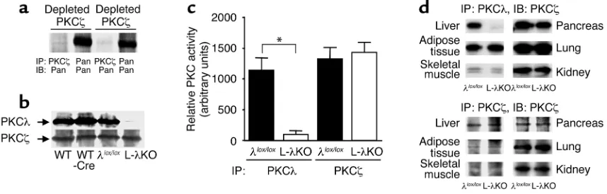
and PKCζ(6). We first investigated the amounts of PKCλand PKCζin liver homogenates of PKCλ+/+(WT) mice. After three sequential immunoprecipitations with antibodies to PKCλor to PKCζ, the correspon-ding isoforms of aPKC were almost completely deplet-ed from the homogenates (Figure 1a). aPKC protein, detected by antibodies that recognize both PKCλand PKCζ, was present both in the PKCλ-depleted and the PKCζ-depleted homogenates, suggesting that liver con-tains both PKCλand PKCζ. The relative abundance of each isoform was comparable. The amount of PKCλin the liver of PKCλlox/lox, Alb-Cre (L-λKO) mice was markedly reduced compared with that in the liver of WT, PKCλ+/+, Alb-Cre(WT-Cre), and PKCλlox/lox(λlox/lox) animals (Figure 1b). In contrast, the hepatic abundance of PKCζwas similar among all four genotypes of mice. Given the similarity in the hepatic expression level of PKCλin WT, WT-Cre, and λlox/loxmice, which indicates that neither the insertion of the loxP sequences in the PKCλ gene nor the expression of Cre recombinase alone affected the abundance of PKCλ, we performed subsequent experiments with L-λKO and λlox/loxmice. The kinase activity of PKCλ, but not that of PKCζ, was
greatly reduced in the liver of L-λKO mice compared with that in the liver of λlox/loxmice (Figure 1c). The abundance of PKCλin other tissues, including skeletal muscle, adipose tissue, the pancreas, the lung, and the kidney, did not differ between L-λKO and λlox/loxmice (Figure 1d). The abundance of PKCζin the two geno-types of mice was similar in all tissues we examined. The gross appearance and histology of the liver, includ-ing the arrangement of hepatocytes in plates and the structure of hepatic lobules, appeared normal in L-λKO mice (data not shown). The serum levels of aspartate aminotransferase, alanine aminotransferase, alkaline phosphatase, lactate dehydrogenase, and albu-min also did not differ significantly between L-λKO and λlox/loxmice (data not shown). These results indicate that the lack of PKCλdid not affect the development or general function of the liver.
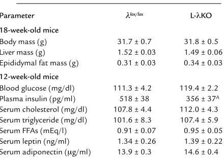
[image:4.576.79.508.458.595.2]Metabolic characteristics of L-λKO mice. The body mass, mass of the liver, and mass of epididymal fat tissue of L-λKO mice were similar to those of λlox/loxmice (Table 1). The serum concentrations of triglyceride, choles-terol, FFAs, leptin, and adiponectin in the randomly fed state also did not differ between mice of the two geno-types. Although the blood glucose concentration in the randomly fed state was similar in the two types of mice, the plasma concentration of insulin in L-λKO mice was significantly lower than that in λlox/loxmice. Blood glu-cose concentrations during a gluglu-cose tolerance test were similar in both L-λKO and λlox/loxmice (Figure 2a). Again, however, the increase in plasma insulin concen-tration induced by glucose intake was smaller in L-λKO mice than in λlox/loxanimals (Figure 2b). Moreover, the glucose-lowering effect of exogenously administered
Figure 1
The abundance and kinase activity of PKCλand PKCζin mice with liver-specific PKCλdeficiency. (a) Total homogenates prepared from the liver of 20-week-old PKCλ+/+mice were subjected to three sequential immunoprecipitations with antibodies to PKCλ(depleted PKCλ) or to
PKCζ(deplete PKCζ). The resultant supernatants were subjected to immunoprecipitation with antibodies to PKCλ, to PKCζ, or with anti-bodies that recognize both PKCλand PKCζ(Pan), and the precipitates were subjected to immunoblot analysis with antibodies that recog-nize both PKCλand PKCζ(Pan). (b) Total homogenates prepared from the liver of 18-week-old PKCλ+/+(WT); PKCλ+/+, Alb-Cre(WT-Cre);
PKCλlox/lox(λlox/lox); or PKCλlox/lox, Alb-Cre(L-λKO) mice were subjected to immunoprecipitation with antibodies to PKCλor to PKCζ, and the
resulting precipitates were subjected to immunoblot analysis with corresponding antibodies. (c) Total homogenates prepared from the liver
of λlox/loxor L-λKO mice were subjected to immunoprecipitation (IP) with antibodies to PKCλor to PKCζ, and the resulting precipitates were
assayed for kinase activity. (d) Extracts of the indicated tissues prepared from λlox/loxor L-λKO mice were subjected to immunoprecipitation
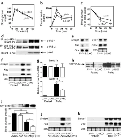
insulin was exaggerated in L-λKO mice (Figure 2c). These observations indicated that the insulin sensitiv-ity of L-λKO mice was increased.
Altered hepatic gene expression in L-λKO mice. Early events of hepatic insulin signaling, including phosphorylation of insulin receptor substrate-1 (IRS-1), IRS-2, and Akt, did not differ between L-λKO and λlox/loxmice after bolus injection of insulin (Figure 2d). We have previ-ously shown that PKCλcontributes to PI3K-dependent gene expression induced by growth factors (7). We there-fore examined L-λKO mice for the hepatic expression of genes that are regulated by insulin. The abundance of mRNA’s for glucokinase (GCK), PKC-1, and G6PC in the liver of randomly fed animals was similar in L-λKO and λlox/loxmice (Figure 2e). However, the amount of transcripts encoding SREBP-1, a transcription factor that regulates the expression of genes important in triglyceride synthesis (19, 20), as well as the amount of those encoding fatty acid synthase, the gene for which is regulated by SREBP-1 (19, 20), were reduced by approximately 50% in the liver of L-λKO mice.
The expression of Srebp1 and its target genes in the liver is induced when mice are refed after starvation (21, 22), a treatment that also results in an increase in the circulat-ing insulin concentration. The increases in the hepatic expression of Srebp1, Fas, and the gene for stearoyl-CoA desaturase-1 (SCD-1; another target of SREBP-1) (19) induced by refeeding were inhibited by about 50%, 35%, and 25%, respectively, in L-λKO mice (Figure 2f). Of the two splice variants of Srebp1mRNA (19), only the abun-dance of Srebp1cmRNA, not that of Srebp1amRNA, is increased in the liver in response to insulin or refeeding (22). The induction of Srebp1c expression in the liver in response to refeeding was inhibited by about 50% in L-λKO mice (Figure 2g). Moreover, immunoblot analysis with antibodies specific for SREBP-1c (14) revealed that the increase in the amount of this protein in a nuclear fraction of the liver induced by refeeding was markedly reduced in L-λKO mice (Figure 2h).
We and other investigators have shown that the effect of insulin on the expression of Srebp1is mediat-ed by a PI3K-dependent pathway (15, 23). We therefore next took advantage of the fact that systemic infusion of adenoviral vectors results in liver-specific expression of exogenous genes (5). Infusion of AxCAMyr-p110, but not of AxCALacZ, resulted in the expression of the Myr-p110 protein in the liver of both L-λKO and λlox/lox mice (Figure 2i); expression of Myr-p110 was not detected in skeletal muscle or adipose tissue (data not shown). Infusion of AxCAMyr-p110, but not of AxCALacZ, also resulted in a reduction in the blood glucose concentration of both L-λKO and λlox/loxmice in the fasted state, suggesting that activation of hepat-ic PI3K signaling lowers blood glucose concentration. However, the glucose-lowering effect of AxCAMyr-p110 was greater in L-λKO mice than in the control animals, consistent with our observation that the glu-cose-lowering effect of exogenously administered insulin was exaggerated in L-λKO mice.
Although the hepatic expression of Srebp1 andFas
was induced by the infusion of AxCAMyr-p110 in both L-λKO and λlox/loxmice (Figure 2j), the extent of this effect was greatly reduced in the former animals, indi-cating that PKCλfunctions as a downstream effector of PI3K in the induction of Srebp1cexpression in the liver. Activation of the liver X receptor also increases the expression of Srebp1cin the liver (16, 24). The hepatic expression of Srebp1and Fasinduced by gavage with T0901317, a liver X receptor agonist (24), was similar in L-λKO and λlox/loxmice (Figure 2k), suggesting that the lack of PKCλdoes not affect the general machinery responsible for the induction of Srebp1c but rather results in a specific disruption in PI3K-dependent sig-naling that leads to increased expression of this gene.
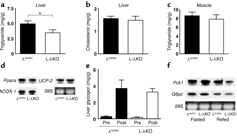
Hepatic lipid content and expression of genes important in
β-oxidation in L-λKO mice. The triglyceride content of the liver was reduced (Figure 3a), whereas the hepatic cho-lesterol content (Figure 3b) and the triglyceride content of skeletal muscle (Figure 3c) were unchanged in L-λKO mice in the randomly fed state compared with the cor-responding values for λlox/loxmice. The synthesis and breakdown (β-oxidation) of triglyceride are the two major determinants of hepatic triglyceride content. The expression of genes that contribute to the oxidation of triglyceride in the liver, including those encoding PPAR-α, acyl-CoA oxidase-1, and uncoupling protein-2, did not differ between L-λKO and λlox/loxmice in the ran-domly fed state (Figure 3d), suggesting that the reduced hepatic triglyceride content of L-λKO mice is attributa-ble to the reduced expression of Srebp1c.
[image:5.576.53.273.77.236.2]Hepatic accumulation of glycogen and the induction of Pck1 and G6pc by starvation/refeeding in L-λKO mice. The hepatic glycogen content in the randomly fed state (data not shown) and the increase in hepatic glycogen content in response to oral glucose intake (Figure 3e) were similar in L-λKO and λlox/loxmice. The expression of Pck1 and G6pcin the liver is inhibited by refeeding after food deprivation. The effect of refeeding on the Table 1
Phenotypic comparison of λlox/loxand L-λKO mice
Parameter λlox/lox L-λKO
18-week-old mice
Body mass (g) 31.7 ± 0.7 31.8 ± 0.5 Liver mass (g) 1.52 ± 0.03 1.49 ± 0.06 Epididymal fat mass (g) 0.31 ± 0.03 0.34 ± 0.03
12-week-old mice
Blood glucose (mg/dl) 111.3 ± 4.2 119.4 ± 2.2 Plasma insulin (pg/ml) 518 ± 38 356 ± 37A
Serum cholesterol (mg/dl) 107.8 ± 4.4 112.0 ± 4.3 Serum triglyceride (mg/dl) 101.6 ± 8.3 107.4 ± 5.9 Serum FFAs (mEq/l) 0.91 ± 0.07 0.95 ± 0.05 Serum leptin (ng/ml) 1.34 ± 0.26 1.39 ± 0.22 Serum adiponectin (µg/ml) 13.9 ± 0.3 14.6 ± 0.4
Figure 2
Glucose and insulin tolerance, insulin signaling, and hepatic gene expression in mice with liver-specific deficiency of PKCλ. (a–c) Blood glucose (a) and plasma insulin (b) concentrations during a glucose-tolerance test in L-λKO and λlox/loxmice at 14 weeks of age, and
blood glucose concentration during an insulin tolerance test at 12 weeks of age (c). Data are mean ± SEM of values from nine to 20 mice. *P < 0.05 vs. the corresponding value for λlox/loxmice (Student’s ttest). (d) Tyrosine phosphorylation of IRS-1 and IRS-2 and
serine phosphorylation of Akt in the liver of λlox/loxor L-λKO mice induced by a bolus injection of insulin. Liver homogenates prepared
2 minutes after administration of insulin (5 U/kg of body mass) or saline were subjected to immunoprecipitation with antibodies to IRS-1 or to IRS-2, and the resulting precipitates were subjected to immunoblot analysis with antibodies to phosphotyrosine (PY). Alternatively, liver homogenates were subjected directly to immunoblot analysis with antibodies specific for phosphorylated Akt (p-Akt). Data are representative of six mice of each genotype. (e–g) Total RNA extracted from the liver of λlox/loxor L-λKO mice (18
weeks of age) in the randomly fed state (n = 8) (e) or after fasting with or without refeeding (n = 4–7) (fand g) was either separately combined and subjected to Northern blot analysis (eand f) or subjected individually to RT-PCR analysis (g) for the indicated mRNA’s. Ethidium bromide staining of 28S rRNA is also shown for Northern analysis. *P < 0.01 (ANOVA). (h) The nuclear fraction of liver homogenates prepared fromλlox/loxor L-λKO mice after fasting with or without refeeding was subjected to immunoblot analysis with
antibodies to SREBP-1c. Data shown are from two mice and are representative of four to six animals. (iand j) Mice (λlox/loxor L-λKO)
16–18 weeks of age (n = 10–16) were injected with AxCAMyr-p110 or AxCALacZ and were subsequently deprived of food for 16 hours. The abundance of Myr-p110 in liver homogenates was then examined by immunoblot analysis with antibodies to Myc (i, upper panel), blood glucose concentration was determined (i, lower panel), and the amounts of Srebp1and FasmRNA’s among separately com-bined total RNA extracted from the liver were evaluated by Northern analysis (j). *P < 0.05, **P < 0.01 (ANOVA). (k) Total RNA extracted from the liver of λlox/loxor L-λKO mice treated with either T0901317 or vehicle was separately combined and subjected to
hepatic expression of these genes was slightly exag-gerated in L-λKO mice compared with that apparent in λlox/loxanimals (Figure 3f). These results thus sug-gested that PKCλsignaling is not required for either the hepatic accumulation of glycogen or for inhibi-tion of the expression of gluconeogenesis genes in the liver. The slight enhancement of refeeding-induced suppression of Pck1and G6pcexpression apparent in L-λKO mice may be related to the increased insulin sensitivity of these animals.
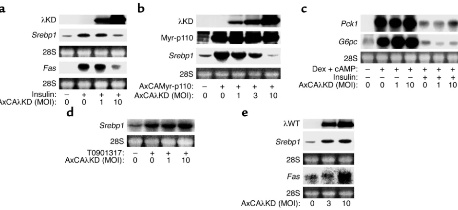
Effects of dominant-negative and WT PKCλon insulin-induced expression of Srebp1 in cultured hepatocytes. To con-firm a causal relation between PKCλdeficiency and the altered hepatic expression of Srebp1in L-λKO mice, we examined the effect of PKCλsignaling on the abun-dance of Srebp1mRNA in primary cultures of rat hepa-tocytes. Incubation of the cells with insulin induced an increase in the amounts of Srebp1and FasmRNA’s (Fig-ure 4a), and this effect was inhibited by adenovirus-mediated expression of λKD, which acts in a domi-nant-negative manner (7, 8). Expression of λKD also inhibited the increase in the amount of Srebp1mRNA induced by Myr-p110 (Figure 4b). In contrast, λKD did not affect either the insulin-induced inhibition of Pck1
and G6pcexpression (Figure 4c) or the expression of
Srebp1induced by T0901317 (Figure 4d), indicating that PKCλsignaling specifically contributes to insulin-induced expression of Srebp1. Moreover, expression of
recombinant WT PKCλincreased the abundance of
Srebp1and FasmRNA’s in the absence of insulin (Fig-ure 4e), indicating that PKCλsignaling is sufficient for the induction of these genes.
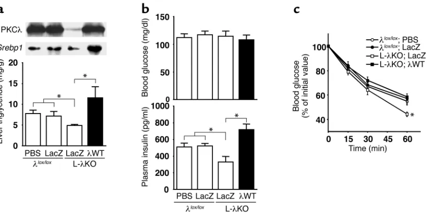
[image:7.576.92.500.394.628.2]Restoration of PKCλexpression reverses the decrease in hepatic lipid content and the increase in insulin sen-sitivity in L-λKO mice. To verify that the altered insulin sensitivity and hepatic triglyceride content of L-λKO mice are attributable to the lack of PKCλin the liver, we restored the hepatic expression of this enzyme in L-λKO animals. Infusion of AxCAλWT into L-λKO mice result-ed in the expression of PKCλin the liver at a level simi-lar to that apparent in λlox/loxmice (Figure 5a). The restoration of PKCλexpression in the liver increased both the abundance of Srebp1 mRNA and the trigly-ceride content in this organ of L-λKO mice. Blood glu-cose concentration was similar in L-λKO mice infused with AxCAλWT or with AxCALacZ and in λlox/loxmice infused with AxCALacZ or with PBS. The plasma insulin concentration of L-λKO mice was increased by infusion of AxCAλWT, but not of AxCALacZ, to an extent similar to that apparent in λlox/loxmice infused with AxCALacZ or with PBS (Figure 5b). Moreover, the enhancement of the glucose-lowering effect of exoge-nously administered insulin apparent in L-λKO mice was also reversed by restoration of PKCλexpression in the liver (Figure 5c). These results thus indicate that the changes in the expression of Srebp1 and in triglyceride
Figure 3
Hepatic lipid and glycogen content and the expression of genes involved in β-oxidation and gluconeogenesis in mice with liver-specific PKCλ
deficiency. (a–c) Triglyceride (a) and cholesterol (b) content of the liver and triglyceride content of hind limb muscle (c) of L-λKO and λlox/lox
mice in the randomly fed state at 18 weeks of age. Data are expressed as mg analyte/g wet tissue and are the mean ±SEM from seven mice. *P < 0.05 (Student’s ttest). (d) Total RNA extracted from the liver of λlox/loxor L-λKO mice (n = 8) at 18 weeks of age and in the randomly
fed state was separately combined and subjected to Northern blot analysis for mRNA’s encoding PPAR-α, acyl-CoA oxidase-1 (ACOX-1), and uncoupling protein-2 (UCP-2). (e) Hepatic glycogen content of λlox/loxor L-λKO mice at 20 weeks of age before (Pre) and 2 hours after
(Post) oral glucose intake. Data are mean ± SEM from four to six mice. (f) Total RNA extracted from the liver of λlox/loxor L-λKO mice (18
content in the liver as well as in whole-body insulin sen-sitivity apparent in L-λKO mice are directly attributable to the lack of PKCλin the liver.
Discussion
On the basis of observations with cultured cells (7, 8), we hypothesized that PKCλ participates in insulin action in vivo as a downstream effector of PI3K. Our present results now demonstrate such a function for PKCλ, at least in the liver. Several of the metabolic effects of insulin in the liver are exerted through the regulation of gene expression. We have previously shown that the regulation by insulin of the expression of Gck, Srebp1c, G6pc, and Pck1in mouse liver is medi-ated by PI3K (5). Of these four genes, each of which participates in the metabolic actions of insulin, we have now revealed that the expression of Srebp1c, a key regulator of fatty acid and triglyceride synthesis (19, 20), is regulated by PKCλacting downstream of PI3K. At present, a signaling pathway that links PKCλand the expression of Srebp1cremains unclear. Cyclohex-imide, an inhibitor of general protein synthesis, has been shown to prevent insulin-induced expression of
Srebp1cin cultured hepatocytes (25), suggesting that de novo protein synthesis is required for this action of insulin. PKCλthus may contribute to induce
expres-sion of such a protein involved in the transcriptional activation of Srebp1c.
The hepatic expression of Srebp1cinduced by refeed-ing or by an active PI3K was markedly, but not com-pletely, prevented in L-λKO mice, suggesting that the induction of Srebp1cis not solely dependent on PKCλ
signaling. Given that the liver expresses both PKCλand PKCζ, PKCζmay be responsible for the residual signal-ing of Srebp1c. A membrane-targeted form of Akt that exhibits higher kinase activity than does WT Akt increases the abundance of Srebp1cmRNA when it is expressed in primary cultured hepatoyctes (23), sug-gesting that Akt, a downstream effector of PI3K, may also contribute to the induction of Srebp1c. However, we have previously shown that the inhibition of endoge-nous Akt activity with the use of a dominant-negative mutant of the kinase did not prevent, but rather aug-mented, insulin-induced expression of Srebp1c(15). Moreover, in ob/ob mice and a mouse model with lipodystrophic diabetes, the abundance of Srebp1c
[image:8.576.56.526.54.270.2]mRNA is increased, whereas insulin-induced phospho-rylation of Akt is markedly reduced in the liver of these animals (26). Mice lacking Akt2, a major isoform of Akt in the liver, have been established (27). The physiologi-cal importance of Akt in the induction of Srebp1cin vivo may be revealed by characterization of the mutant mice. Figure 4
Effects of dominant-negative and WT PKCλon the expression of Srebp1, Fas, Pck1, andG6pcin primary cultured rat hepatocytes. (a, c, and
Atypical PKC isozymes are evolutionarily conserved proteins required for the formation of apical-basal polarity in cells (28), which is important for the struc-tural organization and function of organs. However, the structures of hepatic lobules and hepatocyte plates and the serum parameters of general liver function appeared normal in L-λKO mice. This observation may be attributable to the fact that disruption of the PKCλ
gene was accomplished by Cre recombinase expressed under the control of the promoter of the albumin gene, which is a marker gene of fully differentiated hepato-cytes. A role for PKCλin the development of cellular polarity in the liver might be revealed by characteriza-tion of hepatectomy-induced liver regeneracharacteriza-tion in L-λKO mice, given that the liver regenerates predomi-nantly through the replication of mature hepatocytes under this experimental condition (29).
An unexpected finding of the present study was that L-λKO mice exhibit increased insulin sensitivity. The tissue-specific disruption of a gene important for insulin signaling thus paradoxically resulted in an increase in whole-body insulin sensitivity. A similar phenomenon has been observed with mice lacking the insulin receptor specifically in adipose tissue (30).
Restoration of the hepatic expression of PKCλ
reversed this metabolic phenotype of L-λKO mice, indicating that the lack of PKCλin the liver is indeed responsible for the increased insulin sensitivity of these animals. The precise mechanism that underlies
this phenomenon remains unclear. Evidence suggests that the alteration of fatty acids/triglyceride metabo-lism in insulin’s target tissues is an important deter-minant of insulin sensitivity. The increase in circulat-ing FFAs leads to insulin resistance and the accumulation of triglyceride in skeletal muscle (31), and triglyceride content in the liver or in skeletal mus-cle negatively correlates with insulin sensitivity in humans (32, 33). Leptin and adiponectin, the two major fat-derived hormones, increase insulin sensitiv-ity and concomitantly reduce hepatic triglyceride con-tent (probably by promoting fatty acid oxidation) in an animal model of insulin resistance or in humans with lipodystrophic diabetes (34–36). Moreover, over-expression of lipoprotein lipase in liver or skeletal muscle resulted in an increase in fatty acid metabolites and consequently in the accumulation of triglyceride in the respective tissue, as well as insulin resistance (37). The decrease in the hepatic expression of the lipogenic genes and the subsequent alterations in fatty acid metabolism in L-λKO mice may thus be related to the increased insulin sensitivity of these animals.
TNF-αsecreted from adipose tissue is implicated in the development of obesity-induced insulin resist-ance (38). Atypical PKC is activated by cytokines, including TNF-αand IL-1 (39, 40), and directly phos-phorylates and activates I-κB kinase-β(IKKβ) (41). Administration of salicylic acid derivatives that inhibit IKKβ(42) was shown to increase insulin sen-Figure 5
Effects of adenovirus-mediated restoration of PKCλexpression in the liver of mice with liver-specific PKCλdeficiency on hepatic lipid con-tent, expression of Srebp1in the liver, and insulin sensitivity. (a) Twenty-week-old λlox/loxor L-λKO mice (n = 7–9) were injected with PBS or
with adenoviruses encoding either β-gal (LacZ) or WT PKCλ(λWT), as indicated. Total liver homogenates were subsequently subjected to immunoprecipitation and immunoblot analysis with antibodies to PKCλ(upper panel; data are representative of three experiments). Total RNA extracted from the liver was separately combined and subjected to Northern blot analysis with a probe specific for Srebp1mRNA (mid-dle panel), and hepatic triglyceride content was determined (lower panel; data are shown as mean ± SEM). *P < 0.05 (ANOVA). (band c) Twelve-week-old λlox/loxor L-λKO mice were injected with PBS or with adenoviruses encoding either β-gal or WT PKCλ, after which blood
[image:9.576.88.507.54.267.2]sitivity both in rodent models of diabetes and in human subjects (43, 44). Moreover, heterozygous dis-ruption of the IKKβgene ameliorated the insulin resistance of obese model mice (44). It is therefore possible that the hepatic deficiency of PKCλ in L-λKO mice results in inhibition of a TNF-α/PKCλ/ IKKβsignaling pathway and a consequent increase in whole-body insulin sensitivity.
Transgenic mice that overexpress lipoprotein lipase in skeletal muscle or the liver exhibit an impairment of the insulin-induced increase in PI3K activity associat-ed with IRS-1 or IRS-2, respectively (37). Moreover, administration of salicylic acid derivatives enhanced insulin-induced tyrosine phosphorylation of the insulin receptor in genetically obese animals (44). However, we did not detect changes in the insulin-induced phosphorylation of IRS proteins or of Akt in the liver of L-λKO mice. Although we cannot exclude the possibility that a small increase in the extent of insulin signaling went undetected under our experi-mental conditions, it is possible that the enhancement of insulin action apparent in L-λKO mice occurs at a step other than IRS or Akt phosphorylation. Evidence suggests that PKCζparticipates in a negative feedback pathway of insulin signaling leading to the phospho-rylation of IRS proteins in cultured cells (45). Given that the insulin-induced phosphorylation of IRS pro-teins was not significantly increased in the liver of L-λKO mice, PKCλappears not to participate in such a negative feedback pathway in mouse liver.
In summary, we have shown that, among the vari-ous metabolic actions of insulin, PKCλspecifically contributes to induction of the expression of Srebp1c
and of its target genes important in triglyceride syn-thesis in the liver. Animal models of insulin resist-ance or obesity often manifest increases both in lipid content and in the expression of Srebp1c in the liver (26, 46). Reagents that block PKCλsignaling specifi-cally in the liver might thus prove effective for reduc-ing hepatic Srebp1c expression and consequently hepatic triglyceride content, as well as for ameliorat-ing insulin resistance.
Acknowledgments
We thank D. LeRoith, T. Noguchi, H. Nakajima, N. Iri-tani, D.K. Granner, and K. Murakami for Alb-Cremice, probes for Gck, G6pc, Fas, and Pck1, and for T0901317, respectively. This work was supported by a grant from the Ministry of Education, Culture, Sports, Science, and Technology of Japan (to M. Kasuga and W. Ogawa), a grant-in-aid for the Research for the Future Program from the Japan Society for the Promotion of Science (to M. Kasuga), and a grant from Cooperative Link of Unique Science and Technology for Economy Revitalization (CLUSTER) (to M. Kasuga).
1. DeFronzo, R.A. 1997. Pathogenesis of type 2 diabetes: metabolic and molecular implications for identifying diabetic genes. Diabetes Rev.
5:177–269.
2. Michael, M.D., et al. 2000. Loss of insulin signaling in hepatocytes leads
to severe insulin resistance and progressive hepatic dysfunction. Mol. Cell.
6:87–97.
3. Ogawa, W., Matozaki, T., and Kasuga, M. 1998. Role of binding proteins to IRS-1 in insulin signalling. Mol. Cell. Biochem.182:13–22.
4. Shepherd, P.R., Withers, D.J., and Siddle, K. 1998. Phosphoinositide 3-kinase: the key switch mechanism in insulin signalling. Biochem. J.
333:471–490.
5. Miyake, K., et al. 2002. Hyperinsulinemia, glucose intolerance, and dyslipi-demia induced by acute inhibition of phosphoinositide 3-kinase signaling in the liver. J. Clin. Invest.110:1483–1491. doi:10.1172/JCI200215880. 6. Suzuki, A., Akimoto, K., and Ohno, S. 2003. Protein kinase C λ/ι
(PKC λ/ι): a PKC isoform essential for the development of multicellular organisms. J. Biochem. (Tokyo).133:9–16.
7. Akimoto, K., et al. 1996. EGF or PDGF receptors activate atypical PKCλ
through phosphatidylinositol 3-kinase. EMBO J.15:788–798. 8. Kotani, K., et al. 1998. Requirement of atypical protein kinase Cλfor
insulin stimulation of glucose uptake but not for Akt activation in 3T3-L1 adipocytes. Mol. Cell. Biol.18:6971–6982.
9. Williams, M.R., et al. 2000. The role of 3-phosphoinositide-dependent protein kinase 1 in activating AGC kinases defined in embryonic stem cells. Curr. Biol.10:439–448.
10. Le Good, J.A., et al. 1998. Protein kinase C isotypes controlled by phos-phoinositide 3-kinase through the protein kinase PDK1. Science.
281:2042–2045.
11. Tabuse, Y. 1998. Atypical protein kinase C cooperates with PAR-3 to establish embryonic polarity in Caenorhabditis elegans. Development.
125:3607–3614.
12. Wodarz, A., Ramrath, A., Grimm, A., and Knust, E. 2000. Drosophila atypical protein kinase C associates with Bazooka and controls polarity of epithelia and neuroblasts. J. Cell Biol.150:1361–1374.
13. Yakar, S., et al. 1999. Normal growth and development in the absence of hepatic insulin-like growth factor I. Proc. Natl. Acad. Sci. U. S. A.
96:7324–7329.
14. Yahagi, N., et al. 1999. A crucial role of sterol regulatory element-bind-ing protein-1 in the regulation of lipogenic gene expression by polyun-saturated fatty acids. J. Biol. Chem.274:35840–35844.
15. Matsumoto, M., et al. 2002. Role of the insulin receptor substrate 1 and phosphatidylinositol 3-kinase signaling pathway in insulin-induced expression of sterol regulatory element binding protein 1c and glucoki-nase genes in rat hepatocytes. Diabetes.51:1672–1680.
16. Repa, J.J., et al. 2000. Regulation of mouse sterol regulatory element-binding protein-1c gene (SREBP-1c) by oxysterol receptors, LXRαand LXRβ. Genes Dev.14:2819–2830.
17. Katsurada, A., et al. 1990. Effects of nutrients and hormones on tran-scriptional and post-trantran-scriptional regulation of fatty acid synthase in rat liver.Eur. J. Biochem.190:427–433.
18. Kitamura, T., et al. 1999. Insulin-induced phosphorylation and activa-tion of cyclic nucleotide phosphodiesterase 3B by the serine-threonine kinase Akt. Mol. Cell. Biol.19:6286–6296.
19. Horton, J.D., Goldstein, J.L., and Brown, M.S. 2002. SREBPs: activators of the complete program of cholesterol and fatty acid synthesis in the liver. J. Clin. Invest.109:1125–1131. doi:10.1172/JCI200215593. 20. Shimano, H., et al. 1999. Sterol regulatory element-binding protein-1 as
a key transcription factor for nutritional induction of lipogenic enzyme genes. J. Biol. Chem.274:35832–35839.
21. Horton, J.D., Bashmakov, Y., Shimomura, I., and Shimano, H. 1998. Reg-ulation of sterol regulatory element binding proteins in livers of fasted and refed mice. Proc. Natl. Acad. Sci. U. S. A.95:5987–5992.
22. Shimomura, I., et al. 1999. Insulin selectively increases SREBP-1c mRNA in the livers of rats with streptozotocin-induced diabetes. Proc. Natl. Acad. Sci. U. S. A.96:13656–13661.
23. Fleischmann, M., and Iynedjian, P.B. 2000. Regulation of sterol regula-tory-element binding protein 1 gene expression in liver: role of insulin and protein kinase B/cAkt. Biochem. J.349:13–17.
24. Schultz, J.R., et al. 2000. Role of LXRs in control of lipogenesis. Genes Dev.14:2831–2838.
25. Foretz, M., et al. 1999. ADD1/SREBP-1c is required in the activation of hepatic lipogenic gene expression by glucose. Mol. Cell. Biol.19:3760–3768. 26. Shimomura, I., et al. 2000. Decreased IRS-2 and increased SREBP-1c lead to mixed insulin resistance and sensitivity in livers of lipodystrophic and ob/ob mice. Mol. Cell. 6:77–86.
27. Cho, H., et al. 2001. Insulin resistance and a diabetes mellitus-like syn-drome in mice lacking the protein kinase Akt2 (PKB β). Science.
292:1728–1731.
28. Ohno, S. 2001. Intercellular junctions and cellular polarity: the PAR-aPKC complex, a conserved core cassette playing fundamental roles in cell polarity.Curr. Opin. Cell Biol.13:641–648.
31. Bachmann, O.P., et al. 2001. Effects of intravenous and dietary lipid chal-lenge on intramyocellular lipid content and the relation with insulin sen-sitivity in humans. Diabetes.50:2579–2584.
32. Ryysy, L., et al. 2000. Hepatic fat content and insulin action on free fatty acids and glucose metabolism rather than insulin absorption are asso-ciated with insulin requirements during insulin therapy in type 2 dia-betic patients. Diabetes.49:749–758.
33. Krssak, M., et al. 1999. Intramyocellular lipid concentrations are corre-lated with insulin sensitivity in humans: a 1H NMR spectroscopy study.
Diabetologia.42:113–116.
34. Kakuma, T., et al. 2000. Leptin, troglitazone, and the expression of sterol regulatory element binding proteins in liver and pancreatic islets. Proc. Natl. Acad. Sci. U. S. A.97:8536–8541.
35. Yamauchi, T., et al. 2001. The fat-derived hormone adiponectin reverses insulin resistance associated with both lipoatrophy and obesity. Nat. Med.7:941–946.
36. Petersen, K.F., et al. 2002. Leptin reverses insulin resistance and hepatic steatosis in patients with severe lipodystrophy. J. Clin. Invest.
109:1345–1350. doi:10.1172/JCI200215001.
37. Kim, J.K., et al. 2001. Tissue-specific overexpression of lipoprotein lipase causes tissue-specific insulin resistance. Proc. Natl. Acad. Sci. U. S. A.
98:7522–7527.
38. Uysal, K.T., Wiesbrock, S.M., Mario, M.W., and Hotamisligil, G.S. 1997. Protection from obesity-induced insulin resistance in mice lacking TNF-αfunction. Nature.389:610–614.
39. Muller, G., et al. 1995. PKCζis a molecular switch in signal transduction of TNF-α, bifunctionally regulated by ceramide and arachidonic acid.
EMBO J. 14:1961–1969.
40. Limatola, C., Barabino, C., Nista, A., and Santoni, A. 1997. Interleukin 1-β-induced protein kinase C-ζactivation is mimicked by exogenous phospholipase D. Biochem. J. 321:497–501.
41. Lallena, M.J, Diaz-Meco, M.T., Bren, G., Paya, C.V., and Moscat, J. 1999. Activation of IκB kinase βby protein kinase C isoforms. Mol. Cell. Biol.
19:2180–2188.
42. Yin, M.J., Yamamoto, Y., and Gaynor, R.B. 1998. The anti-inflammatory agents aspirin and salicylate inhibit the activity of IκB kinase-β. Nature.
396:77–80.
43. Hundal, R.S., et al. 2002. Mechanism by which high-dose aspirin improves glucose metabolism in type 2 diabetes. J. Clin. Invest.
109:1321–1326. doi:10.1172/JCI200214955.
44. Yuan, M., et al. 2001. Reversal of obesity- and diet-induced insulin resist-ance with salicylates or targeted disruption of Ikkβ. Science.
293:1673–1677.
45. Liu, Y.F., et al. 2001. Insulin stimulates PKCζ-mediated phosphorylation of insulin receptor substrate-1 (IRS-1). A self-attenuated mechanism to negatively regulate the function of IRS proteins. J. Biol. Chem.
276:14459–14465.