R E S E A R C H A R T I C L E
Open Access
Time course analyses of structural changes
in the infrapatellar fat pad and synovial
membrane during inflammation-induced
persistent pain development in rat knee
joint
Kei Inomata
1, Kunikazu Tsuji
2*, Hiroaki Onuma
1, Takashi Hoshino
1, Mio Udo
1, Masako Akiyama
3, Yusuke Nakagawa
2,
Hiroki Katagiri
1, Kazumasa Miyatake
1, Ichiro Sekiya
4, Takeshi Muneta
1,5and Hideyuki Koga
1Abstract
Background:Osteoarthritis (OA) is a common joint disease in aging societies, which is accompanied by chronic inflammation and degeneration of the joint structure. Inflammation of the infrapatellar fat pad (IFP) and synovial membrane (IFP surface) plays essential roles in persistent pain development in patients with OA. To identify the point during the inflammatory process critical for persistent pain development, we performed a time course histological analysis in a rat arthritis model.
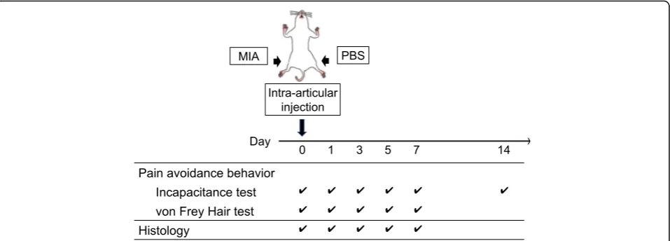
Methods:Wistar rats received single intra-articular injection of monoiodoacetic acid (MIA, 0.2 or 1.0 mg/30μL) in the right knees or phosphate-buffered saline (PBS, 30μL) as a control in the left knees. Pain avoidance behaviors (weight-bearing asymmetry and tactile hypersensitivity of the plantar surface of the hind paw) were evaluated on days 0, 1, 3, 5, 7, and 14 after injection. Histological assessments of the knee joint were performed on days 0, 1, 3, 5, and 7 after MIA injection.
Results:Weight-bearing asymmetry was observed along with the onset of acute inflammation in both the low- (0. 2 mg) and high-dose (1.0 mg) groups. In the low-dose group, weight-bearing asymmetry was completely reversed on day 10, indicating that joint pain seemed to alleviate between days 7 and 10. In contrast, we observed
persistent joint pain after day 10 in the high-dose group. Histological assessments of the high-dose group indicated that the initial sign of inflammatory responses was observed in the perivascular region inside the IFP. Inflammatory cell infiltration from the perivascular region to the parenchymal region of the IFP was observed on day 3 and reached the IFP surface (synovial membrane) on day 7. Extensive fibrosis throughout the IFP was observed between days 5 and 7 after MIA injection.
Conclusion:Our data indicated that acute joint pain occurs along with the onset of acute inflammatory process. Irreversible structural changes in the IFP, such as extensive fibrosis, are observed prior to persistent pain
development. Thus, we consider that this process may play important roles in persistent pain development.
* Correspondence:[email protected]
2Department of Cartilage Regeneration, Tokyo Medical and Dental University,
1-5-45, Yushima, Bunkyo-ku, Tokyo 113-8510, Japan
Full list of author information is available at the end of the article
Background
Osteoarthritis (OA) is one of the most common diseases in aging societies, which is accompanied by chronic joint inflammation, articular cartilage degeneration, and osteophyte formation [1, 2]. Epidemiological analyses in Japan suggested that the number of patients with radio-graphically identifiable knee OA (Kellgren-Lawrence score≥2) was almost 25 million, which is approximately one-fifth of the Japanese population, and almost 30% of them (8 million) have symptoms that affect activities of daily living [3].
The major complaint of patients with knee OA is per-sistent pain, which significantly decreases both, the pa-tients’activities of daily living (ADL) and quality of life (QOL). Therefore, most of the current conservative treatment strategies for knee OA are based on symptom management by anti-inflammatory analgesics and im-provement of joint mobility and flexibility by programed exercise (land or water based), weight control, and edu-cation [4]. However, some patients develop uncontrolled persistent knee pain with disease progression. In these cases, patients are pressed to make decisions for alterna-tive treatments, such as total knee arthroplasty.
Mechanisms of persistent pain development in knee OA have largely not been elucidated. During inflamma-tion or tissue injury, nociceptive pathways are activated by various mediators that sensitize primary afferent nerves in the joint (peripheral sensitization). Over time, these enhanced neuronal activities variegate the plasti-city of second-order neurons in the central nervous sys-tem, making them more responsive to the signals from the periphery. Another important component in the process of persistent pain development is the structural changes in the joint tissues. Previous studies have re-ported that synovial fibrosis contributes to joint pain and stiffness [5–7]. However, it is not clear whether the structural changes of infrapatellar fat pad (IFP) and syn-ovial membrane play important roles in persistent joint pain and to what degree. To answer this question, we performed time course histological assessments in the monoiodoacetic acid (MIA)-induced rat joint inflamma-tion model.
Intra-articular injection of MIA is a well-characterized animal model for inflammation-induced OA [8–11]. By using this model, we have reported two different inflammation-induced articular cartilage degeneration models in rats [12, 13]. One is the low-dose model (0.2 mg), in which acute inflammation was observed within 3 days, peaked at 5 days, then alleviated after 7 days [12, 13]. In this model, we observed slow progression of ar-ticular cartilage degeneration after 28 days without ap-parent synovial inflammation after 14 days [12,13]. The other is the high-dose (1.0 mg) model, in which the on-set of acute inflammation was comparable with that of
the low-dose model; however, obvious irreversible struc-tural changes in the synovial membrane and IFP were observed after 5 days [12, 13]. We have compared the pain avoidance behavior between these two models and found that acute joint pain was observed along with the onset of the acute inflammatory responses in both ex-perimental conditions. In the low-dose model, joint pain was rapidly alleviated after 7 days along with the reso-lution of acute inflammation [13]. In contrast, we noted the development of persistent pain in the high-dose model after 7 days [13]. We considered that these obser-vations strongly suggested the importance of irreversible structural changes in the synovial membrane and IFP between 5 to 7 days during persistent pain development.
Based on this hypothesis, we performed time course histological analyses in the early stage after joint inflam-mation to elucidate which point during the irreversible structural changes in the synovial membrane and IFP is important for persistent pain development. Here, we re-port that extensive fibrotic changes occurred between days 5 and 7 after MIA injection.
Methods Materials
MIA and paraformaldehyde were purchased from Sigma Aldrich (St. Louis, MO, USA). Isoflurane and ethylenedi-aminetetraacetic acid (EDTA) were purchased from Wako Pure Chemical Industries Ltd. (Osaka, Japan).
Animals
Male Wistar rats (Charles River, Tokyo, Japan) at 8 weeks of age, 280–310 g in weight, were subjected to this study. The animals were housed under a 12 h/12 h light/ dark cycle with food and water ad libitum. All proce-dures of this study were conducted in accordance with the protocols approved by the Institutional Animal Care and Use Committee (IACUC) of Tokyo Medical and Dental University.
MIA injection
Analysis of pain avoidance behavior
Severity of joint pain was quantitatively evaluated by weight-bearing asymmetry (incapacitance test, Linton Instrumentation, Norfolk, UK) and tactile hypersensitiv-ity of the plantar surface of the hind paw (von Frey hair test, North Coast Medical, Inc., Gilroy, CA, USA) [9, 13–17]. These tests were performed to verify the repro-ducibility of the experimental model employed in this study with those of our previous report [13].
Weight-bearing asymmetry between the right (MIA side) and left (control side) limbs was measured on days 0, 1, 3, 5, 7, and 14 to reconfirm that pain persistence occurred after 7 days in the high-dose model (Fig. 1). Measurements were done over 100 times for each sam-ple, and the percent weight of the MIA side (right hind limb) was calculated according to the equation below as described previously (n= 6) [13,15].
½Percent of weight on ipsilateral limb¼weight on ipsilateral limb= weight on ipsilateral limbþweight on contralateral limb
ð Þ 100
To verify the reproducible tactile hypersensitivity of the plantar surface of the hind paw was observed during the experimental period in this study, von Frey hair test was performed on days 0, 1, 3, 5, and 7 according to the methods described previously (n = 6, [13, 15]). In these experiments, the elasticity of the von Frey hairs was started from 300 g and gradually decreased until it reached 2 g. Maximum elasticity in which rats did not express any escape behavior was recorded as the paw withdrawal threshold [9,18–20].
Histological and immunohistochemical assessment
The rats were euthanized by carbon dioxide on days 0 (n= 4), 1, 3, 5, and 7 (n = 6, Fig. 1). The whole knee joints were dissected, fixed in 4% paraformaldehyde at pH 7.4 for 7 days, decalcified in 20% EDTA solution at 4 °C for 10 days, and embedded in paraffin wax. Sagittal sections of the whole knee joint were prepared at 5μm. Hematoxylin-eosin (HE) and Masson trichrome (MT) staining were performed as described previously [21,22]. An Olympus BX53 microscope (Olympus, Tokyo, Japan) was used to visualize the histological sections. The % area of fibrosis was measured using ImageJ software (National Institutes of Health, Bethesda, MD, USA).
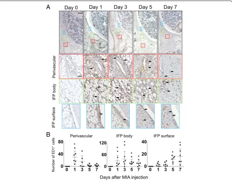
To identify the macrophages that infiltrated into the IFP, immunostaining by ED1 antibody (Abcam, Cam-bridge CamCam-bridgeshire, UK) was performed, as described previously [9, 21, 23]. The number of ED1-positive cells in the IFP was counted using an Olympus BX53 micro-scope (Olympus, Tokyo, Japan).
Semiquantitative evaluation of structural changes in the IFP
Cellularity
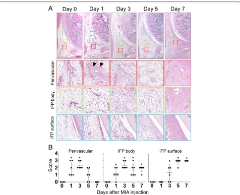
The perivascular region, parenchymal region of the IFP (IFP body), and superficial layer (IFP surface or synovial membrane) were chosen from a section stained with HE (representative pictures are indicated in Fig.3). Cellular-ity of each section was semi-quantitatively evaluated ac-cording to the criteria described in Table 1 and representative pictures were indicated in Additional file1: Figure S1A. Data were indicated as a mean value of 3
[image:3.595.60.539.88.260.2]independent sections (day 0, n= 4; day 1, 3, 5, and 7 samples,n= 6).
Macrophage infiltration
Serial sagittal sections were stained with ED-1 antibody to visualize macrophages that infiltrated in IFP. The three separate areas, perivascular region, parenchymal region of IFP (IFP body), and superficial layer (IFP sur-face or synovial membrane), were chosen from the same section, and the number of macrophages was counted at a magnification of 200 times (representative pictures are indicated in Fig.4). Data were indicated as a mean value of 3 independent sections (day 0,n= 4; day 1, 3, 5, and 7 samples,n= 6).
Fibrosis
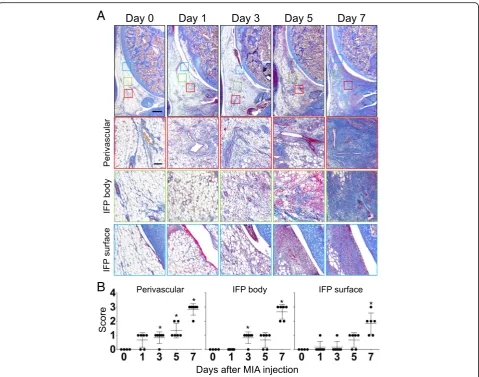
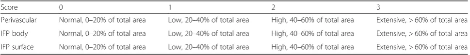
MT staining was performed to visualize collagen fiber deposition in the IFP. The three separate areas (perivas-cular region, parenchymal region of IFP [IFP body], and superficial layer [IFP surface or synovial membrane]) were chosen from the same section, and % area of fibro-sis was measured using ImageJ software (representative pictures are indicated in Fig.5). Data were indicated as a mean value of 3 independent sections (day 0, n = 4; day 1, 3, 5, and 7 samples, n = 6). Semiquantitative evalu-ation was performed according to the criteria described in Table2 and representative pictures were indicated in Additional file1: Figure S1B.
Statistical analysis
Steel’s test, a multiple comparison test for comparing several treatments with a control treatment, was employed to analyze the results of incapacitance test and von Frey Hair test (Fig. 2). Kruskal-Wallis test followed by Steel-Dwass test was employed to analyze the results of histological evaluations between 5 independent time points (Figs. 3, 4, and 5). All statistical analyses were performed using EZR software [24].P-values < 0.05 were considered significant.
Results
Intra-articular injection of high-dose MIA induces persistent knee pain in rats
As described previously [13] and in this study much in detail, intra-articular injection of MIA quickly reduced weight bearing of the ipsilateral limbs on day 1 regard-less of the dose (0.2 mg or 1.0 mg, Fig. 2a). In the low-dose (0.2 mg) group, this reduction continued until days 5 and then gradually alleviated to the pre-experimental levels after day 7. In contrast, weight bearing of the ipsilateral limbs reduced throughout the experimental period in the high-dose (1.0 mg) group (Fig. 2). These data suggest that some biological events that occurred between day 5 and 7 after MIA injection may play important roles during persistent pain develop-ment. Another indication, progressive tactile hypersensi-tivity, which contributes to a cumulative allodynia during inflammation, was also comparable with that of our previous report (Fig.2b) [13]. These may verify the reproducibility of the pain avoidance behavior in our ex-perimental models.
Time course analyses of structural changes in the synovial membrane and IFP
[image:4.595.59.544.100.155.2]Pain avoidance behavior tests indicated that persistent pain generation occurred on day 7 in the high-dose group. We performed histological assessments on days 0, 1, 3, 5, and 7 after MIA injection to identify biological events around the knee joint in the high-dose group (1.0 mg). Figure3presents the cell number changes over time in the perivascular region, IFP body (parenchymal region), and IFP surface (synovial membrane). As shown in this figure, the first sign of the structural change was observed in the perivascular region (Fig.3a, indicated by the arrowhead). We found that the number of cell nuclei stained by hematoxylin immediately increased after MIA injection on day 1 and then reduced after day 5 in the perivascular region. Cell kinetics was semiquantitatively evaluated and plotted in Fig. 3b. Eosin staining, which visualizes both the cytoplasm and extracellular Table 1Semi-quantitative evaluation of structural changes in IFP: Cellularity
Score 0 1 2 3
Perivascular Normal Slightly increased Moderately increased Markedly increased
IFP body Normal Slightly increased Moderately increased Markedly increased
IFP surface ≦Single cell layer 2~3 Cell layers 4~5 Cell layers ≧6 Cell layers (6 or more)
Table 2Semi-quantitative evaluation of structural changes in IFP: Fibrosis
Score 0 1 2 3
Perivascular Normal, 0–20% of total area Low, 20–40% of total area High, 40–60% of total area Extensive, > 60% of total area
IFP body Normal, 0–20% of total area Low, 20–40% of total area High, 40–60% of total area Extensive, > 60% of total area
[image:4.595.56.541.675.732.2]connective tissues, also indicated the surge of cellularity in the perivascular region as early as day 1 after MIA in-jection (Fig.3a). Unlike that of hematoxylin, staining in-tensity of eosin did not decrease after day 5, suggesting the accumulation of extracellular matrices after the surge of cell numbers in the perivascular region. Cell mi-gration kinetics in the perivascular region seemed to be followed by that in the IFP body (Fig. 3a). The number of the cells gradually increased on day 3 and was con-tinuously high until day 7. Interestingly, staining inten-sity of eosin dramatically increased between days 5 and 7 without increasing the number of nuclei (stained by hematoxylin), indicating extensive extracellular matrix
apposition during this period (Fig. 3a). Hyperplastic changes in the IFP surface (synovial membrane) initiated on day 3 after MIA injection and continued throughout the experimental period (Fig. 3a). These data suggest that the inflammatory response may start around the perivascular region and then move to the IFP surface despite the intra-articular injection of MIA. Representa-tive images of the time course changes in the contralat-eral side (intra-articular injection of PBS) are shown in Additional file2: Figure S2A (the top row).
Next, to analyze the changes in inflammatory cell number after intra-articular injection of MIA, immuno-staining of IFP by ED-1 antibody, which identifies mac-rophages, was performed. As presented in Fig. 4, rapid accumulation of macrophages was observed on day 1 in the perivascular region (Fig. 4a, arrows). These macro-phages spread throughout the IFP body on day 3 and reached the IFP surface on day 5 (Fig.4a). Macrophages that infiltrated into the perivascular region and IFP body quickly disappeared after day 5 (Fig. 4a). Semiquantita-tive analyses also support these observations (Fig. 4b). Representative images of the time course changes in the contralateral side (intra-articular injection of PBS) are shown in Additional file 2: Figure S2 (the second row from the top).
Since HE staining suggested extensive extracellular matrix apposition between days 5 and 7 after MIA injec-tion in the high-dose group, we examined collagen fiber accumulation and its time course changes in the IFP. As presented in Fig. 5, fibrotic changes in the IFP started from the perivascular region on day 1. Then, scattered fibrotic areas in the IFP body increased between days 3 and 5. Interestingly, fibrosis rapidly increased between days 5 and 7, and almost all the adipocytes in the paren-chymal region of the IFP disappeared at day 7 (Fig. 5). Representative images of the time course changes in the contralateral side (intra-articular injection of PBS) are shown in Additional file2: Figure S2 (the bottom row).
Discussion
Inflammatory response plays important roles in joint pain development and its persistence. However, it is not clear whether OA-related persistent pain plays an im-portant nociceptive role and to what degree. In this study, we described the time course structural changes in the IFP in the MIA-induced rat knee arthritis model. As described previously, the severity of joint inflamma-tion and articular cartilage degenerainflamma-tion was dependent on the dose of MIA injected. In the low-dose MIA model, joint inflammation was transient, and hyperplas-tic changes in the IFP surface (synovial membrane) were almost completely alleviated on day 14 [12, 13]. In con-trast, IFP inflammation persisted throughout the experi-mental period (28 days) in the high-dose MIA model
A
B
[image:5.595.56.291.87.391.2][12, 13]. In this study, we showed that time course changes in pain avoidance behavior (weight bearing) had a good association with that in IFP inflammation. Weight bearing in the ipsilateral knee joint was signifi-cantly reduced as early as day 1 after MIA injection in-stead of the dose. Immunohistochemical analyses indicated that extensive infiltration of macrophages was observed as early as day 1. In the low-dose MIA model, this pain avoidance behavior continued on day 5 and returned to the pre-experimental levels on day 14, at the time when the hyperplastic changes in the IFP surface were almost completely alleviated. These data suggest that the major cause of acute joint pain is nociception
induced by acute inflammatory response in the IFP. In contrast, weight bearing reduced throughout the experi-mental period in the high-dose model. Thus, we consid-ered that some biological events, those involved in the process of persistent pain development, occur on the first 7 days after MIA injection. Based on these observa-tions, we performed time course histological analyses and showed that extensive collagenous fiber accumula-tion, which causes irreversible structural changes in the parenchymal region of the IFP, occurred between days 5 and 7 after MIA injection.
Results from cellular kinetics studies and immunohis-tochemical analyses are summarized in Fig. 6. The
A
B
[image:6.595.60.537.85.472.2]inflammatory response seemed to initiate around the peri-vascular region and increased cellularity in the parenchy-mal region followed, in which adipocytes were replaced by fibroblastic cells at 5 days (Fig.6a, b, and c). Previous stud-ies suggested that synovial fibroblasts function as sentinel cells that can sense tissue damage in the joint [25]. The process of tissue damage recognition by synovial fibro-blasts is considered to occur through binding of DAMPs (damage-associated molecular patterns) to their receptors, such as TLR4 (Toll-like receptor 4) [25,26]. Engagement of TLR4 activates MyD88 (myeloid differentiation primary response protein 88) signaling pathways within synovial fibroblasts which induce the release of various pro-inflammatory cytokines and chemokines [25,27]. Al-though the underlying molecular pathways have not
identified, we consider that these factors may initiate acute inflammatory responses including macrophage infiltration into the perivascular region at 1 day. These factors may also play important roles to amplify the inflammatory re-sponse in the parenchymal region at 3 days (Fig.6b). We expect that IL1β and TNFα may be included in these process since these molecules have reported to enhance the proliferation of synovial fibroblasts [28, 29]. These time course changes in cell number were observed just before the occurrence of extensive fibrosis in the paren-chymal region of the IFP (Fig. 6c and d). Immunohisto-chemical analyses indicated that infiltration of ED1-positive macrophages in the perivascular and paren-chymal regions of the IFP was observed prior to the in-creased cellularity in each region and disappeared before
A
B
[image:7.595.61.538.87.456.2]A
B
Fig. 5Time course histological changes of IFP (Fibrosis). (a) Masson Trichrome (MT) staining of sagittal section of the right knee joint (Top: Proximal, Left: Anterior). Boxed area in the top panels (Red: perivascular, Green: IFP body, Blue: IFP surface) are enlarged and shown below. Number of samples, day 0: n = 4, days 1, 3, 5, 7: n = 6. Scale bar, top panels: 500μm, Second, third and bottom panels: 100μm. (b) Semi-quantitative evaluation of structural changes in IFP were performed according to the grading criteria described in Table2. Three sections were randomly picked up from each sample and evaluated. Each dot indicated the mean value at each time point. Data were also indicated as mean and SD value. Number of samples in each time point, day 0: n = 4, days 1, 3, 5, 7: n = 6. Kruskal-Wallis test followed by Steel-Dwass test were performed to calculate if the difference was statistically significant. Asterisks indicated that P value was less than 0.05
A
B
C
D
[image:8.595.59.539.86.463.2] [image:8.595.65.536.545.675.2]extensive fibrosis occurred. These data suggest that mac-rophages may play important roles in both proliferation of fibroblastic cells and collagen fiber deposition in the IFP. Although the origin of fibroblastic cells that accumulated in the parenchymal region of the IFP still remained to be elucidated, we consider it feasible that residential fibro-blastic cells in the IFP proliferated before extensive fibrosis occurred. However, it is still possible that fibroblastic cells could migrate from the systemic circulation through the vasculature to the IFP. Our next experimental aim is to elucidate the origin of the fibroblastic cells and roles of macrophages during the structural changes in the IFP.
The direct link between the fibrosis of IFP and joint pain persistence is not fully understood. Our previous study indicated that new calcitonin gene-related peptide-positive nerve fiber formation was observed in the fibrotic regions in the IFP. We predict that IFP fibro-sis may play roles in decreasing pain threshold and in-creasing nociception in the joint [13].
It is reported that not only synovial inflammation but also the changes of bone marrow lesion (BML) are asso-ciated with the fluctuation of knee pain; moreover, pain resolution occurred more frequently when BMLs were smaller-sized, as reported in a human longitudinal study [30]. These data suggest that nociception in the sub-chondral bone may also play significant roles in estab-lishing persistent knee pain. In this study, we focused on the inflammatory process in the IFP and did not con-sider the degenerative process in the articular cartilage since articular cartilage degradation did not extend to the subchondral bone, where the nociceptive receptors are located, on day 5 in our experimental models [13].
Therefore, we analyzed time course structural changes in the IFP during persistent pain development. Our data suggest that irreversible structural changes (fibrosis) were observed between days 5 and 7, which may play critical roles during persistent pain development. We predict that inhibiting the fibrotic changes in the IFP may contribute to prevention of persistent pain develop-ment in patients with OA.
Conclusion
In this study, we showed that acute joint pain occurs along with the onset of acute inflammatory process. Irre-versible structural changes in the IFP, such as extensive fibrosis, are observed prior to persistent pain develop-ment. Thus, we consider that this process may play im-portant roles in persistent pain development.
Additional files
Additional file 1:Figure S1.Semi-quantitative evaluation of structural changes in IFP. Representative images for each grade are indicated. (A) Cellularity, (B) Fibrosis. Scale bar = 100μm. (PDF 4910 kb)
Additional file 2:Figure S2.Representative images of the time course changes in contralateral side (intra-articular injection of PBS). (PDF 4573 kb)
Abbreviations
CGRP:Calcitonin gene-related peptide; EDTA: Ethylenediaminetetraacetic acid; HE: Hematoxylin and eosin; IFP: Infrapatellar fat pad; MIA: Monoiodo-acetic acid; OA: Osteoarthritis; OARSI: Osteoarthritis Research Society International; PBS: Phosphate buffered saline; PFA: Paraformaldehyde
Acknowledgements
The authors thank Ms. Miyoko Ojima for her technical supports. The authors also thank Ms. Miho Okada and Ms. Masayo Tsukamoto for their
administrative supports.
Funding
This study was supported by the Japan Society for the Promotion of Science (JSPS) to KT (25293317, 25460716, 26462287). This study was also supported by the Translational Research Program; Strategic Promotion for Practical Application of Innovative Medical Technology (TR-SPRINT) from the Japan Agency for Medical Research and Development (AMED) to KT (122011A255), the Project for Realization of Regenerative Medicine from AMED to IS (122011A182), and the Project on Elucidation of Chronic Pain from AMED to TM (122013A109). These funding bodies had no involvement in the design of the study, and collection, analysis, and interpretation of data and writing the manuscript.
Availability of data and materials
The datasets used and analyzed during the current study are available from the corresponding author on reasonable request.
Authors’contributions
Substantial contributions to study conception and design: KI, KT, TM. Substantial contributions to acquisition of data: KI, TH, HO. Substantial contributions to analysis and interpretation of data: KI, KT, HO, TH, MU, MA, YN, HK1, KM, IS, TM, HK2. Drafting the article or revising it critically for important intellectual content: KI, KT, MA, TM. All authors read and approved the final manuscript. Final approval of the version of the article to be published: KI, KT, TM, HK2.
Ethics approval
The Institutional Animal Care and Use Committee of Tokyo Medical and Dental University approved this study (approved no. A2017-259A). All animal experiments were conducted according to the institutional guidelines.
Consent for publication Not applicable.
Competing interests
All authors report no conflicts of interest.
Publisher’s Note
Springer Nature remains neutral with regard to jurisdictional claims in published maps and institutional affiliations.
Author details
1Department of Joint Surgery and Sports Medicine, Graduate School, Tokyo
Medical and Dental University, Tokyo, Japan.2Department of Cartilage Regeneration, Tokyo Medical and Dental University, 1-5-45, Yushima, Bunkyo-ku, Tokyo 113-8510, Japan.3Research Administration Unit, Tokyo Medical and Dental University, Tokyo, Japan.4Center for Stem Cell and
Regenerative Medicine, Tokyo Medical and Dental University, Tokyo, Japan.
5National Hospital Organization Disaster Medical Center, Tokyo, Japan.
Received: 3 September 2018 Accepted: 21 December 2018
References
2. Lee MC, Saleh R, Achuthan A, Fleetwood AJ, Förster I, Hamilton JA, Cook AD. CCL17 blockade as a therapy for osteoarthritis pain and disease. Arthritis Res Ther. 2018;20(1):62.
3. Yoshimura N, Muraki S, Oka H, Mabuchi A, En-Yo Y, Yoshida M, Saika A, Yoshida H, Suzuki T, Yamamoto S, et al. Prevalence of knee osteoarthritis, lumbar spondylosis, and osteoporosis in Japanese men and women: the research on osteoarthritis/osteoporosis against disability study. J Bone Miner Metab. 2009;27(5):620–8.
4. McAlindon TE, Bannuru RR, Sullivan MC, Arden NK, Berenbaum F, Bierma-Zeinstra SM, Hawker GA, Henrotin Y, Hunter DJ, Kawaguchi H, et al. OARSI guidelines for the non-surgical management of knee osteoarthritis. Osteoarthr Cartil. 2014;22(3):363–88.
5. Boldt JG, Munzinger UK, Zanetti M, Hodler J. Arthrofibrosis associated with total knee arthroplasty: gray-scale and power Doppler sonographic findings. AJR Am J Roentgenol. 2004;182(2):337–40.
6. Solimeno L, Goddard N, Pasta G, Mohanty S, Mortazavi S, Pacheco L, Sohail T, Luck J. Management of arthrofibrosis in haemophilic arthropathy. Haemophilia. 2010;16(Suppl 5):115–20.
7. Watson RS, Gouze E, Levings PP, Bush ML, Kay JD, Jorgensen MS, Dacanay EA, Reith JW, Wright TW, Ghivizzani SC. Gene delivery of TGF-β1 induces arthrofibrosis and chondrometaplasia of synovium in vivo. Lab Investig. 2010;90(11):1615–27.
8. Koda K, Hyakkoku K, Ogawa K, Takasu K, Imai S, Sakurai Y, Fujita M, Ono H, Yamamoto M, Fukuda I, et al. Sensitization of TRPV1 by protein kinase C in rats with mono-iodoacetate-induced joint pain. Osteoarthr Cartil. 2016;24(7): 1254–62.
9. Nwosu LN, Mapp PI, Chapman V, Walsh DA. Relationship between structural pathology and pain behaviour in a model of osteoarthritis (OA). Osteoarthr Cartil. 2016;24(11):1910–7.
10. Barve RA, Minnerly JC, Weiss DJ, Meyer DM, Aguiar DJ, Sullivan PM, Weinrich SL, Head RD. Transcriptional profiling and pathway analysis of monosodium iodoacetate-induced experimental osteoarthritis in rats: relevance to human disease. Osteoarthr Cartil. 2007;15(10):1190–8.
11. Guzman RE, Evans MG, Bove S, Morenko B, Kilgore K. Mono-iodoacetate-induced histologic changes in subchondral bone and articular cartilage of rat femorotibial joints: an animal model of osteoarthritis. Toxicol Pathol. 2003;31(6):619–24.
12. Udo M, Muneta T, Tsuji K, Ozeki N, Nakagawa Y, Ohara T, Saito R, Yanagisawa K, Koga H, Sekiya I. Monoiodoacetic acid induces arthritis and synovitis in rats in a dose- and time-dependent manner: proposed model-specific scoring systems. Osteoarthr Cartil. 2016;24(7):1284–91.
13. Hoshino T, Tsuji K, Onuma H, Udo M, Ueki H, Akiyama M, Abula K, Katagiri H, Miyatake K, Watanabe T, et al. Persistent synovial inflammation plays important roles in persistent pain development in the rat knee before cartilage degradation reaches the subchondral bone. BMC Musculoskelet Disord. 2018;19(1):291.
14. Bove SE, Calcaterra SL, Brooker RM, Huber CM, Guzman RE, Juneau PL, Schrier DJ, Kilgore KS. Weight bearing as a measure of disease progression and efficacy of anti-inflammatory compounds in a model of monosodium iodoacetate-induced osteoarthritis. Osteoarthr Cartil. 2003;11(11):821–30. 15. Horváth Á, Tékus V, Boros M, Pozsgai G, Botz B, Borbély É, Szolcsányi J,
Pintér E, Helyes Z. Transient receptor potential ankyrin 1 (TRPA1) receptor is involved in chronic arthritis: in vivo study using TRPA1-deficient mice. Arthritis Res Ther. 2016;18:6.
16. Ängeby Möller K, Svärd H, Suominen A, Immonen J, Holappa J, Stenfors C. Gait analysis and weight bearing in pre-clinical joint pain research. J Neurosci Methods. 2018;300:92–102.
17. Cialdai C, Giuliani S, Valenti C, Tramontana M, Maggi CA. Comparison between oral and intra-articular antinociceptive effect of dexketoprofen and tramadol combination in monosodium iodoacetate-induced osteoarthritis in rats. Eur J Pharmacol. 2013;714(1–3):346–51.
18. Fernihough J, Gentry C, Malcangio M, Fox A, Rediske J, Pellas T, Kidd B, Bevan S, Winter J. Pain related behaviour in two models of osteoarthritis in the rat knee. Pain. 2004;112(1–2):83–93.
19. Bove SE, Laemont KD, Brooker RM, Osborn MN, Sanchez BM, Guzman RE, Hook KE, Juneau PL, Connor JR, Kilgore KS. Surgically induced osteoarthritis in the rat results in the development of both osteoarthritis-like joint pain and secondary hyperalgesia. Osteoarthr Cartil. 2006;14(10):1041–8. 20. Orita S, Ishikawa T, Miyagi M, Ochiai N, Inoue G, Eguchi Y, Kamoda H, Arai G,
Toyone T, Aoki Y, et al. Pain-related sensory innervation in monoiodoacetate-induced osteoarthritis in rat knees that gradually
develops neuronal injury in addition to inflammatory pain. BMC Musculoskelet Disord. 2011;12:134.
21. Tang T, Muneta T, Sekiya I. Fibrous change of the infrapatellar fat pad due to strenuous running exercise and its treatment with intraarticular hyaluronan injection in a rat model. J Med Dent Sci. 2008;55(1):163–73. 22. Choi JY, Sim JH, Yeo IL. Characteristics of contact and distance osteogenesis
around modified implant surfaces in rabbit tibiae. J Periodontal Implant Sci. 2017;47(3):182–92.
23. Okuno M, Muneta T, Koga H, Ozeki N, Nakagawa Y, Tsuji K, Yoshiya S, Sekiya I. Meniscus regeneration by syngeneic, minor mismatched, and major mismatched transplantation of synovial mesenchymal stem cells in a rat model. J Orthop Res. 2014;32(7):928–36.
24. Kanda Y. Investigation of the freely available easy-to-use software 'EZR' for medical statistics. Bone Marrow Transplant. 2013;48(3):452–8.
25. Dakin SG, Coles M, Sherlock JP, Powrie F, Carr AJ, Buckley CD. Pathogenic stromal cells as therapeutic targets in joint inflammation. Nat Rev Rheumatol. 2018;14(12):714–26.
26. Crowley T, Buckley CD, Clark AR. Stroma: the forgotten cells of innate immune memory. Clin Exp Immunol. 2018;193(1):24–36.
27. Agarwal S, Misra R, Aggarwal A. Induction of metalloproteinases expression by TLR ligands in human fibroblast like synoviocytes from juvenile idiopathic arthritis patients. Indian J Med Res. 2010;131:771–9. 28. Matsumura E, Tsuji K, Komori K, Koga H, Sekiya I, Muneta T. Pretreatment
with IL-1βenhances proliferation and chondrogenic potential of synovium-derived mesenchymal stem cells. Cytotherapy. 2017;19(2):181–93. 29. Shioda M, Muneta T, Tsuji K, Mizuno M, Komori K, Koga H, Sekiya I. TNFα
promotes proliferation of human synovial MSCs while maintaining chondrogenic potential. PLoS One. 2017;12(5):e0177771. 30. Zhang Y, Nevitt M, Niu J, Lewis C, Torner J, Guermazi A, Roemer F,
![Fig. 2 Pain avoidance behavior tests. (a) Incapacitance test. Weightbearing asymmetry between ipsilateral (MIA side) and contralateral(control side) limbs was measured and the percent weight ofipsilateral limb was calculated as described previously [13]](https://thumb-us.123doks.com/thumbv2/123dok_us/111041.1016193/5.595.56.291.87.391/avoidance-incapacitance-weightbearing-ipsilateral-contralateral-ofipsilateral-calculated-previously.webp)