International Research Journal of Natural and Applied Sciences Vol. 3, Issue 11, November 2016 Impact Factor- 5.46 ISSN: (2349-4077)
© Associated Asia Research Foundation (AARF)
Website: www.aarf.asia Email : editor@aarf.asia , editoraarf@gmail.comINHIBITORY EFFECTS OF NICKEL NITRATE ON BONE
COMPOSITION
Khalid H. Gathwan
Dep. of Basic Sciences, College of Dentistry, University of Baghdad, Baghdad-Iraq.
ABSTRACT
A number of biophysical and biochemical parameters have been made use of, in order to
study the effect of nickel chloride on bone composition of mice. The animals were divided
into two categories- control and experimental. The latter were further subdivided into three
groups I, II and III according to the dose of nickel nitrate (NiNO3) administered to them i.e.
6.8, 13.8 and 30.1 mg/kg body weight, respectively. Femur bones were obtained by
sacrificing the animals thirty days after weaning them once a week. The percentage loss
between the wet weight and dry weight of femur in control animals was found to be 31.5+1.5
.In the three experimental groups I,II and III, the percentage loss was 31.5+1.4, 34.3+2.4
and 37.9+2.8 respectively. The percentage loss between the wet weight in wet water and dry
weight in wet water was 37.1+2.3 in the controls and 37.3+1.9, 38.4+2.2 and 53.1+3.1
respectively in the three experimental groups. The percentage weight loss at 50oC, 200oC,
400oC and 600oC temperature was 31.5+1.3, 51.4+3.2, 53.2+4.1 and 62.5+4.9 respectively
in the control animals. In the group I, the percentage weight loss at these temperatures were
43.4+3.3, 53.3+5.7, 57.5+4.9 and 63.1+5.2; group II animals showed the percentage weight
loss as 52.3+5.3, 54.2+4.9, 58.7+5.7 and 66.0 and the III group showed the loss of
53.1+4.8, 55.1+6.2, 75.0+6.3 and 75.8% respectively .
Introduction
There has been an increasing concern about the entry of potentially harmful substances and trace
elements into the food chain destined for human consumption (1, 2). Heavy metals might be
responsible for a variety of acute and chronic toxic effects in vertebrates (3). Trace metals are
thought to play several roles in synthesis of bone, cross-linking, calcification and diseases of the
connective tissue (4). Various authors have considered bone as a component of extracellular matrix
(5, 6)
. Water constitutes about 26% of bone volume (7) and is believed to facilitate interactions
between the other two phases of the bone extracellular matrix viz., the minerals and the organic
matrix. The organic matter accounts for one-third (30-35%) of dry weight of bone and the rest fit
constituted by inorganic matter (8, 9).
Though the effects of temperature on bone have been investigated, but the mechanism of thermal
interaction is not clearly understood (10, 11). With the different components with change in
temperature the physical properties such as stability and interaction of various component gets
altered (12, 13) and there is also mineral loss under these conditions (14, 15). These findings are
consistent with the existence of membrane receptor which is strongly sensitive to Ni2+ as well as
Ca2+ and Mg2+.
The aim of the present investigation was to determine the effect of NiNO3 both under normal and
altered temperature on the composition of organic and inorganic components of bone.
MATERIAL AND METHODS
Twenty adult male mice Balb/C weighing 30-35 gm. The animals were divided into two groups,
viz., the control and the experimental. The experimental animals were further subdivided into
three groups and were daily administered NiNO3 doses of 6.8 ,13.8 and 30.1 mg/kg of body
weight. The weight of each mice was recorded once a week for one month. They were then
sacrificed and the femur bone was taken out. The bone marrow was flushed out with normal saline
after careful removal of soft tissues. The wet weight and dry weight of bone were taken within 6
hours of sacrifice.
The femur from control and treated mice was crushed to powder form. The powdered samples
prepared from control and treated groups were placed in a crucible and subjected to different
temperatures of 50oC, 200oC, 400oC and 600oC for overnight in a muffle furnace. The weight loss
The significance of the difference between values was estimated by Student’s t-test, p-values of
less than 0.05 were considered to indicate statistically significant differences.
Results and Discussion
The changes in bone composition and thermal effect on the bone collagen matrix have been
studied with three different doses of NiNO3 (6.8, 13.8 and 30.1 mg/kg). There was a decrease in
wet and dry weight of the femur bone in control and experimental groups , the percentage
decrease in control group was found to be 31.5+1.5 , while in the experimental groups, the
decrease was 31.5+1.4 in group I (6.8 mg/kg NiNO3), 34.3+2.3 in group II (13.8 mg/kg NiNO3)
and 37.9+2.8 in group III (30.1 mg/kg NiNO3) , the same observations were made by reheating the
wet and dry samples , the percentage weight loss was 37.1+2.3 in control and 37.3+1.9 (group I),
38.4+2.2 (group II) and 53.1+3.1(group III) in experimental mice (Table 1). The percentage
change in dry weight and dry wet weight indicate that there is an increase in weight loss with
NiNO3 dose administered. The percentage loss was 17.8+1.2 in control, 18.4+1.3 in group I,
32.3+2.5 in group II and 40.0+3.1 in group III (Table.1). The percentage increase in weight loss is
suggestive of low cellular synthesis of bone mass in NiNO3 treated animals as compared to
controls. These results support the earlier findings in lead treated animals observed under various
stimulating conditions and show that there is a low deposition of bone mass (16-21).
The results of thermal analysis of control and experimental samples, the percentage loss of weight
at 50oC was 31.5+1.3 in control and 43.4+3.3, 52.3+5.3 and 53.1+4.8 in the three experimental
groups respectively Table (2) . The percentage loss in the four categories of mice at 200oC was
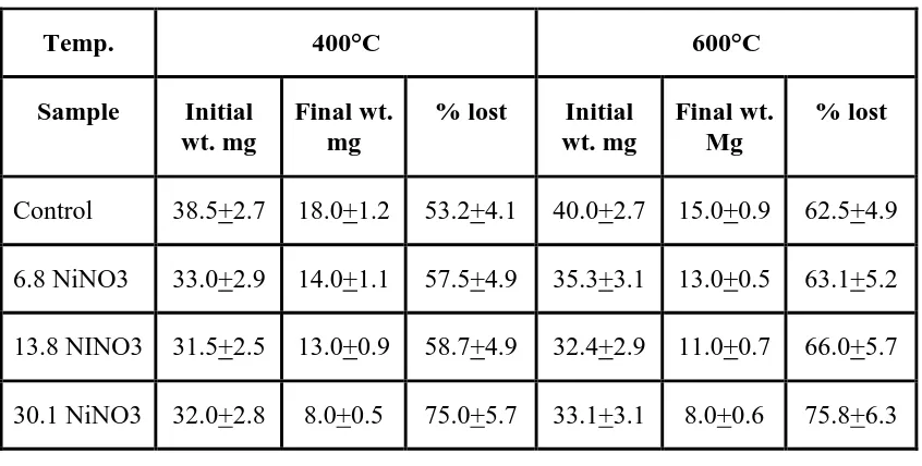
51.4+3.2, 53.3+5.7, 54.2+3.9 and 55.1+6.2,( Table 2A ) at 400oC it was 53.2+4.1, 57.5+4.9,
58.7+4.9 and 75.0+5.7 (Table 2B ) while at 600oC the percentage of weight loss was observed to
be 62.5+4.9, 63.1+5.2, 66.0+5.7 and 75.8+6.3 respectively (Table 3). Thus, it is clear from the
table (Table 2, 3), that the weight lost by the samples increases with the increase in temperature.
Also, the weight loss increases with the increase in NiNO3 dose administered to the animals . This
is suggestive of the fact that the bone material loses its weight because of thermal oxidation of
bone. Temperature affects the thermal stability of bone collagen and the results comply with those
of previous studies (22). At 400 and 600oC, the percentage weight loss was due to evaporation of
water and oxidation of organic content of bone. The loss was significant (p<0.05) in higher doses
Table 1: Variations in wet weight and dry weight of femur in control and experimental
groups
Table 2: Percentage weight lost in control and experimental bone at 50 and 200°C
temperatures.
Temp. 50°C 200°C
Sample Initial
wt. mg
Final
wt. mg
% lost Initial
wt. mg
Final
wt. mg
% lost
Control 38.0+3.3 26.0+2.5 31.5+1.3 35.0+3.2 17.0+1.3 51.4+3.2
6.8 NiNO3 33.0+3.2 23.0+3.7 43.4+3.3 32.0+2.9 15.0+1.1 53.3+5.7
13.8 NINO3 32.0+2.9 21.0+1.5 52.3+5.3 31.0+2.5 14.0+0.9 54.2+3.9
30.1 NiNO3 32.0+3.1 15.0+0.9 53.1+4.8 29.0+1.9 13.0+0.9 55.1+6.2
Sample Wet weight
(mg) (1)
Dry weight (mg) (2)
Wet weight wet water
(mg) (3)
Dry weight wet water
(mg) (4)
% weight (mg) loss between
1&2
% weight (mg) loss between
3 &4
Control 38.0+2.3 26.0+1.7 35.0+2.1 22.0+1.2 31.5+1.5 37.1+2.3
6.8 NiNO3 36.0+3.2 25.0+1.6 33.0+2.3 21.0+1.4 31.5+1.4 37.3+1.4
13.8 NiNO3 32.0+2.4 21.0+1.2 31.0+1.9 20.0+1.3 34.3+2.3 38.4+2.2
[image:4.595.87.505.401.670.2]Table 3: Percentage weight lost in control and experimental bone at 400 and 600°C
temperatures.
References
1. Mailman, R.B., (1980). Introduction to environmental Toxicology. Elsevier, New York.
Pp. 43.
2. Lacher, T.F. Jr. and Goldstein, M. I., (1997). Tropical ecotoxicology status and needs.
Environ. Toxicol. Chem. 16: 100.
3. Parmegianni, L., (1983). Encyclopedia of occupational health and safety. Int. labor.
org., Geneva, Switzerland.
4. Schiffman, F., Corcoran, B.A., and Martin, G.R., (1966). Arch. Biochem. Biophys.,
115:87.
5. Katz, J.L. (1980). The structure and biomechanics of bone in mechanical properties of
biological materials. Cambridge University press, Cambridge. Pp. 137–168.
6. Lakes, R. (1993). Materials with structural hierarchy. Nature, 361: 511-515.
7. Pidaparti, R.M.V.; Chandram, A., Takano, Y., and Turner, C.H., (1996). Bone mineral
lies mainly outside collagen fibrils: Predictions of a composite mode/ for osteonal bone.
J. Biochem., 29: 909–916.
8. Glimcher, M.J. (1959) Molecular biology of mineralized tissues with particular
reference to bone. Rev. Med. Phys. 42: 359–363.
9. Gony, J.K., Arnold, J.S., and Cohn, S.H.(1964). Composition of trabecular and cortical
Temp. 400°C 600°C
Sample Initial
wt. mg
Final wt. mg
% lost Initial
wt. mg
Final wt. Mg
% lost
Control 38.5+2.7 18.0+1.2 53.2+4.1 40.0+2.7 15.0+0.9 62.5+4.9
6.8 NiNO3 33.0+2.9 14.0+1.1 57.5+4.9 35.3+3.1 13.0+0.5 63.1+5.2
13.8 NINO3 31.5+2.5 13.0+0.9 58.7+4.9 32.4+2.9 11.0+0.7 66.0+5.7
10.Cravalho, Ho, E.G. (1972). Heat transfer in biomaterials in biomedical physics and
biomaterials science. HE Stanley (ed) Mit. Press, Cambridge.
11.Behari, J., Rai, D.V. and Jha, R. (1979). The solid state of bone. Calcif. Tiss. Int., 28:
33.
12.Liu, Q., Dewign, J.R., and Van Blitterswijck, C.A. (1998). A study on grafting reaction
of isocynates with HA– Particles. J.Biomed. Mater. Res., 40: 358–364.
13.Rai, D.V., and Singh, K.V. (2000). Effect of mineral loss on the elemental composition
and thermostability of bone collagen. JPAS. 2: 15–18.
14.Rai, D.V., Boop, B. S., and Mangal, P.C. (1990). Bioluminescence in bone, abstract in
XV All India symposium on Biophysics. Feb. Saha Institute of Nuclear Physics. Pp. 57–
58.
15.Vijai, S. S., Christopher, M.R., Bax, B.E., Bax, A.S. M., Towhidul Alam, B.S.,
Moonga, B.M., Christopher, L.H. H and Mone, Z (1993). Activation of the Ca2+
"receptor" on the osteroclast by Ni2+ elicits cytosolic Ca2+ and signals: Evidence for
receptor activation and inactivation intra cellular Ca2+ redistribution, and divalent cation
modulation. Journal of Cell. Physiology. 155: 120–129.
16.Yamaguchi, M., Oishi, H., Suketa,Y. (1987). Stimulators effect of zinc on bone
formation in tissue culture. Biochem. Pharmacol. 36: 4007–4012.
17.Povilles, J.M. (1989). What to expect from the measurement of bone mass. Rev. Rham.
Mal. Osteoortic., 56: 479–485.
18.Deluca, H.F. (1977). Vitamin D endocrine system. "Advances in clinical chemistry."
Academic press, London, 125–174.
19.Hock, J.M. (1986). Stimulation of undermineralized matrix formation by 2,5–
dihydroxy vitamin D3 in long bone of rats. Calciff. Tissue Int., 38: 79–86.
20.Chettle, D.R. (1981). Lead in bone– sampling and quantitation using x–rays. Environ.
Health Perspect., Feb. 91: 49–55.
21.Jornes, P.G., Vandeuyus, F.F., Nayts, G.D. (1991). Bone lead and renal failure.
Nephron, No. 1–4: 494.
22.Kronick, P.L., and Cooke, P. (1996). Thermal stabilization of collagen fibrils by