R E S E A R C H A R T I C L E
Open Access
Lower serum clusterin levels in patients
with erosive hand osteoarthritis are
associated with more pain
Tereza Kropá
č
ková
1,2, Olga
Š
léglová
1,2, Olga R
ůž
i
č
ková
1,2, Ji
ř
í Vencovský
1,2, Karel Pavelka
1,2and Ladislav
Š
enolt
1,2*Abstract
Background:The aims of this study were to analyse the serum concentrations of clusterin (CLU) in patients with hand osteoarthritis (OA) and in healthy controls, to compare CLU levels between patients with erosive and non-erosive disease, and to examine the association of CLU levels with clinical and laboratory parameters. Methods:A total of 135 patients with hand OA (81 with erosive and 54 with non-erosive disease) and 53 healthy individuals were included in this study. All patients underwent clinical and hand joint ultrasound examination. The Australian/Canadian (AUSCAN) hand osteoarthritis index, algofunctional index and a visual analogue scale (VAS) for the measurement of pain were assessed. Serum levels of CLU were measured by an enzyme-linked immunosorbent assay (ELISA).
Results:Serum levels of CLU were significantly lower in patients with hand OA than in control subjects (p< 0.0001). In addition, patients with erosive hand OA had significantly lower CLU levels than those with non-erosive disease (p= 0.044). Negative correlations between CLU levels and pain as assessed by the AUSCAN score and the VAS were found in patients with erosive hand OA (r=−0.275;p= 0.013 andr=−0.220;p= 0.049, respectively).
Conclusion:The present study demonstrates that lower concentrations of CLU are found in hand OA patients than in healthy individuals, especially in those with erosive disease, and that CLU concentrations have a negative association with hand pain.
Keywords:Hand osteoarthritis, Erosive osteoarthritis, Clusterin
Background
Osteoarthritis (OA) of the hands is a degenerative joint disease primarily affecting the interphalangeal and thumb base joints. Hand OA is common among the elderly, espe-cially in women. It may cause pain and disability, and it negatively affects the patients’ quality of life [1, 2]. The erosive form of hand OA is defined radiographically by its subchondral erosion, cortical destruction and subsequent reparative changes, which may include bony ankylosis. Erosive OA typically has an abrupt onset and is accom-panied by local inflammation and worse symptoms than non-erosive disease [3].
Clusterin (CLU), also known as apolipoprotein J, is a pro-tein that is involved in a number of biological processes, in-cluding inflammation and apoptosis. CLU exists in several distinct isoforms that differ in their structure, function and localization. The predominant isoform, secretory clusterin (sCLU), is a heterodimeric glycoprotein that acts as a mo-lecular chaperone [4] and exhibits anti-apoptotic and pro-survival activities [5]. Nuclear clusterin (nCLU) arises via an alternative splicing of theCLUgene leading to exclu-sion of exon II [6] and acts as a pro-apoptotic molecule [7]. Cellular forms of CLU are relatively rare, and their function is still poorly understood, but it does not appear that they affect the apoptotic pathway [8].
Clusterin is produced in many tissues, including ar-ticular cartilage and the synovium. Higher expression of CLU mRNA has been reported in early OA than in nor-mal cartilage [9]. In advanced OA cartilage, CLU mRNA
* Correspondence:senolt@revma.cz
1Institute of Rheumatology, Prague, Czech Republic
2Department of Rheumatology, 1st Faculty of Medicine, Charles University, Prague, Czech Republic
expression was reduced in comparison with that found in early OA. Based on these results, the authors pro-posed a potential role of CLU in the maintenance of ar-ticular cartilage. The upregulated expression of CLU in early OA might reflect an effort of chondrocytes to protect and repair the cartilage tissue while the downregulated CLU mRNA expression and consequently the loss of this protection in the advanced OA cartilage accompanies the final degenerative stage of the disease [9]. Fandridis et al. detected increased expression of sCLU mRNA in advanced OA compared with the expression of that in healthy cartilage. Moreover, they found higher serum sCLU levels in patients with advanced OA than in healthy individuals. Therefore, CLU could be suggested as a bio-marker reflecting cartilage tissue changes [10].
CLU mRNA expression in synovial tissue is decreased in rheumatoid arthritis (RA) compared with its expres-sion in OA or healthy tissue, but the protein levels in synovial fluid are equally present in RA and OA [11]. In cultured fibroblast-like synoviocytes (FLS), CLU inhibits nuclear factor (NF)-κB activation and modulates the expression of genes in the response to tumor necrosis factor (TNF)-αstimulation [11,12]. Recently, sCLU has been shown to inhibit osteoclast proliferation and differ-entiation, and its protective role against bone erosions has been suggested [13].
The aims of this study were therefore to compare the serum levels of CLU between patients with hand OA and healthy subjects and between OA patients with erosive and non-erosive disease and to investigate the as-sociation of CLU levels with measures of disease severity.
Methods Patients
[image:2.595.56.552.426.714.2]A total of 135 patients with hand OA (81 with the ero-sive and 54 with the non-eroero-sive form) and 53 healthy individuals were included in this study. The demo-graphic and clinical characteristics of the subjects are summarized in Table 1. The exclusion criteria for all subjects were the presence of systemic inflammatory dis-ease or cancer; the healthy controls showed no clinical signs of hand OA. All patients fulfilled the American College of Rheumatology (ACR) classification criteria for hand OA [14]; patients with erosive disease had at least one interphalangeal joint with radiographic signs of erosions. Ultrasound of all joints of both hands for the detection of osteophytes and the assessment of power Doppler (PD) and gray scale (GS) synovitis was per-formed by two ultrasonographers using Esaote Mylab 60 equipment (Esaote S.p.A., Genova, Italy) using a linear transducer with a 18 MHz frequency. Synovitis in the GS and PD were scored semiquantitatively (0–3) as described
Table 1Characteristics of patients with hand OA and control subjects
OA patients (n= 135)
Erosive (n= 81)
Non-erosive (n= 54)
Controls (n= 53)
Age, years 66.3 ± 8.3 67.6 ± 8.6* 64.3 ± 7.3 64.6 ± 7.6
Sex, female/male 120/15 74/7 46/8 48/5
CRP, mg/l 3.2 ± 3.9 3.4 ± 4.1 2.9 ± 3.7 3.3 ± 5.1
BMI, kg/m2 27.2 ± 4.2 27.5 ± 4.5 26.8 ± 3.7 NA
Disease duration, years 4.4 ± 5.2 4.4 ± 5.1 4.5 ± 5.5 NA
AUSCAN 22.5 ± 10.5 23.9 ± 11.1* 20.3 ± 9.1 NA
AUSCAN - pain 8.4 ± 4.2 9.0 ± 4.4* 7.5 ± 3.8 NA
AUSCAN - stiffness 1.9 ± 0.9 2.0 ± 0.9 1.9 ± 0.9 NA
AUSCAN - function 12.0 ± 6.4 12.8 ± 6.8 10.9 ± 5.6 NA
Algofunctional index 18.6 ± 5.9 19.5 ± 6.4 17.2 ± 4.8 NA
VAS - pain, mm 44.2 ± 22.7 46.7 ± 24.0 40.3 ± 20.3 NA
US osteophytes, n 12.8 ± 5.1 14.0 ± 4.6** 11.0 ± 5.4 NA
GS synovitis (total) 7.5 ± 8.7 9.3 ± 9.2*** 4.8 ± 7.2 NA
GS synovitis (joint count) 5.5 ± 6.4 6.5 ± 6.6*** 3.9 ± 5.8 NA
PD synovitis (total) 2.0 ± 2.7 2.5 ± 3.1* 1.3 ± 1.9 NA
PD synovitis (joint count) 1.7 ± 2.1 2.1 ± 2.3** 1.1 ± 1.6 NA
Knee OA, n (%) 59 (44) 35 (43) 24 (44) NA
Hip OA, n (%) 40 (30) 27 (33) 13 (24) NA
Knee and hip OA, n (%) 28 (21) 18 (22) 10 (19) NA
earlier [15]. The ultrasonographers were unaware of pa-tient’s clinical examination and laboratory findings. Inter-and intra-observer reliability has recently been published with moderate to very good results [15]. The clinical examinations were performed by qualified rheumatol-ogists. Pain, stiffness and function were assessed by Australian/Canadian (AUSCAN) hand osteoarthritis index [16]. Hand disability was further determined using an algofunctional index [17]. A visual analogue scale (VAS) was used for the assessment of pain. Radiographs of the knees and hips were evaluated for the presence of OA using the Kellgren-Lawrence system in all patients [18]. Written informed consent from each subject was obtained prior to enrolment, and the study was approved by the local ethics committee.
Laboratory measurements
Peripheral blood samples were obtained from all individuals and immediately centrifuged. The serum samples were stored at −80 °C until their analysis. C-reactive protein (CRP) levels were measured turbidimetrically using the Beckman Coulter AU system (Beckman Coulter, Brea, CA, USA). The serum CLU concentrations were analysed by an enzyme-linked immunosorbent assay (ELISA) in compli-ance with the manufacturer’s instructions (BioVendor, Brno, Czech Republic). The samples from the patients and the healthy individuals were analysed together in each ELISA plate. As claimed by the manufacturer, the anti-bodies used in this ELISA are specific for human CLU, the assay detection limit is 5 ng/ml and the detection range is
5–160 ng/ml. The manufacturer’s stated intra-assay and inter-assay coefficients of the variations are 6.2 and 7.8%, respectively. The final absorbance was detected using a Sunrise ELISA reader (Tecan, Salzburg, Austria), with 450 nm as the primary wavelength.
Statistical analysis
The data are presented as the mean and standard deviation (SD) unless stated otherwise. Data were analysed using a GraphPad Prism 6 (GraphPad Software, San Diego, CA, USA). The normal distribution was assessed by the D’ Agos-tino and Pearson omnibus normality test. For the compari-son between groups, the unpaired t-test or Mann-Whitney test were used. Pearson’s and Spearman’s correlation coeffi-cients were calculated to assess the relationship between the CLU levels and other parameters. P-values less than 0.05 were considered statistically significant.
Results
The patients and the control group did not differ in age, gender or CRP levels. However, the patients with erosive OA were older than those with non-erosive disease (p= 0.023). The CRP levels were comparable between both of the groups with hand OA. The AUSCAN total score and its subscale for pain were significantly higher in patients with erosive than in those with non-erosive OA (p= 0.048 and p= 0.032, respectively). Patients with ero-sive OA had more osteophytes (p= 0.003) and higher GS and PD synovitis total scores (p< 0.001 and p= 0.014, respectively) as well as higher number of affected
[image:3.595.60.537.458.703.2]joints (p< 0.001 and p= 0.009, respectively) compared to those with non-erosive disease (Table 1).
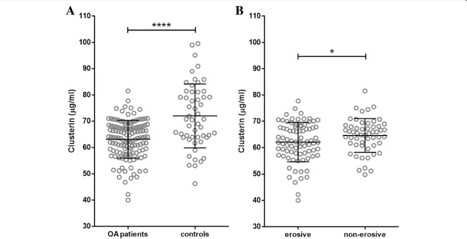
Clusterin levels are lower in patients with hand OA The serum concentrations of CLU were significantly lower in the patients with hand OA than in the healthy subjects (63.12 ± 7.17 vs 72.02 ± 12.19 μg/ml;p< 0.0001) (Fig.1a). After dividing the patients into disease subsets, the difference remained statistically significant for both erosive (p< 0.0001) and non-erosive (p< 0.0001) OA. Moreover, the patients with erosive disease had signifi-cantly lower CLU levels than those with non-erosive OA (62.11 ± 7.51 vs 64.64 ± 6.42 μg/ml; p= 0.044) (Fig. 1b). The CLU levels in hand OA patients were not affected by the concurrent presence of knee and/or hip OA (63.35 vs 62.95μg/ml for knee OA, 62.98 vs 63.18μg/ml for hip OA, 63.47 vs 63.03μg/ml for knee and hip OA; p> 0.05 for all comparisons).
Clusterin levels are inversely associated with pain in patients with erosive hand OA
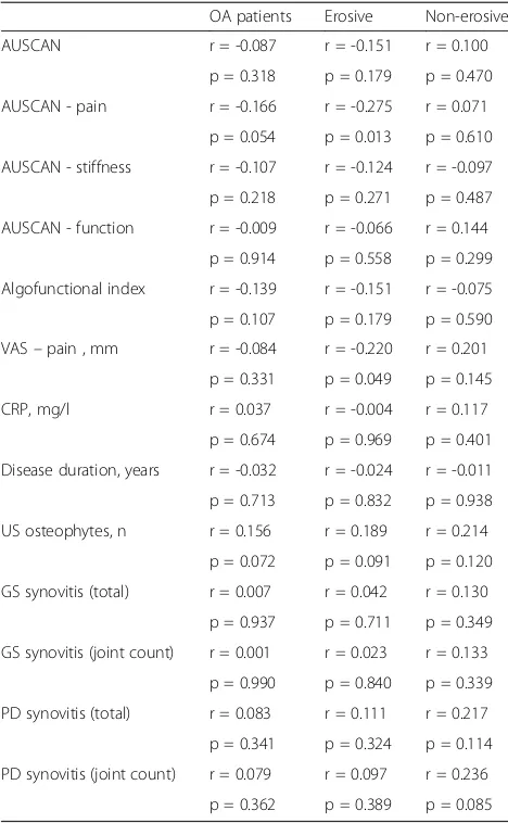
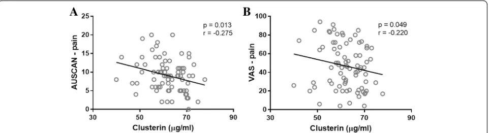
Associations of CLU levels with clinical and laboratory pa-rameters are shown in Table 2. There were no significant correlations between the CLU levels and the algofunctional index and total AUSCAN score in any of the patients with hand OA; however, among those with erosive disease, the CLU levels were negatively correlated with pain as assessed by the AUSCAN and the VAS (r=−0.275; p= 0.013 and r=−0.220;p= 0.049, respectively) (Fig.2). The CLU concentrations were not associated with age, sex or body mass index. There were also no significant correlations be-tween the CLU levels and disease duration, CRP levels, ultrasound-determined synovitis and osteophytes.
Discussion
In this study, we found lower levels of CLU in patients with hand OA than in healthy subjects and an inverse association between CLU levels and pain in patients with erosive disease.
Erosive hand OA is a more severe form of hand OA than non-erosive OA, both clinically and radiographically, and is accompanied by worse pain and disability [19, 20]. In our study, we found significantly higher scores on the AUSCAN and its pain subscale in patients with erosive than in patients with non-erosive OA. Inflammatory signs were also reported to be more frequent in erosive than in non-erosive disease [21,22]. In addition, Punzi et al. [23] found higher CRP levels in erosive OA patients than in those with non-erosive hand OA and suggested CRP as a potential marker of disease activity. However, no differences in the CRP levels between erosive and non-erosive OA were found in other studies [24, 25]. A number of other biomarkers have been studied in hand OA, including markers of inflammation [26]. Recently,
Fioravanti et al. [27] reported significantly higher levels of serum myeloperoxidase (MPO) in patients with hand OA than in healthy controls. Moreover, patients with erosive OA showed significantly elevated MPO levels than those with non-erosive OA, which confirmed the results of a previous study [28] and further supported the involvement of inflammation in the pathogenesis of hand OA, espe-cially in the erosive disease. In the present study, we observed higher ultrasound-determined synovitis total scores as well as the number of affected joints in patients with erosive OA compared to those with non-erosive dis-ease. However, no significant differences in the CRP levels between the two subgroups of patients were found.
[image:4.595.304.538.109.488.2]In this study, we found lower serum levels of CLU in the patients with hand OA than in the healthy controls. Several other studies have investigated CLU in OA [9–11]; however, this is the first study to explore the CLU levels in patients with hand OA. The expression of CLU mRNA
Table 2Correlations between serum CLU levels, clinical and laboratory parameters
OA patients Erosive Non-erosive AUSCAN r = -0.087 r = -0.151 r = 0.100
p = 0.318 p = 0.179 p = 0.470
AUSCAN - pain r = -0.166 r = -0.275 r = 0.071
p = 0.054 p = 0.013 p = 0.610
AUSCAN - stiffness r = -0.107 r = -0.124 r = -0.097
p = 0.218 p = 0.271 p = 0.487
AUSCAN - function r = -0.009 r = -0.066 r = 0.144
p = 0.914 p = 0.558 p = 0.299
Algofunctional index r = -0.139 r = -0.151 r = -0.075
p = 0.107 p = 0.179 p = 0.590
VAS–pain , mm r = -0.084 r = -0.220 r = 0.201
p = 0.331 p = 0.049 p = 0.145
CRP, mg/l r = 0.037 r = -0.004 r = 0.117
p = 0.674 p = 0.969 p = 0.401
Disease duration, years r = -0.032 r = -0.024 r = -0.011
p = 0.713 p = 0.832 p = 0.938
US osteophytes, n r = 0.156 r = 0.189 r = 0.214
p = 0.072 p = 0.091 p = 0.120
GS synovitis (total) r = 0.007 r = 0.042 r = 0.130
p = 0.937 p = 0.711 p = 0.349
GS synovitis (joint count) r = 0.001 r = 0.023 r = 0.133
p = 0.990 p = 0.840 p = 0.339
PD synovitis (total) r = 0.083 r = 0.111 r = 0.217
p = 0.341 p = 0.324 p = 0.114
PD synovitis (joint count) r = 0.079 r = 0.097 r = 0.236
p = 0.362 p = 0.389 p = 0.085
AUSCANAustralian/Canadian,CRPC-reactive protein,GSgray scale,OA
has been reported to be higher in OA than in healthy cartilage [9, 10]. Fandridis et al. [10] also reported higher serum levels of CLU in patients with knee and hip OA than in healthy controls. The reason for these apparently contradictory results may be caused by the different loca-tions of the affected joints. Nevertheless, we did not find any differences in the CLU levels between the hand OA patients with and without knee and/or hip OA. It is also important to note that, in our study, the samples obtained from the patients and the healthy subjects were analysed at the same time, whereas Fandridis et al. used the data from the healthy controls from their previous study and analysed only the patient group [10].
A previous study has shown that CLU mRNA expression in synovial tissue is lower in patients with RA than in patients with OA and healthy individuals [11]. We found significantly lower serum levels of CLU in patients with erosive OA compared with patients with non-erosive hand OA. Bone erosions are present in RA as well as in erosive OA, although their locations differ. The lower expression of CLU in these diseases may be explained by the potentially protective role of CLU against the development of bone erosions [13]. A recent study also suggests a protective function of CLU in inflammation and autoimmune diseases [29], which is in agreement with the study of Newkirk et al. [30] that reported lower serum concentrations of CLU in systemic lupus erythematosus and found negative correla-tions among CLU levels, disease activity and disease symp-toms. In our study, we reported a negative correlation between the CLU levels and hand pain in patients with ero-sive OA. No such association was found in patients with non-erosive disease. Therefore, we can speculate that CLU may play a role in the pathology of erosive hand OA.
This study has several limitations. First, its design was cross-sectional. Therefore, a long-term prospective study is needed to further investigate the role of CLU in hand OA. Second, CLU levels might be affected due to the ef-fects of other involved joints or diseases. However, we did not observe a difference in the CLU levels between
the hand OA patients with and without radiographic hip and/or knee joint involvement.
Conclusions
In conclusion, we demonstrate here for the first time that lower serum levels of CLU are found in patients with hand OA, especially in those with erosive disease, and that a negative association exists between CLU con-centrations and hand pain. These data suggest a possible involvement of CLU in the pathophysiology of erosive hand OA and further support its role in the develop-ment of bone erosions.
Abbreviations
ACR:American College of Rheumatology; AUSCAN: Australian/Canadian; BMI: Body mass index; CLU: Clusterin; CRP: C-reactive protein; ELISA: Enzyme-linked immunosorbent assay; FLS: Fibroblast-like synoviocytes; GS: Gray scale; nCLU: Nuclear clusterin; NF: Nuclear factor; OA: Osteoarthritis; PD: Power Doppler; RA: Rheumatoid arthritis; sCLU: Secretory clusterin; TNF: Tumor necrosis factor; US: Ultrasound; VAS: Visual analogue scale
Acknowledgements
The authors thank Zdena Leopoldová for technical and administrative assistance.
Funding
This study was supported by the Ministry of Health, Czech Republic, research project AZV no. 18–01-00542.
Availability of data and materials
The datasets used and/or analysed during the current study are available from the corresponding author on reasonable request.
Authors’contributions
TK performed the laboratory and statistical analysis, and drafted the manuscript. OŠ, OR, KP and LŠmade clinical assessments and contributed to data acquisition and interpretation. JV and KP revised the manuscript critically for important intellectual content. LŠmade substantial contributions to study concept and design, and revised the final draft of the manuscript. All authors read and approved the final version of the manuscript.
Ethics approval and consent to participate
The study was approved by the local ethics committee of the Institute of Rheumatology in Prague, Czech Republic. Written informed consent was obtained from all participants.
Consent for publication
Not applicable
[image:5.595.59.540.88.219.2]Competing interests
All authors declare no competing interests.
Publisher’s Note
Springer Nature remains neutral with regard to jurisdictional claims in published maps and institutional affiliations.
Received: 28 February 2018 Accepted: 10 July 2018
References
1. Dahaghin S, Bierma-Zeinstra SMA, Ginai AZ, Pols HAP, Hazes JMW, Koes BW. Prevalence and pattern of radiographic hand osteoarthritis and association with pain and disability (the Rotterdam study). Ann Rheum Dis. 2005;64:682–7. 2. Zhang Y, Niu J, Kelly-Hayes M, Chaisson CE, Aliabadi P, Felson DT. Prevalence of
symptomatic hand osteoarthritis and its impact on functional status among the elderly: the Framingham study. Am J Epidemiol. 2002;156:1021–7. 3. Zhang W, Doherty M, Leeb BF, Alekseeva L, Arden NK, Bijlsma JW, et al.
EULAR evidence-based recommendations for the diagnosis of hand osteoarthritis: report of a task force of ESCISIT. Ann Rheum Dis. 2009;68:8–17. 4. Poon S, Easterbrook-Smith SB, Rybchyn MS, Carver JA, Wilson MR. Clusterin
is an ATP-independent chaperone with very broad substrate specificity that stabilizes stressed proteins in a folding-competent state. Biochemistry. 2000;39:15953–60.
5. Trougakos IP, Lourda M, Antonelou MH, Kletsas D, Gorgoulis VG, Papassideri IS, et al. Intracellular clusterin inhibits mitochondrial apoptosis by suppressing p53-activating stress signals and stabilizing the cytosolic Ku70-Bax protein complex. Clin Cancer Res. 2009;15:48–59.
6. Leskov KS, Klokov DY, Li J, Kinsella TJ, Boothman DA. Synthesis and functional analyses of nuclear clusterin, a cell death protein. J Biol Chem. 2003;278:11590–600.
7. Kim N, Yoo JC, Han JY, Hwang EM, Kim YS, Jeong EY, et al. Human nuclear clusterin mediates apoptosis by interacting with Bcl-XL through C-terminal coiled coil domain. J Cell Physiol. 2012;227:1157–67.
8. Prochnow H, Gollan R, Rohne P, Hassemer M, Koch-Brandt C, Baiersdörfer M. Non-secreted clusterin isoforms are translated in rare amounts from distinct human mRNA variants and do not affect Bax-mediated apoptosis or the NF-κB signaling pathway. PLoS One. 2013;8:e75303.
9. Connor JR, Kumar S, Sathe G, Mooney J, O’Brien SP, Mui P, et al. Clusterin expression in adult human normal and osteoarthritic articular cartilage. Osteoarthr Cartil. 2001;9:727–37.
10. Fandridis E, Apergis G, Korres DS, Nikolopoulos K, Zoubos AB, Papassideri I, et al. Increased expression levels of apolipoprotein J/clusterin during primary osteoarthritis. In Vivo (Brooklyn). 2011;25:745–9.
11. Devauchelle V, Essabbani A, De Pinieux G, Germain S, Tourneur L, Mistou S, et al. Characterization and functional consequences of underexpression of clusterin in rheumatoid arthritis. J Immunol. 2006;177:6471–9.
12. Falgarone G, Essabbani A, Dumont F, Cagnard N, Mistou S, Chiocchia G. Implication of clusterin in TNF-αresponse of rheumatoid synovitis: lesson from in vitro knock-down of clusterin in human synovial fibroblast cells. Physiol Genomics. 2012;44:229–35.
13. Choi B, Kang S-S, Kang S-W, Min B-H, Lee E-J, Song D-H, et al. Secretory clusterin inhibits osteoclastogenesis by attenuating M-CSF-dependent osteoclast precursor cell proliferation. Biochem Biophys Res Commun. 2014;450:105–9.
14. Altman R, Alarcón G, Appelrouth D, Bloch D, Borenstein D, Brandt K, et al. The American College of Rheumatology criteria for the classification and reporting of osteoarthritis of the hand. Arthritis Rheum. 1990;33:1601–10. 15. Hurnakova J, Zavada J, Hanova P, Hulejova H, Klein M, Mann H, et al. Serum
calprotectin (S100A8/9): an independent predictor of ultrasound synovitis in patients with rheumatoid arthritis. Arthritis Res Ther. 2015;17:252. 16. Bellamy N, Campbell J, Haraoui B, Gerecz-Simon E, Buchbinder R, Hobby K, et al.
Clinimetric properties of the AUSCAN osteoarthritis hand index: an evaluation of reliability, validity and responsiveness. Osteoarthr Cartil. 2002;10:863–9. 17. Dreiser RL, Maheu E, Guillou GB, Caspard H, Grouin JM. Validation of an
algofunctional index for osteoarthritis of the hand. Rev Rhum Engl Ed. 1995;62:43S–53S.
18. Kellgren JH, Lawrence JS. Radiological assessment of osteo-arthrosis. Ann Rheum Dis. 1957;16:494–502.
19. Addimanda O, Mancarella L, Dolzani P, Punzi L, Fioravanti A, Pignotti E, et al. Clinical and radiographic distribution of structural damage in erosive and nonerosive hand osteoarthritis. Arthritis Care Res. 2012;64:1046–53.
20. Bijsterbosch J, Watt I, Meulenbelt I, Rosendaal FR, Huizinga TWJ, Kloppenburg M. Clinical burden of erosive hand osteoarthritis and its relationship to nodes. Ann Rheum Dis. 2010;69:1784–8.
21. Kortekaas MC, Kwok W-Y, Reijnierse M, Huizinga TWJ, Kloppenburg M. In erosive hand osteoarthritis more inflammatory signs on ultrasound are found than in the rest of hand osteoarthritis. Ann Rheum Dis. 2013;72:930–4. 22. Haugen IK, Mathiessen A, Slatkowsky-Christensen B, Magnusson K, Bøyesen
P, Sesseng S, et al. Synovitis and radiographic progression in non-erosive and erosive hand osteoarthritis: is erosive hand osteoarthritis a separate inflammatory phenotype? Osteoarthr Cartil. 2016;24:647–54.
23. Punzi L, Ramonda R, Oliviero F, Sfriso P, Mussap M, Plebani M, et al. Value of C reactive protein in the assessment of erosive osteoarthritis of the hand. Ann Rheum Dis. 2005;64:955–7.
24. Filková M, Senolt L, Braun M, Hulejová H, Pavelková A, Sléglová O, et al. Serum hyaluronic acid as a potential marker with a predictive value for further radiographic progression of hand osteoarthritis. Osteoarthr Cartil. 2009;17:1615–9.
25. Dolzani P, Assirelli E, Pulsatelli L, Addimanda O, Mancarella L, Peri G, et al. Systemic inflammation and antibodies to citrullinated peptides in hand osteoarthritis. Clin Exp Rheumatol. 29:1006–9.
26. Lennerová T, Pavelka K,Šenolt L. Biomarkers of hand osteoarthritis. Rheumatol Int. 2018;38:725–35.
27. Fioravanti A, Tenti S, Pulsatelli L, Addimanda O. Could myeloperoxidase represent a useful biomarker for erosive osteoarthritis of the hand? Scand J Rheumatol. 2018;1-3. doi:https://doi.org/10.1080/03009742.2017.1386796. [Epub ahead of print]
28. Punzi L, Ramonda R, Deberg M, Frallonardo P, Campana C, Musacchio E, et al. Coll2-1, Coll2-1NO2 and myeloperoxidase serum levels in erosive and non-erosive osteoarthritis of the hands. Osteoarthr Cartil. 2012;20:557–61. 29. Cunin P, Beauvillain C, Miot C, Augusto J-F, Preisser L, Blanchard S, et al.
Clusterin facilitates apoptotic cell clearance and prevents apoptotic cell-induced autoimmune responses. Cell Death Dis. 2016;7:e2215. 30. Newkirk MM, Apostolakos P, Neville C, Fortin PR. Systemic lupus