Summary
Lineage commitment and differentiation into mature cell types are mostly considered to be unidirectional and irreversible processes. However, recent results have challenged this by showing that terminally differentiated cell types can be reprogrammed into other cell types, an important step towards devising strategies for gene therapy and tissue regeneration. In this Review, we summarize recent data on the earliest steps in the development of the mammalian lymphatic vasculature: the specification of lymphatic endothelial cells (LECs). We elaborate on a developmental model that integrates the different steps leading to LEC differentiation and lymphatic network
formation, discuss evidence that suggests that LEC fate is plastic, and consider the potentially far-reaching implications of the ability to convert one cell type into another.
Key words: Prox1, Cell differentiation, Cell plasticity, Endothelial cell, Lymphatics
Introduction
Until recently, lineage commitment and differentiation into a mature cell type were considered to be unidirectional and irreversible processes. This view has dramatically changed after a series of papers by Shinya Yamanaka’s group reported that terminally differentiated somatic cell types can be directly reprogrammed to pluripotency by the addition of four transcription factors (Takahashi and Yamanaka, 2006; Takahashi et al., 2007). This type of approach has also been used to show that three transcription factors are sufficient to reprogram pancreatic exocrine cells into endocrine cells (Zhou et al., 2008). At the single transcription factor level, it was shown that mature B cells can be reprogrammed (induced to dedifferentiate) into functional T cells by switching off the transcription factor Pax5 (Cobaleda et al., 2007), and that lymphatic endothelial cells (LECs) can be reprogrammed into blood endothelial cells (BECs) by switching off the homeobox transcription factor Prox1 (Johnson et al., 2008).
It has also been suggested that during differentiation, certain cell fates are superimposed on pre-existing ones. In this context, transcription factors could repress a pre-existing cell fate and/or activate a new fate. For example, maintaining venous identity requires that COUP-transcription factor II [COUP-TFII; also known as nuclear receptor subfamily 2, group F, member 2 (Nr2f2)] represses Notch signaling in veins, thereby suppressing the expression of arterial-specific genes (You et al., 2005). Also, COUP-TFII gain-of-function in the endothelium downregulates the expression of arterial markers, whereas the loss of COUP-TFII function in venous cells derepresses the arterial program and converts veins into arteries (You et al., 2005).
These findings not only challenge the dogma that terminal differentiation is an irreversible process, but highlight the possibility that reprogramming of differentiated cell types by switching on or off single genes could constitute an important step towards devising strategies to convert one cell lineage into another in gene therapy approaches and tissue regeneration.
In this Review, we summarize some of the key steps leading to the specification and plasticity of the LEC phenotype in mammals.
The lymphatic vasculature
Embryonic development and organ formation require a constant source of blood, which is provided by the blood vascular network. Because of this crucial functional role, there is ample information regarding the genes and mechanisms that participate in angiogenesis (the formation of new blood vessels from pre-existing blood vessels) and vasculogenesis (blood vessel formation by de novo production of endothelial cells), the two main processes in the formation of this vascular system. By contrast, until recently, little was known about the processes that control the formation of the second vascular network present in the bodies of all mammals and, most likely, most vertebrates: the lymphatic system.
The lymphatic vasculature plays key roles in normal and pathological conditions, although its roles differ from those of the blood vasculature. Few life-threatening conditions result from malfunction of the lymphatic vasculature. It is well accepted that the lymphatic network is necessary to drain lymph fluids from extracellular spaces, to absorb lipids from the intestinal tract, to maintain fluid homeostasis and to transport white blood cells and antigen-presenting cells to lymphoid organs. Malfunctions of the lymphatic vasculature can lead to a series of congenital or inherited disorders, such as lymphoedema (imbalance in lymph absorption), which is a disfiguring and disabling disorder often characterized by swelling of the extremities (Witte et al., 2001). The lymphatic vasculature is also the main route for the immune response to infectious agents and for the migration of tumor cells to lymph nodes and to distant organs (Cueni and Detmar, 2008). Recent work also indicates that malfunctions of the lymphatic vasculature could be at least partially responsible for some types of disorders not previously linked to lymphatic malfunction: lymphatic vascular defects were identified as causing late-onset obesity in mice that carried a mutation in one allele of the Prox1transcription factor (Harvey et al., 2005), the activity of which is necessary and sufficient in vivo and in vitro for the specification of LEC fate (Hong et al., 2002; Petrova et al., 2002; Wigle et al., 2002; Wigle and Oliver, 1999). More recently, subcutaneous lymphatic vessels were shown to participate in the regulation of blood pressure during excessive salt intake (Machnik et al., 2009). It thus appears reasonable to speculate that individual variability in the integrity or function of the lymphatic vasculature could, for example, also be responsible for variations in the immune response or in the metastatic spreading of certain tumors. Additional information about the characteristics of the mature lymphatic system can be found in Box 1.
Development 137, 363-372 (2010) doi:10.1242/dev.035360 © 2010. Published by The Company of Biologists Ltd
Endothelial cell plasticity: how to become and remain a
lymphatic endothelial cell
Guillermo Oliver* and R. Sathish Srinivasan
Department of Genetics and Tumor Cell Biology, St Jude Children’s Research Hospital, 262 Danny Thomas Place, Memphis, TN 38105-3678, USA.
*Author for correspondence (guillermo.oliver@stjude.org)
D
E
V
E
LO
P
M
E
N
The last decade has brought a substantial advance in our understanding of the regulatory processes that lead to the formation of a mature and fully differentiated lymphatic vasculature. Several key players that participate in various aspects of the stepwise process (Oliver and Harvey, 2002; Wigle et al., 2002) that leads to the formation of the lymphatic vasculature, including Prox1, Sox18, Vegfr3 (Flt4)/Vegfc, Foxc2, podoplanin, Syk/Slp76 (Lcp2), Ang2 and ephrin B2 (Efnb2), have been identified during the last ten years; their roles have been summarized previously (for reviews, see Adams and Alitalo, 2007; Maby-El Hajjami and Petrova, 2008; Oliver, 2004; Oliver and Alitalo, 2005). Here, we focus on the earliest crucial step of lymphatic vasculature development in mammals, i.e. the specification of the LEC phenotype, centering attention on those few genes that are required for the formation of the entire lymphatic network by acting during the earliest developmental stages. For additional information on other genes that are expressed in embryonic LECs soon after the LEC-specification stage and that affect later aspects of lymphatic vasculature development, such as the separation of the blood and the lymphatic vasculatures, patterning, vessel integrity, remodeling and maturation, and for further details of our current knowledge on the lymphatic vasculature in other organisms, we refer the reader to the available literature (Adams and Alitalo, 2007; Maby-El Hajjami and Petrova, 2008; Oliver, 2004; Oliver and Alitalo,
2005; Yaniv et al., 2006; Hogan et al., 2009; Ny et al., 2005; Kalin et al., 2009). Finally, we also consider the mechanisms that maintain LEC identity and the potential implications of cellular plasticity and reprogramming.
How to become an arterial or venous endothelial cell
As highlighted above, the blood and lymphatic vasculatures share many similarities. The lymphatic vasculature runs in parallel, but develops secondarily to the blood vasculature, and both are lined by endothelial cells (ECs). This tight connection between the two vascular systems is emphasized further by the fact that, as originally proposed in the early 19th century by Sabin (Sabin, 1902) and recently confirmed using a lineage-tracing approach (Srinivasan et al., 2007), the lymphatic vasculature is derived, at least in mammals, from venous ECs. Therefore, a prerequisite for the genesis of the lymphatic network is the formation of the blood vasculature. Early during embryogenesis, mesodermally derived ECs give rise to the primitive embryonic and extra-embryonic vasculature (vasculogenesis). Later, the remodeling of this primary vascular network leads to the formation of new blood vessels from pre-existing vessels by endothelial sprouting and splitting (angiogenesis). Several genes that participate in this complex process have been identified, and functional inactivation or gain-of-function approaches have helped to elucidate their functional roles (see Adams and Alitalo, 2007; De Val and Black, 2009). Mutations in most of these genes affect hematopoiesis, vascular development, vascular remodeling, sprouting, vessel integrity, remodeling, or the number of vessels [for a detailed list, see De Val and Black (De Val and Black, 2009)]. In many instances, some of these defects are so profound that embryos cannot survive beyond embryonic day 10.5 (E10.5). The ETS transcription factor Etv2 (Er71) appears to be one of the key factors necessary for the specification of vascular ECs (Ferdous et al., 2009; Lee et al., 2008). Etv2-null embryos die early during development, and endothelial progenitors and embryonic vessels are not detected (Ferdous et al., 2009; Lee et al., 2008). Because it has been shown that Etv2regulates the activity of other early endothelial genes, including Flk1(Kdr; Vegfr2),Tie2(Tek),Pecamand Tal1, it has been suggested that this transcription factor is at the top of the regulatory cascade that leads to EC specification (Fig. 1) (De Val and Black, 2009; De Val et al., 2008; Ferdous et al., 2009). After ECs develop, and as arterial and venous EC types become specified, blood vessels (arteries and veins) are also formed. During this process, the Notch signaling pathway, and particularly its target transcription factors Hey1 and Hey2, are crucial; Notch signaling is required by ECs to promote arterial cell fate differentiation and to suppress venous fate choices (Fig. 1) (De Val and Black, 2009; Harvey and Oliver, 2004; Kokubo et al., 2005; Lawson et al., 2001; Villa et al., 2001). By contrast, the orphan nuclear receptor COUP-TFII directs differentiation toward a venous EC fate (You et al., 2005). It has been proposed that COUP-TFIIexpression in veins is required to promote venous identity by inhibiting Notch signaling and other key arterial-specification players (Fig. 1) (You et al., 2005). COUP-TFIIloss-of-function mutations lead to the ectopic expression of Notch1in veins (You et al., 2005); conversely, the ectopic expression of COUP-TFII in the developing blood vasculature suppresses arterial gene expression by inhibiting the Notch pathway (You et al., 2005).
These data indicate that the activity of two key players (the Notch pathway and COUP-TFII) is sufficient to specify and modulate the pathways that lead to arterial versus venous cell fate. It also suggests Box 1. The lymphatic system
In mammals, the mature lymphatic system consists of the lymphatic vasculature and the lymphoid organs, such as the lymph nodes, tonsils, Peyer’s patches, spleen and thymus, all of which play an important role in immune responses. The lymphatic vasculature covers most of the body, with the exception of avascular tissues, such as the epidermis, hair, nails, cartilage and cornea, and some vascularized organs, such as the retina and the brain (Cueni and Detmar, 2008).
The blood and lymphatic vasculatures are lined by endothelial cells (ECs). The smaller lymphatic capillaries are different from blood capillaries in that they are blind-ended structures that lack fenestrations, a continuous basal lamina and pericytes, which are the smooth-muscle-like contractile cells that wrap around the outer surface of blood vessels. Lymphatic capillaries are composed of a single-cell layer of overlapping ECs that are interconnected by specialized discontinuous button-like junctions and that contain few intercellular tight junctions or adherens junctions (Baluk et al., 2007). These ECs form loose intercellular valve-like junctions and exhibit large interendothelial pores and anchoring filaments that connect the vessels to the extracellular matrix (Baluk et al., 2007). All of these features make the lymphatic capillaries highly permeable. As the surrounding interstitial pressure changes, the anchoring filaments tighten and relax, causing the lymphatics to expand and fill or contract and push lymph. Under high interstitial pressure, EC junctions open, anchoring filaments extend, and fluid moves into the vessel (Schmid-Schonbein, 2003).
The smaller lymphatic capillaries first drain into pre-collecting lymphatics, which eventually merge into larger secondary collecting lymphatic vessels that contain smooth muscle cells that provide contractile activity to assist lymph flow, possess a continuous basal membrane and ECs that exhibit continuous zipper-like junctions (Baluk et al., 2007). These larger vessels also contain intraluminal, one-way valve-like junctions that prevent retrograde fluid flow (von der Weid and Zawieja, 2004). Tissue fluid in the larger collecting lymphatics drains into the thoracic duct and is then returned to the blood circulation through lymphatic-vasculature connections established at the junction of the jugular and subclavian veins.
D
E
V
E
LO
P
M
E
N
that the fate of these two EC types (arterial and venous) is relatively plastic, as one cell type can easily be converted into the other by the manipulation (gain or loss) of a single gene.
Lymphatic vasculature development: how to become an LEC
As mentioned above, the mammalian lymphatic vasculature is venous derived (Fig. 2). Therefore, a prerequisite for lymphatic vasculature formation should be the presence of a normal embryonic blood vascular network, a proposal supported by the finding that no, or very few, LECs are present in Tie2-Cre;COUP-TFIIf/fconditional mutant mouse embryos, in which venous fate is lost (Srinivasan et al., 2007).
Although a functional cardinal vein is present in mice as early as E8.5, the appearance of the first differentiating LECs around the anterior cardinal vein occurs much later, at ~E9.75 (Srinivasan et al., 2007; Wigle et al., 1999; Wigle and Oliver, 1999). This indicates that the presence of veins, although necessary, is not sufficient to trigger the program leading to LEC differentiation and that not all tissues are competent to respond to signals provided by an inducer. Normally, the differentiation of a specialized cell type starts once a tissue becomes competent (i.e. able) to respond to specific inductive signals (Waddington, 1940); this is not a passive state, but an actively acquired condition. A few years ago, borrowing some of the classic embryological concepts of Xenopus lens cell type
determination (Grainger, 1992), we proposed that, akin to many organ and tissue formation processes, the differentiation of the lymphatic vasculature is a stepwise process (Wigle et al., 2002; Oliver and Detmar, 2002; Oliver, 2004).
Accordingly, in the context of LEC differentiation, competence can be defined as the initial stage during which embryonic venous ECs become responsive to LEC-inductive signals (Oliver, 2004). Once venous ECs receive the appropriate inductive signals, they become committed to an LEC fate, and their developmental potential starts to become restricted. This commitment stage is characterized by an initial labile phase of specification (when a cell is able to differentiate autonomously if placed in a neutral environment) and a second stage of determination (when committed cells will differentiate autonomously even if placed into another tissue context) (Slack, 1991).
How do these general developmental concepts fit with what is currently known about LEC differentiation? Currently, gene inactivation approaches have identified three genes (Prox1,Sox18 and COUP-TFII) as crucial regulators of LEC differentiation in mammals, as, so far, these are the only genes for which functional inactivation results in a complete lack of LECs and, therefore, of the entire lymphatic vasculature (Francois et al., 2008; Srinivasan et al., 2007; Wigle et al., 2002; Wigle and Oliver, 1999).
Below, we summarize the key developmental steps that lead to the formation of a mature LEC, focusing on those key genes that have been shown to be required during the early LEC-specification step. Although the use of these developmental concepts is useful, as it helps us to identify relevant aspects of the molecular events involved in the genesis of the mammalian lymphatic network, this proposed stepwise model remains hypothetical owing to experimental limitations, and should be considered as such.
Competence
In mice, the earliest differentiating LECs are detected at the level of the anterior cardinal vein at ~E9.75 (Srinivasan et al., 2007). Two key findings support this assertion. First, around this stage, a subpopulation of venous ECs (that is somehow induced asymmetrically from within the venous EC population) starts to express the homeobox-containing gene Prox1(Wigle and Oliver, 1999), which, as previously stated, is necessary and sufficient in vivo and in vitro for LEC specification (Hong et al., 2002; Petrova et al., 2002; Wigle et al., 2002; Wigle and Oliver, 1999). Second, soon afterward, Prox1-expressing cells start to exit the veins, following very precise paths to form the initial lymph sacs (Wigle and Oliver, 1999), the embryonic structures from which the entire lymphatic vasculature is eventually derived.
The fact that Prox1expression in the veins (and therefore the initiation of LEC differentiation) does not start concomitantly with venous formation, but instead a few days later, supports the notion that the molecular profile provided by venous ECs at this early stage is not sufficient to trigger Prox1expression and, therefore, LEC differentiation. In this context, one might speculate that apart from inducing venous fate, COUP-TFIIactivity might also be involved in repressing Prox1, such that this gene is not expressed by venous ECs prior to a particular developmental stage (in the mouse, E9.75), thus preventing precocious LEC differentiation. However, a more likely explanation is that the expression of additional gene products is necessary for COUP-TFII-expressing venous ECs to become competent to respond to specific Prox1-inducing signals. What might these additional competence factors be? A likely candidate for this role is the SRY-related HMG-domain transcription factor Sox18 (Fig. 2). Recent work has identified Sox18 as an upstream regulator VEGF
Mesoderm
Etv2
BMP Notch Wnt
Mesodermal angioblast
Endothelial cells
Arterial endothelial cells Venous endothelial cells COUP-TFII
Notch
COUP-TFII
Sox18
[image:3.612.50.297.57.228.2]Lymphatic endothelial cells Prox1 Notch
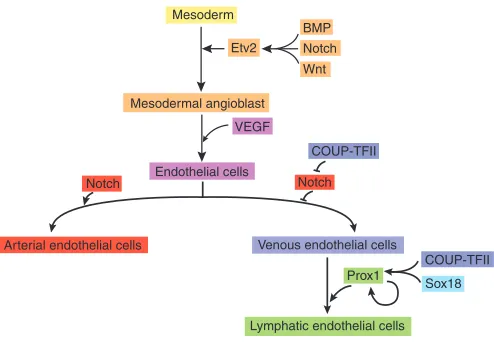
Fig. 1. The main steps and key regulators in blood and lymphatic vasculature development.The development of the embryonic vasculature requires the differentiation of endothelial cells (ECs), as depicted schematically here. Within the mammalian embryo, the first step in this process is the formation of endothelial precursors
(angioblasts) from mesodermal progenitors during gastrulation. The ETS transcription factor Etv2 is at the top of the pathway that leads to EC differentiation, and the Notch, BMP and Wnt signaling pathways regulate its expression. VEGF signaling is also important during the differentiation of angioblasts into ECs. Subsequently, Notch signaling is required to promote arterial endothelial cell identity, and the expression of the orphan nuclear receptor COUP transcription factor II (COUP-TFII) promotes venous endothelial cell fate by downregulating Notch signaling. Upon its activation in a subpopulation of the venous endothelial cells, the SRY-related HMG-domain transcription factor Sox18 cooperates with COUP-TFII to activate expression of the homeobox transcription factor Prox1. Prox1expression is sufficient to specify lymphatic endothelial cell (LEC) fate. Later on, Prox1expression becomes independent of external stimuli, as it regulates its own expression and maintains LEC identity.
D
E
V
E
LO
P
M
E
N
of Prox1expression in the anterior cardinal vein (Francois et al., 2008). In humans, mutations in SOX18 lead to hypotrichosis-lymphedema-telangiectasia, a syndrome characterized by swelling of the extremities and attributable to a defective lymphatic vasculature (Irrthum et al., 2003). In mice, depending on the genetic background, the functional inactivation of this gene leads to mild-to-severe phenotypic alterations that can result in embryonic lethality because of defective blood vessels; in some backgrounds, Sox18mutant mice also lack a lymphatic vasculature (Francois et al., 2008). In this latter case, Francois et al. (Francois et al., 2008) showed that in addition to its expression in arterial endothelial cells (Pennisi et al., 2000), Sox18 expression is also detected in a subpopulation of ECs located in the anterior cardinal vein starting at ~E9.0, approximately half a day before Prox1(Fig. 2). Using a variety of in vivo and in vitro approaches, Francois et al. showed that Sox18 is a direct in vivo activator of Prox1expression in venous ECs (Francois et al., 2008). Therefore, in Sox18-null embryos, Prox1 expression is not induced in venous ECs, LEC specification is defective, and the formation of the lymphatic vasculature is arrested (Francois et al., 2008). Sox18expression in differentiating LECs and in forming lymphatic vessels is detected up to ~E14.5 (Francois et al., 2008). The loss of Sox18expression after this stage indicates a transient requirement during early LEC differentiation stages, perhaps solely to induce Prox1.
To accommodate the available data summarized above, two possible explanations could be considered. The first is that Sox18 is not the factor that collaborates with COUP-TFIIin providing venous ECs with the competence to respond to specific LEC-inductive signals. If this is the case, the search for such a factor remains open. The ideal candidate gene fulfilling this function should start to be expressed in a subpopulation of venous ECs at ~E9.0, it should induce Prox1expression by itself or in combination with other factors, its functional inactivation should result in the absence of LECs, and it would probably not be expressed in embryonic arteries. The second possibility is that Sox18 is actually the competence factor required to initiate LEC differentiation; however, if so, why is Prox1expression not equally induced in embryonic arteries that normally express Sox18? One plausible interpretation is that an arterial-specific gene, or group of genes, is responsible for inhibiting the Prox1induction mediated by Sox18; therefore, LEC specification cannot take place in arteries. In this scenario, a likely candidate for this inhibitory effect is Notch signaling. As previously mentioned, the Notch signaling pathway plays a crucial role in arterial differentiation (Krebs et al., 2000; Lawson et al., 2001), as Notch receptors are expressed in arterial ECs where, in addition to promoting arterial fate, they suppress venous identity (Lawson et al., 2001). Accordingly, Notch signaling could inhibit the activation of Prox1by Sox18 in arterial ECs by directly repressing Prox1, some upstream co-activator of Prox1(e.g.
Embryonic veins (>E8.5)
Competence (E9.0)
Commitment (E9.75-E14.5) Specification(E9.75-E12.5)
Determination (E12.5-E14.5)
Nrp2, Pdpn, COUP-TFII, Vegfr3
Differentiation and maturation (E14.5-postnatal)
Efnb2, Foxc2, Ang2, Aspp1,T-synthase
Vegfc
LEC sprouting Lymphatic plexus remodeling and maturation Formation of lymph sacs
Lymphovenous separation (Slp76, Syk, Plcg2)
COUP-TFII
Sox18
Prox1
Venous endothelial cell (EC)
Venous EC competent for lymphatic EC (LEC) fate
Specified LECs Key
[image:4.612.51.488.57.328.2]Mesenchyme
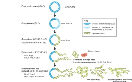
Fig. 2. Stepwise development of the mammalian lymphatic vasculature. In mice, embryonic veins start to develop under the control of COUP-TFII at ~E8.5. A few hours later, at ~E9.0, the LEC competence factor Sox18starts to be expressed in a subpopulation of these venous endothelial cells. Expression of Sox18 and COUP-TFII appears sufficient to induce Prox1expression at ~E9.75 in a subset of competent venous endothelial cells. The commitment towards LEC differentiation is initiated upon Prox1expression, with Prox1-expressing venous progenitors acquiring an LEC fate. Subsequently, during determination, these Prox1-expressing cells become further committed to the LEC lineage by migrating away from the veins under the influence of mesenchymal Vegfc signals. As LECs migrate away from the veins, the activity of SH2 domain-containing leukocyte protein of 76 kDa (Slp76), spleen tyrosine kinase (Syk) and phospholipase C, gamma 2 (Plcg2) are required for lymphovenous separation. These migrating LECs start to form the first primitive lymph sacs and to express additional LEC markers, such as neuropilin 2 (Nrp2), forkhead box C2 (Foxc2), podoplanin (Pdpn), angiopoietin 2 (Ang2), ephrin B2 (Efnb2), protein phosphatase 1 regulatory (inhibitor) subunit 13B (Aspp1) and core 1 synthase glycoprotein-N-acetylgalactosamine 3-beta-galactosyltransferase 1 (T-synthase). Later, during the differentiation and maturation steps, LECs sprout and migrate from the lymph sacs to form the whole lymphatic vascular network. Their remodeling and maturation give rise to the entire lymphatic network of capillaries and collecting vessels.
D
E
V
E
LO
P
M
E
N
COUP-TFII), or both. Alternatively, and in line with the explanation proposed above, in veins some other factor might cooperate with Sox18 in the induction of Prox1in venous ECs, with COUP-TFII being the likely candidate (R.S.S., unpublished) (Fig. 1). The conditional deletion of COUP-TFIIfrom Prox1-expressing venous ECs results in mutant mouse embryos that are almost completely devoid of LECs and lymphatic vasculature (R.S.S., unpublished), indicating that COUP-TFII activity is indeed crucial during the earliest stages of LEC differentiation.
In the future, the generation of new animal models that put these possibilities to the test should help conclusively determine which factor or factors are responsible for imparting lymphatic competence to the embryonic veins. In the meantime, the available data suggest that Sox18is a likely candidate to fulfill this function and that, in cooperation with COUP-TFII, it induces Prox1expression in a subset of venous ECs (Figs 1 and 2). How this subset is selected locally is not yet known; however, it can be speculated that some secreted factor (or factors) located in the vicinity of the veins is responsible for inducing Sox18and Prox1expression on one side of the vein, or for repressing their expression on the opposite side. This polarized expression is probably relevant in determining the ultimate location of the forming primitive lymph sacs, as only specified LECs (Prox1+Sox18+) located on one side of the vein will eventually migrate away from the veins (see below) (Wigle and Oliver, 1999; Wigle et al., 2002; Srinivasan et al., 2007).
Specification
Once venous ECs acquire the competence to respond to specific inductive signals, they start to become committed to an LEC fate (Fig. 2). The initial phase of this process is the specification of the LEC phenotype (Fig. 2). As mentioned above, during the competence stage, the activity of Sox18 and COUP-TFII appears to be required to induce Prox1 expression in venous LEC progenitors (Fig. 1). There is ample evidence in support of the proposal that Prox1 expression is necessary and sufficient to specify the LEC phenotype in venous BECs (Wigle et al., 2002; Hong et al., 2002; Petrova et al., 2002). The functional inactivation of Prox1in mice has revealed that in addition to its role in the lymphatic vasculature, it is crucial for the development of many other organs and tissues, such as the lens, retina, liver, pancreas and heart (Dyer et al., 2003; Risebro et al., 2009; Sosa-Pineda et al., 2000; Wang et al., 2005; Wigle et al., 1999). Prox1-null embryos die at ~E14.5, and, in most genetic backgrounds, heterozygous pups die at birth, most likely because of heart abnormalities and the presence of chylothorax (the abnormal accumulation of lymphatic fluid or chyle in the pleural cavity) and chylus ascites (the abnormal extravasation of milky chyle into the peritoneal cavity), which are caused by a defective lymphatic vasculature (Harvey et al., 2005; Wigle and Oliver, 1999).
Two initial clues suggested a vital role for Prox1in the formation of the mammalian lymphatic vasculature: first, its expression in developing LECs, but not in BECs, made Prox1the first LEC-specific marker (Wigle and Oliver, 1999); and second, functional inactivation of Prox1in mice generated the first animal model that completely lacked a lymphatic vasculature (Wigle and Oliver, 1999). Further work determined that the absence of the lymphatic network was directly caused by the lack of LECs. Through the use of additional LEC and BEC markers, it was then shown that LEC differentiation was defective in Prox1-null embryos, and also that Prox1activity was necessary to specify an LEC phenotype in a subset of venous ECs (Wigle et al., 2002). This strongly supported the original proposal of Florence Sabin (Sabin, 1902) that the
lymphatic vasculature in mammals is venous derived. Subsequently, it was shown that Prox1activity is sufficient to promote an LEC phenotype in cultured human umbilical vein endothelial cells (HUVECs) (Hong et al., 2002; Petrova et al., 2002). Together, these in vitro and in vivo findings established Prox1as an early-acting instructive factor in mammalian LEC differentiation, being necessary and sufficient to divert venous ECs towards the LEC phenotype (Oliver and Detmar, 2002; Wigle et al., 2002). The initial, restricted expression of Prox1 in a subpopulation of venous ECs that would otherwise remain blood vascular ECs has focused current research on this stage of tissue specialization in an effort to determine the cellular and molecular basis of the commitment to the lymphatic pathway and the intrinsic character of the LEC precursor state. Although no in vivo targets of Prox1 during the presumed specification step have yet been identified, it is likely that this step requires the concomitant downregulation of venous BEC markers and the upregulation of LEC-specific markers. In this context, Prox1 could function as a binary transcriptional switch, turning the BEC program off and the LEC program on (Oliver and Srinivasan, 2008).
As venous ECs become specified to the LEC fate, they start to move away from the veins, following specific migratory paths (Fig. 2) (Wigle and Oliver, 1999). These migrating LECs eventually form the primitive lymph sacs (Wigle and Oliver, 1999), embryonic structures that develop from LEC fusion and dilation into surrounding mesenchyme tissue and from which most of the lymphatic network is subsequently derived (Fig. 2).
The initiation of LEC migration from the veins could be considered as the first morphological indication that the growth of the lymphatic vasculature, known as lymphangiogenesis, has begun. In mammals, one of the genes that control the migration of these Prox1-expressing LEC progenitors away from the veins is vascular endothelial growth factor C (Vegfc), which is also expressed by vascular smooth muscle cells and mesenchymal cells near veins (Fig. 2) (Kukk et al., 1996). In Vegfc-null mouse embryos, LEC specification still takes place (Prox1expression is normal in venous LEC progenitors), but the embryos lack a lymphatic vasculature because of the failed migration of Prox1-expressing LECs from the embryonic veins (Karkkainen et al., 2004). Interestingly, the analysis of Vegfcheterozygous mice has revealed a threshold effect, whereby a reduction in Vegfc levels lead to cutaneous lymphatic hypoplasia and lymphedema (Karkkainen et al., 2004). Further support for a role of Vegfc in the regulation of lymphangiogenesis comes from a study that has identified missense mutations in the gene coding for its receptor, VEGFR3, in human patients with hereditary lymphedema (Karkkainen et al., 2000).
A detailed analysis of Prox1-null embryos suggested that the initiation of LEC migration from the veins is independent of Prox1 activity, as Prox1-null ECs are still able to migrate away from the veins (Wigle et al., 2002; Wigle and Oliver, 1999). However, the migratory path of these ECs is abnormal, which indicates that some required guidance signal (or the ability to interpret it) is defective in the mutant ECs (Wigle and Oliver, 1999). Therefore, it is likely that once Prox1 expression is initiated by venous ECs, the expression of some receptor-ligand signaling system (e.g. Vegfr3/Vegfc) is required to guide the subsequent polarized budding of LECs towards the ligand source, thereby ensuring the appropriate cell numbers and precise location of the primary lymph sacs. Vefgr3 is normally expressed in developing LECs; however, its expression is lost in Prox1-null embryos (Wigle et al., 2002). The fact that in the context of Prox1-null mutant embryos, ECs managed to migrate away from the veins (Wigle et al., 2002; Wigle and Oliver, 1999), argues not
D
E
V
E
LO
P
M
E
N
only that the initiation of this process is independent of Prox1 activity, but also that the loss of Vegfr3 expression is most likely responsible for the abnormal migration.
Genetic lineage-tracing experiments have revealed that the specification and migration of LECs are dynamic processes in which new LECs are continuously being generated from Prox1-expressing venous ECs, while already specified LECs are quite far along the morphogenetic process of migrating distally from the veins (Srinivasan et al., 2007). In mice, this process lasts for several days, from ~E9.75 to E14.5. Interestingly, the finding that in Prox1-null embryos, the Prox1promoter is initially active in migrating ECs, but is shut off a few days later, suggests that the migrating Prox1 -expressing LECs provide a positive autoregulatory signal back to the veins to maintain Prox1expression and thus the continued generation of an appropriate number of LEC precursors (Fig. 1) (Wigle et al., 2002; Oliver and Detmar, 2002). This putative autoregulation of Prox1promoter activity might be a relatively simple and sensitive mechanism to help control the number of LECs produced by the veins at any specific developmental stage.
The specification step is considered to be labile. As development progresses, recently specified migrating LECs start to express additional lymphatic markers as they become further committed towards an LEC fate; therefore, their developmental potential becomes restricted. Accordingly, the process of LEC specification in mice might start once Prox1expression is detected in a subpopulation of venous ECs, at ~E9.75. As development proceeds, the balance between BEC and LEC fates becomes more biased towards that of LECs, such that at ~E13.5 the newly specified LEC fate is on its way to being terminally differentiated. This could explain why the expression of genes that are also involved in venous/arterial formation is no longer detected or is no longer necessary after E14.5; at around that stage, Sox18expression is no longer observed in developing LECs (Francois et al., 2008) and COUP-TFIIactivity is no longer required (R.S.S., unpublished) (see below).
As specified LECs move away from the veins and start aggregating to form the primary lymph sacs, the forming lymphatics should become separated from the blood vasculature (Fig. 2). Work conducted in the last few years has identified the signaling molecules Slp76, Syk and Plcg2 as key players in this process, as their functional inactivation in mice results in a blood-filled lymphatic phenotype (Abtahian et al., 2003; Ichise et al., 2009).
Determination
The second step of the commitment phase is the determination of LEC fate, a process by which the developmental potential of the specified LECs becomes further restricted by acquiring, in a stepwise manner, all the additional molecular features that are typical of fully differentiated LECs (Fig. 2). During this process, the regulatory cascade initiated by Prox1during the initial specification step begins to unfold, and differentiating Prox1-expressing LECs progressively incorporate the expression of additional gene products. Consequently, the LEC transcriptome depends on the developmental stage, although it could be argued that in mice this determination step occurs approximately between E13.5 and mid-gestation (~E16.5).
As discussed above, the functional inactivation of genes required during the competence or specification steps should result in a lack of LECs. By contrast, the mutation or functional inactivation of genes that participate in the LEC-determination phase should not arrest LEC formation, but instead result in different types of lymphatic vasculature defects as a consequence of alterations in later aspects of lymphatic development, such as mispatterning, abnormal
blood/lymphatic connections, hypertrophy, lack of valves or a reduction in the number of lymphatics. On this basis, several genes that probably participate in the determination phase have been identified in the last few years (Fig. 2). As several reviews discussing their expression and function are available (Adams and Alitalo, 2007; Maby-El Hajjami and Petrova, 2008; Oliver and Srinivasan, 2008), we briefly highlight just a few of these genes.
Expression of Vegfr3can be considered as a reliable indicator of the progression of LEC determination (Fig. 2). During early development, this gene is normally widely expressed by venous ECs and LECs; however, whereas its expression in LECs continues even into adulthood, it is downregulated in venous ECs concomitant with LEC fate determination (Kaipainen et al., 1995; Wigle et al., 2002). As mentioned above, missense mutations in VEGFR3 have been identified in patients with hereditary lymphedema (Karkkainen et al., 2000).
Another candidate indicator of the progression of the determination phase is neuropilin 2 (Nrp2), a receptor for class III semaphorins that can also interact with Vegfr2 and Vegfr3 and that is expressed in veins at ~E10.0 (Neufeld et al., 2002). At ~E13.0, while the formation of the lymph sacs is progressing, Nrp2 expression is maintained in LECs and is turned off in venous ECs (Yuan et al., 2002). In addition, the functional inactivation of Nrp2 results in reduced LEC proliferation and fewer lymphatic capillaries (Yuan et al., 2002).
Another likely player in LEC determination is the mucin-type transmembrane glycoprotein podoplanin (Pdpn; T1a) (Schacht et al., 2003). Expression of this gene is first detected in LECs at ~E12.5, and Pdpn–/– pups die soon after birth with severe lymphedema, which results from highly abnormal lymphatic vascular patterning and function (Schacht et al., 2003). In addition, Pdpn–/–pups appear to have defects in the establishment of proper connections between deep and superficial lymphatic vessels (Schacht et al., 2003).
As mentioned above, COUP-TFII activity is necessary at the competence stage to initiate the program that leads to LEC differentiation, most likely by cooperating with Sox18 in the activation of Prox1 expression. In addition to its venous expression, COUP-TFIIis expressed in embryonic and postnatal LECs (R.S.S., unpublished) (Lee et al., 2009; Yamazaki et al., 2009), and direct genetic testing (conditional inactivation) shows that its activity is also required during the LEC fate-determination phase (R.S.S., unpublished). In vivo data have shown that the conditional inactivation of COUP-TFIIat time points later than E11.5, but prior to E13.5, results in mispatterned, blood-filled lymphatic vessels (R.S.S., unpublished). Importantly, after E13.5, COUP-TFIIactivity in LECs is no longer required (R.S.S., unpublished). Molecular studies in cultured ECs have suggested that some of the functions of Prox1 during embryonic lymphangiogenesis could be mediated by direct protein-protein interaction with COUP-TFII (Lee et al., 2009; Yamazaki et al., 2009). Therefore, it is possible that some of these later roles of COUP-TFII during LEC determination are mediated via its interaction with Prox1.
Differentiation and remodeling
In mammals, the lymphatic vasculature begins to spread throughout most of the developing embryonic tissues by a process of extensive sprouting from the primary lymph sacs during mid-gestation and while primary LEC determination and commitment are progressing (Fig. 2). These growing lymphatic vasculature branches are not yet fully mature or terminally differentiated (Oliver, 2004). Only near the time of birth do lymphatic vessels express the complete profile
D
E
V
E
LO
P
M
E
N
of markers found in mature, differentiated lymphatics (Oliver, 2004; Oliver and Alitalo, 2005; Saharinen et al., 2004). Several genes that participate in the later aspects of developmental lymphangiogenesis have been identified in recent years (Fig. 2 and see below) (for reviews, see Adams and Alitalo, 2007; Cueni and Detmar, 2008; Maby-El Hajjami and Petrova, 2008; Oliver and Alitalo, 2005; Oliver and Srinivasan, 2008). Notably, mouse mutants for several of these genes, namely the PDZ-interacting domain of Efnb2(Makinen et al., 2005), the forkhead transcription factor Foxc2 (Petrova et al., 2004), the ligand for the endothelial Tie2 receptor tyrosine kinase angiopoietin 2 (Angpt2; also known as Ang2or Agpt2) (Gale et al., 2002), fasting-induced adipose factor [Fiaf; also known as angiopoietin-like 4 (Angptl4)] (Backhed et al., 2007), apoptosis stimulating protein of p53 (Aspp1; Ppp1r13b) (Hirashima et al., 2008) and T-synthase (C1galt1) (Fu et al., 2008), all exhibit lymphatic phenotypes that can be related to alterations in their end-stage remodeling or maturation, such as blood-filled lymphatics, abnormal recruitment of pericytes by the lymphatic capillaries, lack of valves, abnormal patterning, hyperplasia, chylothorax, a reduced number of lymphatic capillaries and edema.
How to remain an LEC: LEC fate plasticity and maintenance
Above, we have summarized the current knowledge regarding the program that leads to the terminal differentiation of mammalian LECs. During this process, committed Prox1-expressing LEC
progenitors in embryonic veins activate and repress, respectively, the expression of selected LEC and BEC genes, and, as they become terminally differentiated, their developmental potential gradually becomes restricted. Recent work has revealed that the mature LEC phenotype is a surprisingly plastic, reprogrammable condition that depends on constant Prox1 activity for its maintenance (Fig. 3). For example, the conditional downregulation of Prox1 during embryonic, postnatal, or adult stages is sufficient to reprogram (dedifferentiate) LECs back into BECs (Fig. 3B,E) (Johnson et al., 2008). Consequently, the identity of the mutant lymphatic vessels was also partially reprogrammed as they acquired some features typical of the blood vasculature and even became filled with blood. Short interfering RNA (siRNA)-mediated downregulation of Prox1 in cultured LECs showed that the reprogramming of LECs to BECs is a Prox1-dependent, cell-autonomous process. These results led to the proposal that the final LEC state is constantly and actively programmed by the presence of the Prox1 transcription factor, which acts as a binary BEC suppressor/LEC identity maintainer, similar to its role during earlier stages of specification (as described above). Simply switching off Prox1 expression allows a dedifferentiation reprogramming cascade to take place, and the cells that lose Prox1 expression reacquire BEC status (Fig. 3) (Johnson et al., 2008). Therefore, LECs appear to be one of the few known differentiated cell types that require the constant expression of a particular ‘instructor’ gene (in this case Prox1) to maintain their phenotypic identity.
Loss of Notch signaling or gain of COUP-TFII activity
Loss of COUP-TFII activity
or gain of Notch signaling Loss of Prox1
Loss or gain of function phenotypes
+ Prox1 NormaI blood
vascular network
A B
[image:7.612.53.489.370.651.2]C D E
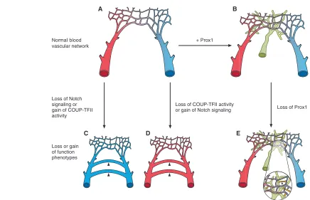
Fig. 3. Endothelial cell identity is plastic and reversible.(A)The normal blood vascular network consists of arteries (red) and veins (blue) that connect only through the common capillary network. (B)Upon expression of Prox1in venous endothelial cells, LECs originate from the embryonic veins and form the third independent vascular network, the lymphatics (green). (C)In arteries, the loss of Notch signaling or the ectopic gain of COUP-TFII expression promotes a fate change, such that the arteries acquire a venous identity and abnormal shunts are observed (arrowheads). (D)In veins, Notch signaling is ectopically activated in the absence of COUP-TFII; consequently, the veins acquire an arterial fate and exhibit abnormal shunts (arrowheads). (E)The loss of Prox1expression by LECs results in their dedifferentiation towards a BEC phenotype, such that the lymphatic vasculature partially acquires features of the blood vasculature and establishes abnormal connections with blood vessels (arrowheads in
inset). This scheme was modified with permission from Fig. 1 of Adams and Alitalo (Adams and Alitalo, 2007).
D
E
V
E
LO
P
M
E
N
Apart from the example of Prox1in LECs, not much is known about how cell types maintain their terminally differentiated fate. In general, cell type differentiation is considered to be a unidirectional, irreversible and terminal process. However, recent work has challenged these views by showing that individual transcription factors can reprogram differentiated somatic cells (Takahashi et al., 2007), that mature B lymphocytes can be reprogrammed back into functional T cells by switching off expression of the transcription factor Pax5(Cobaleda et al., 2007), and that a subpopulation of CD11b+macrophages has lymphatic endothelial characteristics and might be involved in the induction of pathological lymphangiogenesis in the cornea (Maruyama et al., 2005). Conversely, in ECs, arteriovenous identity can be reprogrammed by the loss or ectopic activation of Notch signaling or of COUP-TFII (Fig. 3A,C,D) (Roca and Adams, 2007; You et al., 2005), and lymphatic identity can be acquired upon the forced expression of Prox1in BECs maintained in culture (Hong et al., 2002; Petrova et al., 2002).
These findings raise some interesting questions. First, what, in functional terms, is the meaning of terminal differentiation? Second, why is the expression of Prox1in LECs constantly required? How does it work molecularly; is it at the level of chromatin structure or epigenetic landmarks? Third, is LEC-to-BEC dedifferentiation a process that takes place in normal or pathological settings in vivo?
The examples described above of certain cell types (B cells, LECs and venous/arterial BECs) that are capable of dedifferentiating when key transcriptional regulators (e.g. Pax5 or Prox1) are downregulated suggest that the concept of terminal differentiation might need to be revisited. It could be that, in certain settings, the fate of at least some differentiated cell types could be altered or reversed by the specific silencing or ectopic expression of some key regulators, as discussed below.
In this context, it is possible that the expression of important regulators of cell type specification, such as Prox1, is constantly required to maintain the fate of a differentiated cell type, and that even subtle alterations in expression levels might allow significant variability in the stability of cell fate, or in the fidelity of the associated gene expression profile, with consequences for the associated cellular and physiological behavior.
An obvious question is whether or not the ability of a differentiated cell type to interconvert flexibly between differentiated cell fates provides any selective advantage that could, for example, facilitate certain regenerative responses. Currently, however, there are no available data to support the possibility that changes in cell fate via transdifferentiation or dedifferentiation might occur in normal physiological conditions.
In the case of Prox1, one might speculate that under normal conditions, its constant expression by LECs helps these cells to maintain their differentiated phenotype independently of signals from the tissue environment. By contrast, in a pathological setting (e.g. inflammation or tumorigenesis), an abnormal environment (e.g. a tumor) could, by altering the normal levels of Prox1expression, trigger the dedifferentiation of LECs into BECs. Indeed, BEC/LEC lineage switches have been observed in various human malignancies. In angiosarcomas, intratumoral capillaries express a mixed blood/lymphatic phenotype (Breiteneder-Geleff et al., 1999), and, in some tumors, newly formed blood vessels exhibit structural heterogeneity and dysfunction (Abramsson et al., 2003). The plasticity of differentiated LECs might help explain the lineage switches that have been reported for some types of malignancies. Thus, LEC dedifferentiation, as promoted by the downregulation of Prox1, could be one of the mechanisms that operate during
tumorigenesis (Johnson et al., 2008). Furthermore, it is possible that in certain conditions that require the rapid additional supply of blood, the angiogenetic formation of new vessels might be too slow and might not satisfy tissue metabolic requirements quickly enough. Perhaps, the dedifferentiation of a nearby lymphatic network into blood vessels, with Prox1 downregulation as the triggering event, could provide additional, more rapid access to nutrients and oxygen (Johnson et al., 2008).
Conclusions
Work in the last decade has advanced our knowledge of lymphatic biology and reinvigorated interest in this particular topic. A number of genes that play crucial roles during lymphatic development have been identified, and valuable mouse models have become available. In addition, recent work in other model organisms (e.g. zebrafish, frog, chicken) has started to highlight similarities and differences between species in the mechanisms that lead to the formation of the lymphatic network and also to provide us with valuable in vivo and in vitro tools with which to quickly evaluate the functional role of newly identified lymphangiogenic gene products and compounds (Ny et al., 2005; Yaniv et al., 2006; Wilting et al., 2006; Hogan et al., 2009; Kalin et al., 2009).
A fascinating topic with a potentially important impact on translational research is EC plasticity. Recent progress in stem cell research has revealed how adult somatic cells can be converted back to a pluripotent state (iPS) using exogenous factors. Indeed, iPS cells can potentially differentiate into any cell type and could be used in therapeutic and regenerative medicine. However, despite their popularity, a number of hurdles related to the safety and mechanisms of iPS cell delivery will need to be overcome before iPS cells become widely used in therapeutic applications. As recently highlighted by J. Rossant (Rossant, 2009), another major impact of iPS research relates to the established concept that developmental pathways are irreversible. It is likely that in years to come, and as more data accumulate on iPS and cellular plasticity, this concept will be re-evaluated.
In the case of ECs, the available data suggest that the phenotype of the three main types of ECs (arterial, venous and lymphatic) can be superimposed, or reverted to, by subtle alterations in the combination or in the expression levels of a few key regulators (e.g. Notch signaling, COUP-TFIIand Prox1) (Fig. 3). This change in cell fate appears to be sufficient to promote alterations in vessel identity and function, and this plasticity could have extremely versatile applications during normal and pathological vessel growth. We can speculate that in the future, the in vivo reprogramming of differentiated ECs could be an easier and potentially safer alternative to the use of iPS cells in order to stimulate the growth or regression of blood or lymphatic vessels in various human pathologies.
Acknowledgements
We thank Drs Chris Wright and Natasha Harvey for critical reading of this manuscript and for helpful comments and suggestions, and Betsy Williford and Julie Groff from Biomedical Communications for their expert help in the design of the figures. This project was supported in part by the NIH (R01-HL073402), Cancer Center Support, and the American Lebanese Syrian Associated Charities (ALSAC). Deposited in PMC for release after 12 months.
Competing interests statement
The authors declare no competing financial interests.
References
Abramsson, A., Lindblom, P. and Betsholtz, C.(2003). Endothelial and nonendothelial sources of PDGF-B regulate pericyte recruitment and influence vascular pattern formation in tumors. J. Clin. Invest. 112, 1142-1151. Abtahian, F., Guerriero, A., Sebzda, E., Lu, M. M., Zhou, R., Mocsai, A.,
Myers, E. E., Huang, B., Jackson, D. G., Ferrari, V. A. et al.(2003).
D
E
V
E
LO
P
M
E
N
Regulation of blood and lymphatic vascular separation by signaling proteins SLP-76 and Syk. Science299, 247-251.
Adams, R. H. and Alitalo, K.(2007). Molecular regulation of angiogenesis and lymphangiogenesis.Nat. Rev. Mol. Cell Biol. 8, 464-478.
Backhed, F., Manchester, J. K., Semenkovich, C. F. and Gordon, J. I.(2007). Mechanisms underlying the resistance to diet-induced obesity in germ-free mice.
Proc. Natl. Acad. Sci. USA104, 979-984.
Baluk, P., Fuxe, J., Hashizume, H., Romano, T., Lashnits, E., Butz, S., Vestweber, D., Corada, M., Molendini, C., Dejana, E. et al.(2007). Functionally specialized junctions between endothelial cells of lymphatic vessels.
J. Exp. Med. 204, 2349-2362.
Breiteneder-Geleff, S. S. A., Kowalski, H., Horvat, R., Amann, G., Kriehuber, E., Diem, K., Weninger, W., Tschachler, E., Alitalo, K. and Kerjaschki, D. (1999). Angiosarcomas express mixed endothelial phenotypes of blood and lymphatic capillaries: podoplanin as a specific marker for lymphatic endothelium.
Am. J. Pathol. 154, 385-394.
Cobaleda, C. J. W. and Busslinger, M.(2007). Conversion of mature B cells into T cells by dedifferentiation to uncommitted progenitors. Nature449, 473-477.
Cueni, L. N. and Detmar, M.(2008). The lymphatic system in health and disease.
Lymphat. Res. Biol. 6, 109-122.
De Val, S. and Black, B. L.(2009). Transcriptional control of endothelial cell development. Dev. Cell16, 180-195.
De Val, S., Chi, N. C., Meadows, S. M., Minovitsky, S., Anderson, J. P., Harris, I. S., Ehlers, M. L., Agarwal, P., Visel, A., Xu, S. M. et al.(2008).
Combinatorial regulation of endothelial gene expression by ets and forkhead transcription factors. Cell135, 1053-1064.
Dyer, M. A., Livesey, F. J., Cepko, C. L. and Oliver, G.(2003). Prox1 function controls progenitor cell proliferation and horizontal cell genesis in the mammalian retina. Nat. Genet. 34, 53-58.
Ferdous, A., Caprioli, A., Iacovino, M., Martin, C. M., Morris, J., Richardson, J. A., Latif, S., Hammer, R. E., Harvey, R. P., Olson, E. N. et al.(2009). Nkx2-5 transactivates the Ets-related protein 71 gene and specifies an
endothelial/endocardial fate in the developing embryo. Proc. Natl. Acad. Sci.
USA106, 814-819.
Francois, M., Caprini, A., Hosking, B., Orsenigo, F., Wilhelm, D., Browne, C., Paavonen, K., Karnezis, T., Shayan, R., Downes, M. et al.(2008). Sox18 induces development of the lymphatic vasculature in mice. Nature456, 643-647.
Fu, J., Gerhardt, H., McDaniel, J. M., Xia, B., Liu, X., Ivanciu, L., Ny, A., Hermans, K., Silasi-Mansat, R., McGee, S. et al.(2008). Endothelial cell O-glycan deficiency causes blood/lymphatic misconnections and consequent fatty liver disease in mice. J. Clin. Invest. 118, 3725-3737.
Gale, N. W., Thurston, G., Hackett, S. F., Renard, R., Wang, Q., McClain, J., Martin, C., Witte, C., Witte, M. H., Jackson, D. et al.(2002). Angiopoietin-2 is required for postnatal angiogenesis and lymphatic patterning, and only the latter role is rescued by Angiopoietin-1. Dev. Cell3, 411-423.
Harvey, N. L. and Oliver, G.(2004). Choose your fate: artery, vein or lymphatic vessel? Curr. Opin. Genet. Dev. 14, 499-505.
Harvey, N. L., Srinivasan, R. S., Dillard, M. E., Johnson, N. C., Witte, M. H., Boyd, K., Sleeman, M. W. and Oliver, G.(2005). Lymphatic vascular defects promoted by Prox1 haploinsufficiency cause adult-onset obesity. Nat. Genet. 37, 1072-1081.
Hirashima, M., Sano, K., Morisada, T., Murakami, K., Rossant, J. and Suda, T. (2008). Lymphatic vessel assembly is impaired in Aspp1-deficient mouse embryos. Dev. Biol. 316, 149-159.
Hogan, B. M., Bos, F. L., Bussmann, J., Witte, M., Chi, N. C., Duckers, H. J. and Schulte-Merker, S.(2009). Ccbe1 is required for embryonic lymphangiogenesis and venous sprouting. Nat. Genet. 41, 396-398. Hong, Y. K., Harvey, N., Noh, Y. H., Schacht, V., Hirakawa, S., Detmar, M.
and Oliver, G.(2002). Prox1 is a master control gene in the program specifying lymphatic endothelial cell fate. Dev. Dyn. 225, 351-357.
Ichise, H., Ichise, T., Ohtani, O. and Yoshida, N.(2009). Phospholipase Cgamma2 is necessary for separation of blood and lymphatic vasculature in
mice. Development136, 191-195.
Irrthum, A., Devriendt, K., Chitayat, D., Matthijs, G., Glade, C., Steijlen, P. M., Fryns, J. P., Van Steensel, M. A. and Vikkula, M.(2003). Mutations in the transcription factor gene SOX18 underlie recessive and dominant forms of hypotrichosis-lymphedema-telangiectasia. Am. J. Hum. Genet. 72, 1470-1478. Johnson, N. C., Dillard, M. E., Baluk, P., McDonald, D. M., Harvey, N. L.,
Frase, S. L. and Oliver, G.(2008). Lymphatic endothelial cell identity is reversible and its maintenance requires Prox1 activity. Genes Dev. 22, 3282-3291.
Kaipainen, A., Korhonen, J., Mustonen, T., van Hinsbergh, V. W., Fang, G. H., Dumont, D., Breitman, M. and Alitalo, K.(1995). Expression of the fms-like tyrosine kinase 4 gene becomes restricted to lymphatic endothelium during development. Proc. Natl. Acad. Sci. USA92, 3566-3570.
Kalin, R. E., Banziger-Tobler, N. E., Detmar, M. and Brandli, A. W.(2009). An in vivo chemical library screen in Xenopus tadpoles reveals novel pathways involved in angiogenesis and lymphangiogenesis. Blood114, 1110-1122.
Karkkainen, M. J., Ferrell, R. E., Lawrence, E. C., Kimak, M. A., Levinson, K. L., McTigue, M. A., Alitalo, K. and Finegold, D. N.(2000). Missense mutations interfere with VEGFR-3 signalling in primary lymphoedema. Nat. Genet. 25, 153-159.
Karkkainen, M. J., Haiko, P., Sainio, K., Partanen, J., Taipale, J., Petrova, T. V., Jeltsch, M., Jackson, D. G., Talikka, M., Rauvala, H. et al.(2004). Vascular endothelial growth factor C is required for sprouting of the first lymphatic vessels from embryonic veins. Nat. Immunol. 5, 74-80.
Kokubo, H., Miyagawa-Tomita, S., Nakazawa, M., Saga, Y. and Johnson, R.
L.(2005). Mouse hesr1 and hesr2 genes are redundantly required to mediate
Notch signaling in the developing cardiovascular system. Dev. Biol. 278, 301-309.
Krebs, L. T., Xue, Y., Norton, C. R., Shutter, J. R., Maguire, M., Sundberg, J. P., Gallahan, D., Closson, V., Kitajewski, J., Callahan, R. et al.(2000). Notch signaling is essential for vascular morphogenesis in mice. Genes Dev. 14, 1343-1352.
Kukk, E., Lymboussaki, A., Taira, S., Kaipainen, A., Jeltsch, M., Joukov, V. and Alitalo, K.(1996). VEGF-C receptor binding and pattern of expression with VEGFR-3 suggests a role in lymphatic vascular development. Development122, 3829-3837.
Lawson, N. D., Scheer, N., Pham, V. N., Kim, C. H., Chitnis, A. B., Campos-Ortega, J. A. and Weinstein, B. M.(2001). Notch signaling is required for arterial-venous differentiation during embryonic vascular development.
Development128, 3675-3683.
Lee, D., Park, C., Lee, H., Lugus, J. J., Kim, S. H., Arentson, E., Chung, Y. S., Gomez, G., Kyba, M., Lin, S. et al.(2008). ER71 acts downstream of BMP, Notch, and Wnt signaling in blood and vessel progenitor specification. Cell Stem Cell2, 497-507.
Lee, S., Kang, J., Yoo, J., Ganesan, S. K., Cook, S. C., Aguilar, B., Ramu, S., Lee, J. and Hong, Y. K.(2009). Prox1 physically and functionally interacts with COUP-TFII to specify lymphatic endothelial cell fate. Blood113, 1856-1859. Maby-El Hajjami, H. and Petrova, T. V.(2008). Developmental and pathological
lymphangiogenesis: from models to human disease. Histochem. Cell Biol. 130, 1063-1078.
Machnik, A., Neuhofer, W., Jantsch, J., Dahlmann, A., Tammela, T., Machura, K., Park, J. K., Beck, F. X., Muller, D. N., Derer, W. et al.(2009). Macrophages regulate salt-dependent volume and blood pressure by a vascular endothelial
growth factor-C-dependent buffering mechanism. Nat. Med. 15, 545-552.
Makinen, T., Adams, R. H., Bailey, J., Lu, Q., Ziemiecki, A., Alitalo, K., Klein, R. and Wilkinson, G. A.(2005). PDZ interaction site in ephrinB2 is required for the remodeling of lymphatic vasculature. Genes Dev. 19, 397-410.
Maruyama, K., Ii, M., Cursiefen, C., Jackson, D. G., Keino, H., Tomita, M., Van Rooijen, N., Takenaka, H., D’Amore, P. A., Stein-Streilein, J. et al. (2005). Inflammation-induced lymphangiogenesis in the cornea arises from CD11b-positive macrophages. J. Clin. Invest. 115, 2363-2372.
Neufeld, G., Kessler, O. and Herzog, Y.(2002). The interaction of Neuropilin-1 and Neuropilin-2 with tyrosine-kinase receptors for VEGF. Adv. Exp. Med. Biol.
515, 81-90.
Ny, A., Koch, M., Schneider, M., Neven, E., Tong, R. T., Maity, S., Fischer, C., Plaisance, S., Lambrechts, D., Heligon, C. et al.(2005). A genetic Xenopus laevis tadpole model to study lymphangiogenesis. Nat. Med. 11, 998-1004. Oliver, G.(2004). Lymphatic vasculature development. Nat. Rev. Immunol. 4,
35-45.
Oliver, G. and Detmar, M.(2002). The rediscovery of the lymphatic system: old and new insights into the development and biological function of the lymphatic vasculature. Genes Dev. 16, 773-783.
Oliver, G. and Harvey, N.(2002). A stepwise model of the development of lymphatic vasculature. Ann. New York Acad. Sci. 979, 159-165.
Oliver, G. and Alitalo, K.(2005). The lymphatic vasculature: recent progress and paradigms. Annu. Rev. Cell Dev. Biol. 21, 457-483.
Oliver, G. and Srinivasan, R. S.(2008). Lymphatic vasculature development: current concepts. Ann. New York Acad. Sci. 1131, 75-81.
Pennisi, D., Gardner, J., Chambers, D., Hosking, B., Peters, J., Muscat, G., Abbott, C. and Koopman, P.(2000). Mutations in Sox18 underlie
cardiovascular and hair follicle defects in ragged mice. Nat. Genet.24, 434-437. Petrova, T. V., Makinen, T., Makela, T. P., Saarela, J., Virtanen, I., Ferrell, R. E.,
Finegold, D. N., Kerjaschki, D., Yla-Herttuala, S. and Alitalo, K.(2002). Lymphatic endothelial reprogramming of vascular endothelial cells by the Prox-1 homeobox transcription factor. EMBO J. 21, 4593-4599.
Petrova, T. V., Karpanen, T., Norrmen, C., Mellor, R., Tamakoshi, T., Finegold, D., Ferrell, R., Kerjaschki, D., Mortimer, P., Yla-Herttuala, S. et al.(2004). Defective valves and abnormal mural cell recruitment underlie lymphatic vascular failure in lymphedema distichiasis. Nat. Med. 10, 974-981.
Risebro, C. A., Searles, R. G., Melville, A. A., Ehler, E., Jina, N., Shah, S., Pallas, J., Hubank, M., Dillard, M., Harvey, N. L. et al.(2009). Prox1
maintains muscle structure and growth in the developing heart. Development
136, 495-505.
Roca, C. and Adams, R. H.(2007). Regulation of vascular morphogenesis by
Notch signaling. Genes Dev. 21, 2511-2524.
D
E
V
E
LO
P
M
E
N
Rossant, J.(2009). Reprogramming to Pluripotency: From frogs to stem cells. Cell
138, 1047-1050.
Sabin, F.(1902). On the origin of the lymphatics system from the veins and the development of the lymph hearts and the thoracic duct in the pig. Am. J. Anat.
1, 367-391.
Saharinen, P., Tammela, T., Karkkainen, M. J. and Alitalo, K.(2004). Lymphatic vasculature: development, molecular regulation and role in tumor metastasis and inflammation. Trends Immunol. 25, 387-395.
Schacht, V., Ramirez, M. I., Hong, Y. K., Hirakawa, S., Feng, D., Harvey, N., Williams, M., Dvorak, A. M., Dvorak, H. F., Oliver, G. et al.(2003). T1alpha/podoplanin deficiency disrupts normal lymphatic vasculature formation
and causes lymphedema. EMBO J. 22, 3546-3556.
Schmid-Schonbein, G. W.(2003). The second valve system in lymphatics.
Lymphat. Res. Biol. 1, 25-29.
Slack, J. M. W.(1991). From Egg to Embryo: Regional Specification in Early Development. New York: Cambridge University Press.
Sosa-Pineda, B., Wigle, J. T. and Oliver, G.(2000). Hepatocyte migration during liver development requires Prox1. Nat. Genet. 25, 254-255.
Srinivasan, R. S., Dillard, M. E., Lagutin, O. V., Lin, F. J., Tsai, S., Tsai, M. J., Samokhvalov, I. M. and Oliver, G.(2007). Lineage tracing demonstrates the venous origin of the mammalian lymphatic vasculature. Genes Dev. 21, 2422-2432.
Takahashi, K. and Yamanaka, S.(2006). Induction of pluripotent stem cells from mouse embryonic and adult fibroblast cultures by defined factors. Cell126, 663-676.
Takahashi, K., Tanabe, K., Ohnuki, M., Narita, M., Ichisaka, T., Tomoda, K. and Yamanaka, S.(2007). Induction of pluripotent stem cells from adult human fibroblasts by defined factors. Cell131, 861-872.
Villa, N., Walker, L., Lindsell, C. E., Gasson, J., Iruela-Arispe, M. L. and Weinmaster, G.(2001). Vascular expression of Notch pathway receptors and ligands is restricted to arterial vessels. Mech. Dev. 108, 161-164.
von der Weid, P. Y. and Zawieja, D. C.(2004). Lymphatic smooth muscle: the motor unit of lymph drainage. Int. J. Biochem. Cell Biol. 36, 1147-1153.
Waddington, C. H.(1940). Organisers and Genes. Cambridge: Cambridge University Press.
Wang, J., Kilic, G., Aydin, M., Burke, Z., Oliver, G. and Sosa-Pineda, B.(2005). Prox1 activity controls pancreas morphogenesis and participates in the production of ‘secondary transition’ pancreatic endocrine cells. Dev. Biol. 286, 182-194. Wigle, J. T. and Oliver, G.(1999). Prox1 function is required for the development
of the murine lymphatic system. Cell98, 769-778.
Wigle, J. T., Chowdhury, K., Gruss, P. and Oliver, G.(1999). Prox1 function is crucial for mouse lens-fibre elongation. Nat. Genet. 21, 318-322.
Wigle, J. T., Harvey, N., Detmar, M., Lagutina, I., Grosveld, G., Gunn, M. D., Jackson, D. G. and Oliver, G.(2002). An essential role for Prox1 in the induction of the lymphatic endothelial cell phenotype. EMBO J. 21, 1505-1513. Wilting, J., Aref, Y., Huang, R., Tomarev, S. I., Schweigerer, L., Christ, B.,
Valasek, P. and Papoutsi, M.(2006). Dual origin of avian lymphatics. Dev. Biol.
292, 165-173.
Witte, M. H., Bernas, M. J., Martin, C. P. and Witte, C. L.(2001). Lymphangiogenesis and lymphangiodysplasia: from molecular to clinical lymphology. Microsc. Res. Tech.55, 122-145.
Yamazaki, T., Yoshimatsu, Y., Morishita, Y., Miyazono, K. and Watabe, T. (2009). COUP-TFII regulates the functions of Prox1 in lymphatic endothelial cells through direct interaction. Genes Cells14, 425-434.
Yaniv, K., Isogai, S., Castranova, D., Dye, L., Hitomi, J. and Weinstein, B. M. (2006). Live imaging of lymphatic development in the zebrafish. Nat. Med. 12, 711-716.
You, L. R., Lin, F. J., Lee, C. T., DeMayo, F. J., Tsai, M. J. and Tsai, S. Y.(2005). Suppression of Notch signalling by the COUP-TFII transcription factor regulates vein identity. Nature435, 98-104.
Yuan, L., Moyon, D., Pardanaud, L., Breant, C., Karkkainen, M. J., Alitalo, K. and Eichmann, A.(2002). Abnormal lymphatic vessel development in
neuropilin 2 mutant mice. Development129, 4797-4806.
Zhou, Q., Brown, J., Kanarek, A., Rajagopal, J. and Melton, D. A.(2008). In vivo reprogramming of adult pancreatic exocrine cells to beta-cells. Nature455, 627-632.