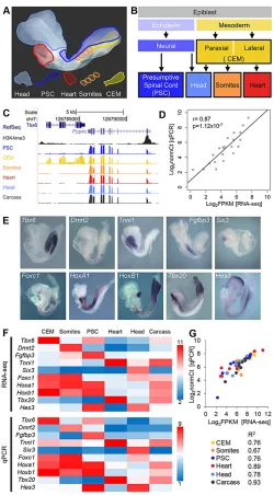
The tissue specific transcriptomic landscape of the mid gestational mouse embryo
Full text
Figure


Related documents
19% serve a county. Fourteen per cent of the centers provide service for adjoining states in addition to the states in which they are located; usually these adjoining states have
To identify the physiological targets of PieE during infection, we established a new purification method for which we created an A549 cell line stably expressing the Escherichia
Further experiences with the pectoralis myocutaneous flap for the immediate repair of defects from excision of head and
Using unbiased metabolomics, we discovered that Vibrio cholerae mutants genetically locked in a low cell density (LCD) QS state are unable to alter the pyruvate flux to convert
The realization of European Economic and Monetary Union has made it obvious that certain fundamental principles of the EC Treaty have be- come part of Germany's
(c) Expression of rtxBDE is induced by zebrafish larva coloniza- tion. Results of a  -galactosidase assay done using lysates of bacterial cells growing planktonically in E3 medium
It was decided that with the presence of such significant red flag signs that she should undergo advanced imaging, in this case an MRI, that revealed an underlying malignancy, which
Also, both diabetic groups there were a positive immunoreactivity of the photoreceptor inner segment, and this was also seen among control ani- mals treated with a