RESEARCH ARTICLE
Embryonic cholecystitis and defective gallbladder contraction
in the
Sox17
-haploinsufficient mouse model of biliary atresia
Hiroki Higashiyama1,*, Aisa Ozawa1,*, Hiroyuki Sumitomo1,*, Mami Uemura1,2,*, Ko Fujino1, Hitomi Igarashi1, Kenya Imaimatsu1, Naoki Tsunekawa1, Yoshikazu Hirate2, Masamichi Kurohmaru1, Yukio Saijoh3,
Masami Kanai-Azuma2and Yoshiakira Kanai1,‡
ABSTRACT
The gallbladder excretes cytotoxic bile acids into the duodenum through the cystic duct and common bile duct system. Sox17
haploinsufficiency causes biliary atresia-like phenotypes and hepatitis in late organogenesis mouse embryos, but the molecular and cellular mechanisms underlying this remain unclear. In this study, transcriptomic analyses revealed the early onset of cholecystitis in
Sox17+/−embryos, together with the appearance of ectopic cystic
duct-like epithelia in their gallbladders. The embryonic hepatitis showed positive correlations with the severity of cholecystitis in individualSox17+/−embryos. Embryonic hepatitis could be induced
by conditional deletion ofSox17in the primordial gallbladder epithelia but not in fetal liver hepatoblasts. The Sox17+/− gallbladder also
showed a drastic reduction in sonic hedgehog expression, leading to aberrant smooth muscle formation and defective contraction of the fetal gallbladder. The defective gallbladder contraction positively correlated with the severity of embryonic hepatitis in Sox17+/−
embryos, suggesting a potential contribution of embryonic cholecystitis and fetal gallbladder contraction in the early pathogenesis of congenital biliary atresia.
KEY WORDS: Gallbladder, Biliary atresia,Sox17,Shh
INTRODUCTION
The gallbladder is a flexible organ of the biliary tract that regulates bile storage and discharge by the contractile movement of its smooth muscles. Malformation of the gallbladder and bile ducts can cause disease, including cholesterol gallstones, chronic inflammation and biliary atresia (reviewed by Portincasa et al., 2004, 2008; Asai et al., 2015). Congenital biliary atresia is a rare condition in newborn infants (Kohsaka et al., 2002; Mieli-Vergani and Mieli-Vergani, 2009) that causes inflammation in the bile ducts and liver due to the blockage of bile flow (cholestasis). It is usually characterized by an aberrant gallbladder of reduced length, with irregular walls and indistinct mucosal lining, that appears to be closely associated with neonatal cholestasis associated with severe inflammation of the intrahepatic ducts
(Desmet, 1992; Mack and Sokol, 2005; Mieli-Vergani and Vergani, 2009).
The etiology of human biliary atresia remains unclear. It is speculated to be caused by either environmental factors, such as viral infections or toxin exposure in genetically susceptible individuals, or by developmental errors during the specification and morphogenesis of bile duct epithelia (reviewed by Mack and Sokol, 2005; Mieli-Vergani and Vergani, 2009; Nakamura and Tanoue, 2013; Davenport, 2016). Whether a result of internal or external factors, epithelial defects and/or injury of the extrahepatic bile ducts may be associated with the onset of human biliary atresia by the neonatal stage (Mack and Sokol, 2005; Davenport, 2016).
The extrahepatic biliary structures (gallbladder, cystic duct, hepatic ducts and common bile duct) originate from the biliary primordium (Spence et al., 2009; Uemura et al., 2010, 2013), which expresses SRY-box 17 (Sox17), a core regulator of endoderm determination in mice and humans (Tam et al., 2003). Formation of the intrahepatic duct is regulated cooperatively bySox9andSox4
(Poncy et al., 2015), albeit that their roles in the extrahepatic duct remain unclear. In a previous study (Uemura et al., 2013), perinatal lethality was observed in∼90% ofSox17heterozygous (Sox17+/−)
mice and the embryos displayed defective development of the gallbladder including defective bile duct epithelial wall and a biliary atresia-like phenotype (an abnormal accumulation of luminal decidual cells in the bile duct), together with severe embryonic hepatitis after the first biliary excretion into the fetal duodenum at ∼16.5 days post coitum (dpc). This timing of biliary atresia-like symptoms in mouse embryos is consistent with the hypothesis of early onset of biliary atresia in human fetuses (Tan et al., 1994; reviewed by Davenport, 2016). In human fetuses, bile acid synthesis occurs during the early organogenesis stages, during which bile starts to be excreted into the intestine near the end of the first trimester (Nakagawa and Setchell, 1990 and references therein). Furthermore, recent studies of naturally occurring outbreaks of sheep biliary atresia revealed that biliatresone, a causative toxin for biliary atresia, reduces Sox17 expression levels in the bile duct epithelia, and that silencingSox17mimics the effects of biliatresone on the bile duct epithelia (Lorent et al., 2015; Waisbourd-Zinman et al., 2016). Together with the extensive anatomical similarity of the extrahepatic biliary tracts and the associated blood vessels, nerves and smooth muscles between mice and humans (Higashiyama et al., 2016), the Sox17+/− mouse embryo provides a useful
experimental model with which to study the initial pathogenesis of biliary atresia.
In this study, we demonstrate the early onset of cholecystitis in the cystic duct-like gallbladder ofSox17+/−mouse embryos, in which
the reduction in sonic hedgehog (Shh) expression leads to defective contraction of the smooth muscles.
Received 27 November 2016; Accepted 12 April 2017
1Department of Veterinary Anatomy, The University of Tokyo, Yayoi 1-1-1,
Bunkyo-ku, Tokyo 113-8657, Japan.2Center for Experimental Animals, Tokyo Medical and
Dental University, Yushima 1-5-45, Bunkyo-ku, Tokyo 113-8510, Japan.
3Department of Neurobiology and Anatomy, The University of Utah, Salt Lake City,
UT 84132-3401, USA.
*These authors contributed equally to this work and are joint first authors
‡Author for correspondence (aykanai@mail.ecc.u-tokyo.ac.jp)
H.H., 1324-8139; A.O., 0000-0002-0506-5909; H.S., 0000-0003-0916-7846; M.U., 0000-0003-0036-3103; Y.K., 0000-0003-2116-7806
DEVEL
O
RESULTS
SOX9-positive cystic duct-like epithelia in theSox17+/−
primordial gallbladder
In wild-type embryos, the fetal gallbladder forms a pseudostratified columnar epithelium with epithelial folds by 17.5 dpc, while the cystic duct consists of a single-layered cuboidal epithelium with few epithelial folds throughout the fetal stages (Fig. 1A). TheSox17+/− gallbladder shows severe hypotrophy and a single-layered cuboidal epithelium with few epithelial folds (Fig. 1A) (see also Uemura et al., 2013), similar in appearance to the cystic duct epithelia of wild-type embryos at the same stage (17.5 dpc). This is consistent with morphometric data showing a significant reduction in epithelial height, cavity and/or epithelial area of the Sox17+/−
gallbladder, such that these values are similar to those of wild-type cystic ducts (Fig. 1B).
SOX17 is highly expressed in the developing gallbladder and cystic duct epithelia. It is enriched in the distal region of the developing gallbladder epithelium. This is in contrast to the
restricted distribution of SOX9-positive epithelial cells to the proximal region of the cystic duct (Fig. 1C; Fig. S1), thus showing a mostly non-overlapping pattern between distal SOX17-positive and proximal SOX9-positive epithelial cells in the gallbladder of wild-type embryos. In the Sox17+/− gallbladder, the SOX9-positive
domain expands into the distal region in the gallbladder (Fig. 1D), despite the reduced proliferation in the proximal cystic duct domain of the Sox17+/− versus wild-type gallbladder (Fig. S2). qPCR analysis confirms thatSox9expression is significantly increased in
Sox17+/−gallbladder compared with wild type at 15.5 dpc (Fig. 1E).
Considering our previous observation of hypoplasia and
[image:2.612.49.354.286.737.2]deciduation in theSox17+/−gallbladder epithelia (Uemura et al., 2013), these data suggest that the gallbladder epithelial cells might be replaced with the ectopic SOX9-positive cystic duct-like epithelial cells. In addition, such elevated expression of Sox9in theSox17+/−gallbladder correlates with the subsequent expression ofSox4, which can act redundantly withSox9in intrahepatic duct formation (Poncy et al., 2015) (Fig. S3).
Fig. 1.Sox17+/−gallbladder epithelium exhibits cystic
duct-like phenotypes.(A) Transverse sections [Hematoxylin and Eosin (HE) staining] of the gallbladder and cystic duct from wild-type (wt) andSox17+/−mouse embryos at 17.5 dpc. The levels of the sections are indicated in A′(arrowheads). Insets show high magnification images of the epithelium. (B) Quantitative analyses show a significant reduction in epithelial height and epithelial/cavity areas of the gallbladder inSox17+/− embryos at 17.5 dpc. *P<0.05, **P<0.01, ANOVA followed by Tukey’s test. (C) Anti-SOX17 and anti-SOX9 double immunohistochemistry in the primordial gallbladder and cystic duct regions of the wild-type embryo at 13.5 dpc. (D)Sox17+/−embryos display ectopic SOX9-positive cells in the distal edge of the gallbladder at 13.5 and 15.5 dpc. Bracket indicates the SOX9-positive domain. (E) qPCR analysis indicates a significant increase inSox9mRNA in theSox17+/−gallbladder at 15.5 dpc. *P<0.05, Student’s
t-test. In B and E, sample number is indicated within each bar. cd, cystic duct; duo, duodenum; gb, gallbladder; hd, hepatic duct; pd, pancreatic duct; pv, portal vein; D, distal; P, proximal. Scale bars: 100 µm.
DEVEL
O
Onset of cholecystitis inSox17+/−gallbladder during fetal
stages
To examine the transcriptomic changes in theSox17+/−gallbladder,
microarray analyses were conducted using gallbladder (distal) and cystic duct (proximal) segments of the Sox17+/− and wild-type
gallbladder primordia at 15.5 dpc, before the first secretion of bile fluid from the fetal liver (Uemura et al., 2013) (Fig. 2A,B). The transcriptomic analysis identified 279 upregulated and 501 downregulated genes in Sox17+/− gallbladder compared with
wild-type littermates at 15.5 dpc (n=4; Table S1). Gene set enrichment analysis (GSEA) confirmed Sox9 among the top 50 enriched/upregulated genes in theSox17+/−gallbladder (Fig. S4). Moreover, these data were compared with those of gallbladder-specific or cystic duct-gallbladder-specific genes from the same littermates [5361 or 4886 genes (n=2) expressed at higher or lower levels, respectively, in the gallbladder segment compared with the cystic duct segment of wild-type embryos]. Of the downregulated genes in the Sox17+/− gallbladder, 207/279 (74.4%) overlap with the gallbladder-specific genes that we identified (Fig. 2A, Table 1).
Furthermore, of the upregulated genes, 221/501 (44.1%) overlap with the cystic duct-specific gene list (Fig. 2B, Table 1). This is consistent with data showing the appearance of SOX9-positive cystic duct-like epithelia in theSox17+/−gallbladder during the late fetal stages (Fig. 1).
GSEA showed an enrichment of genes in the category‘definitive hemopoiesis’(Fig. S4). Gene ontology (GO) analysis confirmed that the 280 upregulated genes, other than the 221 cystic duct-specific genes, are involved in‘myeloid cell differentiation’(Klf1,
[image:3.612.47.384.294.737.2]Tal1,Epb42,Ahsp,Prdx3,Psen2andTrim10) (Fig. 2B), suggesting the enrichment of immature hematopoietic cells (e.g. inflammation) in the fetal gallbladder region even at 15.5 dpc. Key marker genes of several hepatobiliary and digestive tract disorders were also upregulated in this dataset, including olfactomedin 4 (Olfm4) (Liu et al., 2010; Gersemann et al., 2012), ATP-binding cassette, subfamily B4 (Abcb4) (Fickert et al., 2004; Esten Nakken et al., 2007; Baghdasaryan et al., 2011; Gordo-Gilart et al., 2015) and polycystic kidney and hepatic disease 1 (Pkhd1) (Nakamura et al., 2010; Hartley et al., 2011). We therefore examined the expression
Fig. 2. Global expression profiles and cholecystitis of fetalSox17+/−gallbladders.
(A,B) Microarray analysis identifies 279 downregulated genes (blue circle in A) and 501 upregulated genes (red circle in B) in theSox17+/− gallbladder at 15.5 dpc, as compared with gallbladder from wild-type (wt) littermates. Among these downregulated or upregulated genes, 207 (74.4%) or 221 (44.1%) were shared with the 5361 gallbladder-specific or 4886 cystic duct
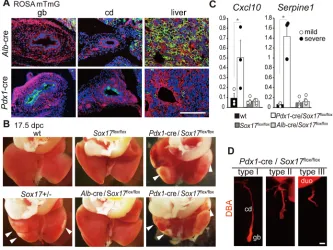
(cd)-specific genes, respectively, in wild-type embryos at the same stage. (C,D) qPCR analyses showing the upregulated profiles of the biliary disease marker genesOlfm4,Abcb4andPkhd1(C) and the inflammatory marker genesCxcl10and Serpine1(D) inSox17+/−gallbladder at 17.5 dpc. They-axis is fold change in expression level of each gallbladder or liver sample relative to those of the wild type (mean value set to 1). Each solid or open circle indicates a sample from an embryo with (severe phenotype) or without (mild phenotype) gross anatomical hepatic lesions, respectively. Bar charts show the average mean values of all (severe and mild) samples. The expression level of each marker gene in theAlb-cre/Sox17+/+andAlb-cre/
Sox17flox/+embryos at the same stage is also
shown. *P<0.05, **P<0.01, Mann-Whitney U-test. (E) Spearman rank correlation analysis of the association between gallbladder (y-axis) and liver (x-axis) phenotypes for the indicated genes in Sox17+/−embryos at 17.5 dpc. Both axes represent fold change in expression relative to wild-type littermates (mean value set to 1). InSox17+/− embryos, significant correlations between the two tissues were detected in both the severe phenotype and all (mild and severe phenotype) samples for Cxcl10andSerpine1mRNA levels, and in only the severe phenotype samples forOlfm4.
DEVEL
O
levels of these three disease markers, in addition to two inflammatory markers, namely Cxcl10 (Leonhardt et al., 2006) and Serpine1(Bessho et al., 2014), in both gallbladder and liver samples isolated from Sox17+/−embryos with or without hepatic
lesions (severe or mild phenotype group) at 17.5 dpc (Fig. 2C,D; Fig. S5A).
qPCR analysis showed a significant increase in the expression of
Olfm4,Abcb4 and Cxcl10 in theSox17+/− gallbladder compared with the wild-type gallbladder (Fig. 2C,D, left two bars in gb), in addition to high expression levels of bothOlfm4andCxcl10in the severely affected livers (Fig. 2C,D, solid circles inʻliver’). Serpine1
expression appeared to be higher, albeit not significantly, in both the gallbladder and liver of some severe phenotype Sox17+/−
embryos. Among these four genes, we found significant positive correlations in the increased expression levels of Olfm4 (only severe samples), Cxcl10 and Serpine1 (both severe and mild samples) between gallbladder and liver samples (Spearman test; Fig. 2E; see also Fig. S5B), suggesting a positive correlation in phenotypic severity between the gallbladder (i.e. cholecystitis)
and intrahepatic regions (i.e. hepatitis) in Sox17+/− embryos. Interestingly,Abcb4expression was increased only in gallbladder and not liver tissues (Fig. 2C; Fig. S5A), suggesting a potentially useful marker specific for extrahepatic cholestasis (Schaap et al., 2009).
Pkhd1, which encodes the ciliary protein fibrocystin, was upregulated in several mild phenotype samples (without any gross anatomical hepatic lesions) (Fig. 2C; Fig. S5A). In Sox17+/−
gallbladders, we found a positive correlation betweenPkhd1and
Olfm4 expression levels independent of gross anatomical hepatic lesions, in addition to the negative correlation betweenPkhd1and
Abcb4expression levels (Fig. S5C).
In addition, qPCR analysis of several fetal myeloid cell markers, namelyTal1(Kelliher et al., 1996), Klf1(McConnell and Yang, 2010) and Prdx3(Park et al., 2016), revealed a tendency for an increase in their expression levels in the Sox17+/− gallbladder
[image:4.612.53.566.76.448.2]region, together with a positive correlation among these three genes (Fig. S6A,B). Moreover, immunohistochemical analysis revealed that Gr1 (LY6G)-positive or F4/80 (ADGRE1)-positive spherical
Table 1. Top 15 of the 207 downregulated (gallbladder-specific) or the 221 upregulated (cystic duct-specific) genes inSox17+/−gallbladders at
15.5 dpc
Description Gene Fold change
Downregulated genes gb (+/−)<gb (wt)
(reduced levels)
cd (wt)<gb (wt) (gb-specific genes) Cbp/p300-interacting transactivator with Glu/Asp-rich
carboxy-terminal domain 1
Cited1 −9.1 (−10.3,−6.3,−12.7,−10.1) −5.8 ( −5.9,−7.6)
Sonic hedgehog Shh −9.0 ( −9.4,−7.0,−13.4, −8.7) −11.3 (−16.1,−6.4)
Maltase-glucoamylase Mgam −5.6 (−11.0,−6.2, −5.0, −3.2) −2.9 ( −1.4,−4.6) Pro-melanin-concentrating hormone Pmch −4.7 ( −3.5,−6.8, −4.5, −4.7) −5.2 ( −5.0,−4.8) Cytochrome P450, family 2, subfamily f, polypeptide 2 Cyp2f2 −4.1 ( −3.2,−4.9, −3.4, −3.0) −3.9 ( −1.5,−5.6) S100 calcium-binding protein G S100g −4.0 ( −4.4,−6.5, −3.7, −2.8) −3.5 ( −3.2,−2.1) Hedgehog-interacting protein Hhip −3.5 ( −3.4,−2.7, −3.8, −4.8) −3.5 ( −4.0,−3.7) Rho GTPase activating protein 36 1100001E04Rik(Arhgap36) −3.4 ( −3.3,−5.3, −2.4, −2.6) −6.7 ( −2.8,−4.9) Janus kinase and microtubule interacting protein 2 Jakmip2 −3.4 ( −7.5,−3.5, −2.8 −2.5) −1.8 ( −2.5, 2.1) Cytochrome c oxidase, subunit VIIa 1 Cox7a1 −3.1 ( −2.4,−3.1, −4.0, −3.7) −3.1 ( −2.9,−3.8) Solute carrier family 14 (urea transporter), member 1 Slc14a1 −3.1 ( −2.9,−3.0, −2.6, −4.4) −2.3 ( −1.3,−3.2) ADAM-like, decysin 1 Adamdec1 −3.1 ( −2.9,−4.3, −2.4, −4.1) −3.8 ( −3.3,−4.7)
SRY-box 17 Sox17 −3.1 ( −2.4,−2.8, −3.9, −3.5) −3.4 ( −3.4,−2.8)
Cholecystokinin A receptor Cckar −3.0 ( −1.7,−2.6, −4.9, −3.3) −6.6 (−12.1,−3.7) Cytochrome P450, family 4, subfamily x, polypeptide 1 Cyp4x1 −2.9 ( −3.1,−4.8, −1.7, −2.8) −2.6 ( −1.5,−1.6)
Upregulated genes gb (+/−)>gb (wt)
(elevated levels)
cd (wt)>gb (wt) (cd-specific genes) Extracellular proteinase inhibitor Expi(Wfdc18) 4.2 ( 4.5, 3.3, 2.8, 6.2) 2.3 ( 1.5, 5.2)
RIKEN cDNAB130021B11 gene B130021B11Rik 3.5 ( 2.4, 2.0, 1.5, 2.2) 3.6 ( 2.2, 2.3)
Polycystic kidney and hepatic disease 1 Pkhd1 3.3 ( 2.8, 1.0, 1.6, 5.7) 1.6 ( 2.1,−1.4)
Carboxylesterase 3 Ces3 3.1 ( 4.6, 2.0, 2.1, 3.7) 5.1 ( 9.3, 4.1)
Claudin 8 Cldn8 3.0 ( 4.7, 1.7, 2.2, 4.3) 2.1 ( 2.6, 3.1)
Apolipoprotein A-II Apoa2 2.9 ( 1.3,−1.0, 10.5, 2.4) 11.9 ( 44.7, 8.2)
DNA segment, Chr 4, Brigham&Women’s Genetics 0951 expressed, mRNA
D4Bwg0951e(Lurap1l) 2.8 ( 3.3, 2.8, 1.9, 3.5) 1.7 ( 1.9, 1.3)
Insulin-like growth factor binding protein 1 Igfbp1 2.8 ( 4.0, 1.1, 4.9, 3.0) 4.6 ( 8.9, 9.0)
TAO kinase 3 Taok3 2.7 ( 4.5,−1.2, 1.4, 1.5) 3.2 ( 2.1,−2.0)
Transcription elongation factor A (SII)-like 7 RP23-105O4.2(Tceal7) 2.7 ( 21.3, 1.6, 1.4, −1.1) 2.0 ( 1.3, 1.9) Solute carrier family 34 (sodium phosphate), member 2 Slc34a2 2.6 ( 2.7, 2.0, 2.8, 4.9) 4.4 ( 3.6, 6.3) Lectin, galactose binding, soluble 3 Lgals3 2.6 ( 1.5, 1.2, 14.4, 2.1) 2.8 ( 22.4, 3.2)
Pregnancy zone protein Pzp 2.5 ( 1.6,−1.3, 9.9, 3.1) 12.2 ( 43.2, 15.9)
Reelin Reln 2.4 ( 2.6, 1.8, 1.7, 4.4) 5.0 ( 4.4, 6.7)
Golgi apparatus protein 1 Glg1 2.4 ( 2.4,−1.1, 3.1, 1.2) 1.8 ( 1.7,−1.3)
Microarray expression analyses were performed using the presumptive gallbladder (gb, distal) and cystic duct (cd, proximal) segments of the primordial gallbladder ofSox17+/−(+/−) and wild-type (wt) embryos at 15.5 dpc. Fold changes that are in bold are from quadruplicate or duplicate microarray data generated by AltAnalyze; the numbers in parentheses indicate the fold changes in each individual. Each value in parenthesis represents one relative value of two hybridization signals, i.e. A/a, B/b, C/c and D/d (‘+/−versus wt’or‘cd versus gb’) of four array experiments. Mean value in bold is (A+B+C+D)/(a+b+c+d) for each probe generated by AltAnalyze. Gallbladder-specific or cystic duct-specific genes were defined as exhibiting a difference of at least 1.5-fold between the gallbladder and cystic duct regions, respectively, of wild-type embryos at 15.5 dpc.
DEVEL
O
myeloid-like cells appeared to be enriched in the surrounding mesenchymal region of the Sox17+/− gallbladder, albeit with no direct infiltration into the epithelial layers of the Sox17+/−
gallbladder (Fig. S6C).
Fetal liver phenotypes induced by conditional deletion of Sox17in liver hepatoblasts or primordial gallbladder epithelia
Sox17is expressed not only in the primordial gallbladder, but also in a population of hepatoblast progenitor cells at early somite stages (Okada et al., 2012). To specify the contribution of reducedSox17
activity in the liver or gallbladder in the onset of embryonic hepatitis, we examined the liver phenotypes ofSox17floxembryos
with a conditional deletion ofSox17using albumin (Alb)-cre and
Pdx1-cre mice. In contrast toAlb-cre-dependent recombination of theROSAmTmGallele in liver hepatoblasts (Fig. 3A),Pdx1-cre was
able to induce recombination in approximately half of the fetal gallbladder and cystic duct epithelia by 17.5 dpc (Fig. 3A), in addition to its efficient deletion of Sox17 in the pancreas and duodenum, with both tissues showing no detectable Sox17
expression at these stages (Spence et al., 2009; Uemura et al., 2010). First we examined the liver and gallbladder phenotypes ofAlb -cre/Sox17flox/+ embryos, and these showed no developmental
defects in liver and gallbladder development (data not shown).
qPCR analysis confirmed no elevated expression of the
inflammatory marker genes in gallbladder or liver of Alb-cre/ Sox17flox/+embryos at 17.5 dpc (Fig. 2C,D, two righthand bars in
each group), suggesting no appreciable link to hepatitis of the
reduced Sox17 expression in the liver hepatoblasts. TheAlb-cre/ Sox17flox/floxembryos also showed normal liver and gallbladder
phenotypes (Fig. 3B).
By contrast,Pdx1-cre/Sox17flox/floxembryos showed severe gross
anatomical hepatic lesions in some embryos (4/55 embryos; Fig. 3B), together with elevatedCxcl10 andSerpine1 expression in the affected livers (Fig. 3C). Moreover, thePdx1-cre/Sox17flox/flox
embryos showed varying degrees of shortening of the gallbladder and cystic duct (Fig. 3D), with hepatitis observed in fetuses with normal (type I, 3/34 embryos in Fig. 3D) and mild (type II, 1/10 embryos) phenotypes, but no hepatitis in individuals with a complete lack of the gallbladder (type III, 0/11 embryos). Moreover, histopathological and qPCR analyses confirmed that
Pdx1-cre/Sox17flox/flox embryos show biliary atresia-like
phenotypes similar to those of Sox17+/− embryos, such as
epithelial deciduation, bile duct stenosis/atresia and reduced Shh
expression (Fig. S7) (Uemura et al., 2013; this study). These data suggest that the hepatitis inSox17+/−embryos is caused by reduced
Sox17activity in the primordial gallbladder, and is not associated with the liver hepatoblasts.
SeveralShh-related genes are downregulated in theSox17+/−
gallbladder
[image:5.612.139.472.389.639.2]Among the 207 downregulated genes in theSox17+/−gallbladder at 15.5 dpc (Fig. 2A, Table 1; Fig. S4), we focused on the altered expression ofShh and of the Shh-responsive geneHhip. Whole-mountin situhybridization analysis of wild-type embryos at 11.5-13.5 dpc revealed thatShh, as well asSox17, was expressed in the
Fig. 3. Embryonic hepatitis can be induced by conditionalSox17deletion in primordial gallbladder but not in liver hepatoblasts.(A) Fluorescent images of gallbladder (gb), cystic duct (cd) and liver at 17.5 dpc, showing a liver-specific or gallbladder-specific fluorescent switch (from red to green) inAlb-cre/ ROSAmTmGorPdx1-cre/ROSAmTmGembryos, respectively. ThePdx1-cre/ROSAmTmGembryo shows recombination in approximately half of the gallbladder and
cystic duct epithelia by 17.5 dpc. (B) Caudal views of the fetal liver at 17.5 dpc. Peripheral inflammation of the liver lobules (arrowheads) is evident in twoPdx1-cre/ Sox17flox/floxembryos (top, type I; bottom, type II; see D), but not in theAlb-cre/Sox17flox/floxembryo. Normal healthy livers (from wild-type andSox17flox/flox
embryos) and a typical defective liver (Sox17+/−embryo) are also shown. (C) qPCR analyses showing upregulation of the inflammatory marker genesCxcl10and
Serpine1in threePdx1-cre/Sox17flox/floxlivers with appreciable gross anatomical lesions (solid circles). Expression data of wild type,Sox17flox/floxandAlb-cre/
Sox17flox/floxare also shown. *P<0.05, Mann-Whitney U-test. (D) DBA staining showing phenotypic variation of the gallbladder and cystic duct in individualPdx1
-cre/Sox17flox/floxembryos: type I (normal length of the gallbladder and cystic duct, 34/55 embryos), type II (half-length, 10/55) and type III (lack of the gallbladder
and cystic duct, 11/55). duo, duodenum. Scale bars: 200 µm.
DEVEL
O
epithelium of the distal portion of gallbladder (Fig. 4A). This is in contrast to the lack of positive signals forIhh, the paralog ofShh, in the gallbladder (Fig. 4A). Ptch1, Gli1 and Hhip were highly expressed in mesenchymal tissues adjacent to the gallbladder epithelium. Analysis using the Shh+/−(GFP) line confirmed
expression of Shh in the bile duct epithelium of both the gallbladder and cystic duct, but not in the hepatic ducts, at 16.5 dpc (Fig. 4B). These expression data suggest a potential epithelial-mesenchymal interaction for SHH signaling in developing gallbladders of mid-to-late organogenesis stage embryos.
Next, we examined the expression levels ofShhand related genes using qPCR. BothShhandHhipwere significantly downregulated in theSox17+/−gallbladder (P<0.05), as compared with the
wild-type gallbladder at 15.5 dpc (Fig. 4C). The expression levels of
Gli1,Gli3and Ptch1were not affected, whereasGli2expression tended to be reduced, albeit not significantly, in the Sox17+/− gallbladders (Fig. 4C).
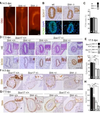
Full activity of eitherSox17orShhis required for proper formation of smooth muscle layers in the developing gallbladder
The liver and gallbladder phenotypes were examined in wild-type,
Shh+/−andShh−/−embryos at 14.5-17.5 dpc. The gross anatomical
and histopathological analyses appeared normal in theShh+/−livers, which were similar to those of wild-type littermates. Shh−/−
embryos, by contrast, were headless and exhibited severe growth retardation in the liver, albeit without any sign of hepatic inflammation (Fig. S8). Whole-mount DBA (Dolichos biflorus
agglutinin) staining for the bile duct revealed a shortened gallbladder and cystic duct, accompanied by a reduction in size of the whole liver inShh−/−embryos (14.5 dpc, Fig. 5A). However, in
contrast to the shortened gallbladder length, transverse sectioning analysis revealed that the Shh−/− gallbladder epithelium was
histologically normal, and SOX17-positive cells were present and
indistinguishable from those in wild type (Fig. 5B). The proportion of PCNA-positive or Ki67-positive cells among total cells showed no appreciable differences in the gallbladder epithelia among wild-type,Shh+/−andShh−/−embryos at 14.5 dpc (Fig. 5B,C; Fig. S9). Cumulatively, these data suggest that there are no appreciable defects in epithelial proliferation ofShh−/−gallbladder primordia, which is in sharp contrast to the hypoplasia of the Sox17+/− gallbladder epithelium at the same stages (Uemura et al., 2013).
Next, we examined the gallbladder phenotypes of Sox17+/−;
Shh+/−double-heterozygous embryos at 14.5-17.5 dpc (Fig. 5D-H).
Histopathological analyses revealed no appreciable changes in
epithelial morphology or anti-SOX9 or anti-SOX17
immunostaining intensity betweenSox17+/−andSox17+/−;Shh+/− gallbladders, except for one severe sample showing luminal cell debris in the presumptive gallbladder region (1/4Sox17+/−;Shh+/− embryos; Fig. 5D, right; Fig. S10). Quantitative data also confirmed that there were no significant alterations in epithelial height between
Shh+/−andShh+/+gallbladders in either theSox17+/−or wild-type
background (Fig. 5E).
In contrast to the lack of appreciable defects in theShhmutant gallbladder epithelium, the formation of smooth muscle layers was considerably affected by the reduced dosage of Sox17 and Shh
(Fig. 5F,G). Anti-αsmooth muscle actin (SMA) staining of wild-type embryos revealed that SMA-positive cells were detectable within the distal mesenchymal region of the gallbladder at ∼11.5 dpc, when they expand in a distal-to-proximal manner, leading to the formation of smooth muscle layers surrounding the gallbladder region, but not the cystic duct region, by 15.5 dpc (Fig. S11). In theSox17+/−,Shh+/−andShh−/−embryos, anti-SMA staining showed reduced signal intensities in the gallbladder mesenchyme at 14.5 dpc (Fig. 5F), in which SMA-positive cells appear to be located randomly and discontinuously in the mesenchymal region around the epithelium (Fig. 5F, insets). At later stages, theShh+/−gallbladders showed proper formation of the
[image:6.612.49.386.60.338.2]SMA-positive smooth muscle layer, similar to that of the wild-type Fig. 4. Expression profiles ofShhand its downstream genes in the developing
gallbladder.(A) Whole-mountin situhybridization (top) and section (bottom; at the level of the gallbladder, indicated by the dotted line) images, showing the expression ofSox17,ShhandShh -related genes in wild-type embryos at 11.5 dpc. Sox17andShhare highly expressed in the gallbladder epithelia, in contrast to the
mesenchymal enrichment forPtch1,Gli1andHhip. (B) Whole-mount GFP fluorescence (left) and anti-GFP-stained sections (three right images) of
Shh+/−(GFP)and wild-type embryos at 16.5 dpc,
showing GFP/Shh-positive epithelial signals in the gallbladder and cystic duct, but not in the hepatic ducts. Ducts are outlined. (C) qPCR analyses showing expression levels ofShhand related genes inSox17+/−embryos. *P<0.05, Student’s
t-test. Sample number is indicated within each bar. cd, cystic duct; duo, duodenum; gb, gallbladder; hd, hepatic duct. Scale bars: 100 µm.
DEVEL
O
gallbladder, in which SMA-positive cells completely surround the gallbladder epithelia (Fig. 5F, left two images). BothSox17+/−and
Sox17+/−;Shh+/−embryos showed severely affected smooth muscle
layers, in which SMA-positive cells appeared reduced and patchy in the surrounding mesenchyme (Fig. 5G, right two images). Morphometric analyses using anti-SMA staining revealed that the signal density was significantly reduced in the gallbladder mesenchyme ofShh+/−,Sox17+/−orSox17+/−;Shh+/−embryos at 14.5 dpc, compared with that of wild-type embryos (Fig. 5H). Moreover, anti-SMA signals were more severely affected in
Sox17+/−;Shh+/− gallbladders than in Shh+/− gallbladders, albeit
with no significant difference. These data suggest that the formation of smooth muscle layers inShh+/−orSox17+/−gallbladders is both
delayed and defective, and this phenotype appears to be more severe inSox17+/−;Shh+/−gallbladders.
Fully activeSox17is required for proper formation of contractile smooth muscle layers of the gallbladder by the perinatal stage
To examine the contractile ability of the smooth muscle layers in the fetal gallbladder, we analyzed the network of smooth muscle cells in wild-type and Sox17+/− embryos at 17.5 dpc by whole-mount anti-SMA staining (Fig. 6). In the wild-type gallbladder, most of the smooth muscle fibers run in a circular direction (Fig. 6A), whereas in Sox17+/− gallbladders the smooth muscle cells appear to be
distributed randomly and irregularly (Fig. 6B), with a swirling
pattern distinct from that seen in the wild-type circular smooth muscle layer. This is consistent with the fragmented pattern of the smooth muscle layer observed in transverse sections (Fig. 5G).
Next we examined KCl-induced contraction (the relative changes in the maximum luminal diameter) of the fetal gallbladder in wild-type and Sox17+/− embryos with or without gross anatomical hepatic lesions (severe or mild phenotype group) at 17.5 dpc (Fig. 6C,D). In wild-type gallbladders, the KCl-induced contraction level was 87.4±1.9% (n=33; Fig. 6D). However, in theSox17+/− gallbladder, KCl treatment caused no appreciable contraction: 94.2±2.9% (n=11) and 99.4±4.4% (n=12) in the mild and severe phenotype groups, respectively (Fig. 6D). In particular, the contraction level of the severe phenotype group was significantly reduced in theSox17+/−gallbladders compared with wild type.
To examine non-induced muscle contraction in the fetal gallbladder at the perinatal stagein vivo, we isolated the liver and gallbladder from wild-type andSox17+/−embryos at 17.5 dpc and measured the rate of
[image:7.612.50.390.53.436.2]autonomous gallbladder contraction under a dissection microscope for 10 min at room temperature. In wild-type gallbladders, the average rate of circular contractile movement was 0.48±0.28 times/min (n=5; Movie 1). This is in contrast to the rare and longitudinal muscle contraction in Sox17+/−gallbladders at 0.09±0.06 times/min (n=5; Movie 2). These data show that the defective formation of the circular smooth muscle layer affects muscle contractility in the Sox17+/− gallbladder. This might contribute to the onset of cholestasis and biliary atresia in theSox17+/−mouse model.
Fig. 5.Sox17andShhcoordinately regulate the proper formation of smooth muscles in the gallbladder.(A) Whole-mount DBA staining of wild-type,Shh+/−andShh−/−liver with gallbladder and cystic duct. (B) Anti-SOX17 and anti-PCNA staining of serial transverse sections of wild-type andShh−/−gallbladder at 14.5 dpc. (C) Quantitative analysis of the PCNA index ( percentage PCNA-positive cells among total cells) in the gallbladder epithelium shows no differences among wild-type,Shh+/−andShh−/− gallbladders at 14.5 dpc. (D) HE and anti-SOX9 staining in transverse sections of fetal gallbladders of wild-type,Shh+/−,Sox17+/−and
Sox17+/−;Shh+/−embryos at 17.5 dpc.
(E) Quantitative data from wild-type andSox17+/− embryos at 17.5 dpc show no appreciable alteration in epithelial height of the gallbladder upon loss of oneShhallele. (F,G) Anti-SMA staining on transverse sections of the gallbladder inSox17+/−,Shh+/−,Shh−/−andSox17+/−;Shh+/− embryos at 14.5 dpc (F) and 17.5 dpc (G). SMA-positive cells were patchy in the gallbladder mesenchyme and severely reduced in theShh−/− gallbladder at 14.5 dpc. At 17.5 dpc, proper formation of the SMA-positive muscle layer was evident inShh+/−embryos, whereas SMA-positive signals remained patchy inSox17+/−,
Shh+/−andSox17+/−;Shh+/−gallbladders. (H) Quantitative data of SMA-positive signals show a gradual reduction in theShh+/−,Sox17+/− andSox17+/−;Shh+/−gallbladders. Insets are higher magnification images of SMA-positive cells in the gallbladder epithelia (B,D) and its surrounding mesenchyme (F,G). *P<0.05, **P<0.01, ANOVA followed by Tukey’s test. Sample size is indicated within each bar. Scale bars: 100 µm.
DEVEL
O
Ectopic SHH signaling rescues the defective formation of smooth muscle layers in theSox17+/−gallbladderin vitro Using anin vitroculture system in which each gallbladder is held within a single cylindrical groove of an agarose gel (Fig. 7A,B), we carried out a rescue experiment using recombinant SHH-soaked beads, which can weakly to moderately induce upregulation and
downregulation of endogenous Gli2 and Shh expression,
respectively, in the 13.5 dpc gallbladder explants (Fig. S12). Wild-type gallbladder explants elongated in a distal-to-proximal manner to form a tubular structure along the cylindrical space within the agarose gel. Furthermore, these gallbladders displayed a well-developed pseudostratified columnar epithelium with epithelial folds (n=14/17; Fig. 7A,C, left), in addition to well-developed SMA-positive smooth muscle layers surrounding a SOX9-negative gallbladder epithelium (n=11/14; Fig. 7D,F, left). By contrast, the control (PBS) explants ofSox17+/−gallbladders displayed a
single-layered cuboidal epithelium without any epithelial fold formation (n=12/12; Fig. 7C). The SMA-positive smooth muscle layer
developed poorly around the SOX9-positive gallbladder
epithelium (n=10/11; Fig. 7D), recapitulating the defective phenotypes of theSox17+/−gallbladder in vivo. Interestingly, the addition of SHH-soaked beads rescued several of the Sox17+/−
gallbladder phenotypes, resulting in a well developed smooth muscle layer organized around a pseudostratified epithelium (n=10/ 13; Fig. 7C,D, right two images). Morphometric analysis confirmed
that the epithelial height and area of the luminal cavity in the
Sox17+/− gallbladder were comparable to those of wild-type explants following SHH treatment (n=11; Fig. 7G). These findings suggest that reduced SHH signaling results in defective smooth muscle formation and epithelial fold formation in the
Sox17+/−gallbladder.
In addition, ectopic SHH signaling had no appreciable effect on the percentage of PCNA-positive or Ki67-positive cells in the
Sox17+/− gallbladder epithelium (Fig. 7E,H; Fig. S13), or on the
number of ectopic SOX9-positive epithelial cells in theSox17+/− gallbladder (epithelial area in Fig 7G, Fig. 7F,I). The Sox17+/−
gallbladder explants showed shredding of some epithelial cells even
in vitro(Fig. S14A; see also Fig. 7C, right, inset). However, the remaining Sox17+/− epithelial layer exhibits proper subcellular localization of both E-cadherin and ZO-1 (TJP1) even in the region near the damaged site (Fig. S14A). Moreover, the expression levels ofCxcl10andOlfm4, although notSerpine1, showed a tendency to increase in theSox17+/−explants as compared with the wild-type explants (Fig. S14B), but such increased expression, at least of
Olfm4, could not be rescued by SHH-soaked beads (Fig. S14B). These data suggest that most defective phenotypes in theSox17+/− gallbladder epithelium are SHH independent. The present results are summarized in Fig. S15.
DISCUSSION
First, we demonstrated the presence of ectopic SOX9-positive cystic duct-like epithelia in the Sox17+/− gallbladder, together with the cooperatively increased expression of Sox4, which functions redundantly withSox9in intrahepatic ducts (Poncy et al., 2015).
Sox9is highly expressed in the cystic duct epithelium, in addition to the intrahepatic, pancreatic and common bile ducts (Furuyama et al., 2011). One possible explanation for the ectopicSox9/Sox4-positive cystic duct epithelia in the proximal gallbladder region is an expansion of the cystic duct region toward the gallbladder domain as a consequence of the reduced proliferation and luminal deciduation of theSox17+/−gallbladder epithelial cells (Uemura et al., 2013). However, the reduced epithelial proliferation is also observed in the cystic duct domain of the Sox17+/− gallbladder primordium, in addition to there being lower proliferative activity in the proximal cystic duct region than in the SOX17-positive distal region (Uemura et al., 2013). Although SOX17 and SOX9/SOX4 have similar sequence specificity in their DNA-binding HMG box domains (Kanai et al., 1996; Mertin et al., 1999), they have distinct characteristics forβ-catenin binding and dimerization (Wilson and Koopman, 2002; Sinner et al., 2007; Kamachi and Kondoh, 2013). Hence, the cystic duct-like phenotype in the proximal Sox17+/− gallbladder might be caused partially by the appearance of ectopic SOX9/SOX4-positive cells in the bile duct epithelia, instead of by the loss of SOX17-positive cells. Further studies are required to more precisely define the hierarchy downstream of SOX17 or SOX9/SOX4 in the specification of the gallbladder and cystic duct epithelia of wild-type andSox17+/−embryos.
[image:8.612.65.280.58.284.2]By the perinatal stage, cystic duct-like Sox17+/− gallbladders showed increased expression of the inflammatory/cholestasis-associated markersAbcb4andOlfm4, in addition toCxcl10, a key chemokine gene characteristic of the early immune response (Fig. 2C,D). The expression levels of several fetal myeloid cell markers also showed a tendency to increase in the Sox17+/− gallbladders, together with the enrichment of Gr1-positive or F4/80-positive myeloid-like cells in the gallbladder mesenchymal region (Fig. S6), suggesting fetal cholecystitis inSox17+/−embryos.Abcb4 encodes a lipid translocator for phosphatidylcholine, which Fig. 6. Defective contraction of smooth muscles inSox17+/−gallbladder.
(A,B) Whole-mount anti-SMA staining of wild-type andSox17+/−gallbladders at 17.5 dpc. (Top) Confocalz-projection from the area of the gallbladder indicated by the dashed box (left). (Bottom) Orthogonal projection in thexz plane made at the level of the arrowhead (above). The orientation of the discontinuous SMA-positive cells was randomized in theSox17+/−gallbladder, in contrast to the dense SMA-positive fibers arranged mainly in a circular direction in the wild-type gallbladder. D, distal; P, proximal. (C,D) Dissection microscope images of the fetal gallbladder before (top) and after (bottom) KCl treatment (C) and KCl-induced contraction levels (relative changes in luminal diameter; D) of wild-type and theSox17+/−gallbladder with (severe) or without (mild) gross anatomical hepatic lesions at 17.5 dpc. KCl treatment reduced luminal diameter considerably in the wild-type but not in theSox17+/− gallbladder, with the severe phenotype group being particularly unaffected. Number of samples (gallbladders) are indicated. **P<0.01, ANOVA followed by Tukey’s test. Scale bar: 100 µm.
DEVEL
O
transfers bile into bile salt micelles for the protection of the apical surface of the bile duct epithelia (Fickert et al., 2004; Esten Nakken et al., 2007; Baghdasaryan et al., 2011; Oude Elferink and Paulusma, 2007). Abcb4 null mice display severe sclerosing or chronic cholangitis (Fickert et al., 2004; Esten Nakken et al., 2007; Baghdasaryan et al., 2011). Since a significant elevation ofABCB4
was also reported in human patients with extrahepatic cholestasis (Schaap et al., 2009),Abcb4upregulation inSox17+/−gallbladders
in vivo might be caused partially by its adaptive response to minimize cellular damage by decreasing bile salt toxicity. Olfm4
encodes a secreted protein with one olfactomedin-like domain (also known as an intestinal stem cell marker; Gersemann et al., 2012) and is upregulated in inflammatory bowel diseases such as ulcerative colitis and Crohn’s disease (Liu et al., 2010; Gersemann et al., 2012). It is also possible that the in vivo
upregulation of these bile duct disease markers is an effort to compensate for the defective barrier of the bile duct epithelia in the
Sox17+/−gallbladder.
Genetic analysis demonstrated that embryonic hepatitis could be induced by a conditional Sox17 deletion in the primordial
gallbladder, but not in the liver hepatoblasts (Fig. 3; Fig. S7). The expression analysis of inflammatory markers also showed a positive correlation between cholecystitis and the severity of embryonic hepatitis inSox17+/−embryos (Fig. 2D,E). These data suggest that the cholecystitis causes the biliary atresia and subsequent hepatic inflammation inSox17+/−embryos. It is also possible that, at the late organogenesis stages of the extrahepatic bile ducts, the inflammatory responses of the bile duct epithelia and its surrounding mesenchymal tissues cause severe defects in both bile duct morphogenesis and epithelial barrier function, leading to increased bile leakage and inflammatory responses in the fetal gallbladder by 17.5 dpc.
[image:9.612.50.375.57.502.2]The present genetic andin vitrorescue experiments revealed that SHH signaling may be crucial for the proper formation of smooth muscles downstream ofSox17in the normal development of the gallbladder during the late organogenesis stages. HH signaling in the endodermal epithelium plays a major role in the mesenchymal development of the digestive tract. Loss of HH signaling has also been shown to decrease cell proliferation in the underlying mesenchyme, resulting in thinner walls of the stomach and Fig. 7. Defective smooth muscle development of Sox17+/−gallbladders is rescued by exogenous
SHH.(A,B) Three day organ culture of wild-type or Sox17+/−gallbladder primordium initiated at 13.5 dpc (A). The gallbladder and bead are held inside a single cylindrical groove of agarose gel as schematically represented in B. D, distal; P, proximal. (C-F) HE, anti-SMA, anti-PCNA or anti-SOX9 staining on transverse sections of wild-type orSox17+/− gallbladder explants with PBS-soaked or SHH-soaked beads. Insets are higher magnification images of the epithelium or the signals. (G-I) Quantitative morphometric analysis of wild-type andSox17+/− gallbladder explants cultured with PBS-soaked or SHH-soaked beads. Exogenous SHH signaling rescued the defects in epithelial height and cavity area, but did not exert any significant influence on epithelial area (G), PCNA index (H) or relative SOX9-positive cell numbers (I) within the gallbladder epithelium. *P<0.05, **P<0.01, ANOVA followed by Tukey's test. Sample number is indicated with each bar. Scale bars: 100 µm.
DEVEL
O
intestine (Mao et al., 2010; reviewed by van den Brink, 2007). Moreover, SHH signaling enhances smooth muscle formation in the intestine, lung, and urinary bladder via other molecules, including Ptch and Gli family members (Apelqvist et al., 1997; Li et al., 2004; Caubit et al., 2008; DeSouza et al., 2013), suggesting that smooth muscle cells might be a target of SHH signaling. These previous reports help to explain the delayed and aberrant formation of smooth muscle layers in the developingSox17+/−gallbladder, and they are also consistent with several previous reports showing deficient smooth muscle layers in the fetal gallbladder caused by Foxf1
haploinsufficiency, a downstream mediator of HH signaling, in mouse organogenesis stage embryos (Kalinichenko et al., 2002; Madison et al., 2009).
It was recently suggested that the deformation, inflammation and repair processes of the bile ducts are closely associated with SHH signaling. For example, Jung et al. (2015) reported that SHH pathway activation was observed, especially in the cholangiocytes of the peribiliary glands, in human patients with biliary atresia. In pathological studies, some have reported that Hh expression is considerably increased in fibrotic damaged biliary diseases in response to injury (Omenetti et al., 2008; Omenetti and Diehl, 2011; Cui et al., 2013; Hu et al., 2015). It has also been speculated that the SHH signaling pathway regulates the epithelial-mesenchymal transition of cholangiocytes (Omenetti et al., 2008; Jung et al., 2015). The effects of excess SHH signaling encompass defective hepatobiliary ducts in a zebrafish model (Cui et al., 2013; Tang et al., 2016), suggesting that excess SHH signaling can have detrimental effects on the proper development and maintenance of the biliary duct. The current phenotype of reduced SHH signaling in the defective gallbladder might be explained by the distinct roles of SHH signaling during developmental and early pathogenic stages (i.e. smooth muscle layer formation and tubulogenesis) versus late pathogenic and inflammatory processes (i.e. transdifferentiation of damaged mesenchymal cells into smooth muscle cells for tissue repair). It is also possible that SHH signaling must be maintained at an appropriate level, since both excess and reduced SHH signaling may possibly lead to defective formation and maintenance of the biliary duct system.
Finally, the present study has shown defective gallbladder contraction inSox17+/−perinatal embryos (Fig. 6; Movies 1 and 2). In particular,Sox17+/−gallbladders with hepatic lesions have severe
defects in contraction, suggesting a potential contribution of the defective muscle layers to the onset of cholestasis in theSox17+/−
embryos soon after the first biliary excretion into the fetal duodenum. Since well-developed smooth muscle layers are restricted to the gallbladder wall, in contrast to poor formation of the smooth muscle layers around the cystic and common bile ducts (Higashiyama et al., 2016 and references therein), the defective gallbladder contraction directly affects the flow of bile from the fetal gallbladder into the duodenum in theSox17+/−embryos. This symptom is also consistent with the ultrasonographic diagnostic features for human biliary atresia, such as the ʻnon-contractile’ gallbladder with its reduced length, irregular wall and indistinct mucosal lining (Kanegawa et al., 2003). Further studies are required to establish the contribution of fetal gallbladder contraction in the onset of biliary atresia-like syndrome in individualSox17+/−embryos.
MATERIALS AND METHODS
Animal care and use
Animal experiments were performed in strict accordance with the Guidelines for Animal Use and Experimentation established by the University of Tokyo (approval ID: P13-763, P14-877), the Tokyo
Medical and Dental University (approval ID: 0140007A, 0150259C2, 0160024C2, 0170248C2) and the University of Utah (approval ID: 14-01003). TheSox17+/−embryos at F9-10 generation were obtained from
wild-type females [C57BL6 (B6) strain; Clea, Japan] mated with the
Sox17+/−male mice (Kanai-Azuma et al., 2002), which were intercrossed
and maintained at F8-9 backcross generation to the B6 strain. In this mating system, theSox17+/−embryos (F9-10) show∼70% neonatal lethality, while
the remaining survivors can be grown until adulthood without any signs of hepatitis, albeit with a small gallbladder. Sox17+/−(GFP), Sox17 flox/flox
(B6×129sv background; 13.5-17.5 dpc) (Kim et al., 2007),Alb-cre (Postic et al., 1999),Pdx1-cre (Hingorani et al., 2003) andShh+/−(GFP)(Harfe et al.,
2004) mice were also used in this study.
Histology, immunohistochemistry and whole-mountin situ hybridization
Tissues were fixed in 4% paraformaldehyde in PBS for 12 h at 4°C. For sections, the fixed samples were embedded in paraffin and serially sectioned (4 µm thick). For comparative analyses of transverse sections of the gallbladder, sections at the level of maximum diameter (at least four sections in each individual sample) were used. Immunohistochemistry was conducted by a standard protocol (see the supplementary Materials and Methods) and in situ hybridization was performed as described by Hiramatsu et al. (2009). Details of reagents and methods are provided in the supplementary Materials and Methods.
Measurement of length, area and signal density
For length and area measurements, ImageJ 1.48V software (National Institutes of Health, USA) was used. For measurement of signal density of anti-SMA-stained sections, we conducted grid analyses of the HRP-positive reactions. A grid (each box=10×10 µm) was overlaid on the image of the stained section and the signal density (d) calculated by d=b/(a+b), where (a) the signal passed through more than two sides of the box and (b) the signal was fragmented or passed through one side of the box.
RNA extraction, microarray and qPCR analyses
The Sox17+/− and wild-type gallbladders at 15.5 dpc were used for
microarray expression analysis according to the method developed by Huang et al. (2009a,b). GSEA was carried out according to Subramanian et al. (2005). qPCR analysis was used to determine marker gene expression levels relative toGapdh. Details of these methods are provided in the supplementary Materials and Methods.
Monitoring the contractile movement of fetal gallbladder Whole livers, including the gallbladder and biliary tract (17.5 dpc), were maintained in 10% fetal calf serum-DMEM (Sigma) at 37°C, and prepared for time-lapse imaging using a dissection microscope (Olympus SZX16) equipped with a video recording system (Olympus DP71 camera; imaged every 2 s for a total of 10 min). Each gallbladder was separated from the liver and imaged before and after 45 mM KCl treatment for 10 min. Maximum luminal diameter of the gallbladder before and after KCl was measured using ImageJ 1.48V, and the relative change in maximum luminal diameter estimated as an indication of gallbladder contraction.
Organ culture
The gallbladder was isolated from the fetal liver using a dissection microscope at 13.5 dpc. To apply the pressure that would normally be exerted by the liver and to promote directional growth, the gallbladders were placed in a groove of a 1.5% agarose gel plate prepared using a stainless steel needle of 0.26 mm diameter, which is similar to the liver gap around the gallbladder at 16.5 dpc (see Fig. 7B). Beads soaked in mouse recombinant SHH (1 mg/ml; SRP6004, Sigma) or PBS were placed on the distal tip of the gallbladder and cultured in 10% fetal calf serum-DMEM at 37°C for 72 h.
Statistical analyses
All quantitative data are represented as mean±s.e.m. Student’st-test, Mann– Whitney U-test and ANOVA tests were used to determine overall differences between two groups or among more than two groups. Where differences existed, Tukey’s test was also used to compare each value with
DEVEL
O
every other value. The correlation between genes/groups in both gallbladder and liver samples was estimated using Spearman’s rank correlation test.
P<0.05 was considered statistically significant.
Acknowledgements
We thank Drs A. Asai, S. Elliott, K. Nakamura and A. Tanoue for critical reading of the manuscript; Prof. Dr S. J. Morrison for providingSox17+/−(GFP)mice; and
Dr M. Kawasumi, Y. Kuroda, Ms Y. Uchiyama and I. Yagihashi for helpful support.
Competing interests
The authors declare no competing or financial interests.
Author contributions
Conceptualization: H.H., N.T., Y.K.; Methodology: H.H., A.O., H.S., M.U., K.I., Y.S.; Software: A.O., H.S.; Validation: H.H., A.O., H.S., M.U.; Formal analysis: H.H., A.O., H.S., M.U., H.I.; Investigation: H.H., A.O., H.S., M.U., N.T.; Resources: H.H., A.O., M.U., K.F., H.I., Y.H., M.K., Y.S., M.K.-A.; Data curation: H.H., A.O., H.S., M.U., K.F., Y.H., Y.S.; Writing - original draft: H.H.; Writing - review & editing: H.H., M.K., M.K.-A., Y.K.; Visualization: H.H., K.I.; Supervision: M.K., Y.K.; Project administration: Y.K.; Funding acquisition: Y.K.
Funding
This work was supported mainly by grants from the Ministry of Education, Culture, Sports, Science, and Technology of Japan to Y.K. (S-24228005) and M.K.-A. (C-24500485). This work was also supported by a grant from the National Institute of Child Health and Human Development to Y.S. (R01 HD066121). Deposited in PMC for release after 12 months.
Data availability
Microarray data are available at NCBI Gene Expression Omnibus under accession number GSE74576.
Supplementary information
Supplementary information available online at
http://dev.biologists.org/lookup/doi/10.1242/dev.147512.supplemental
References
Apelqvist, Å., Ahlgren, U. and Edlund, H. (1997). Sonic hedgehog directs
specialised mesoderm differentiation in the intestine and pancreas.Curr. Biol.7, 801-804.
Asai, A., Miethke, A. and Bezerra, J. A.(2015). Pathogenesis of biliary atresia:
defining biology to understand clinical phenotypes. Nat. Rev. Gastroenterol. Hepatol.12, 342-352.
Baghdasaryan, A., Claudel, T., Gumhold, J., Silbert, D., Adorini, L., Roda, A.,
Vecchiotti, S., Gonzalez, F. J., Schoonjans, K. and Strazzabosco, M.(2011).
Dual farnesoid X receptor/TGR5 agonist INT-767 reduces liver injury in the
Mdr2−/−(Abcb4−/−) mouse cholangiopathy model by promoting biliary HCO
3 −
output.Hepatology54, 1303-1312.
Bessho, K., Mourya, R., Shivakumar, P., Walters, S., Magee, J. C., Rao, M.,
Jegga, A. G. and Bezerra, J. A.(2014). Gene expression signature for biliary
atresia and a role for interleukin-8 in pathogenesis of experimental disease.
Hepatology60, 211-223.
Caubit, X., Lye, C. M., Martin, E., Core, N., Long, D. A., Vola, C., Jenkins, D.,
Garratt, A. N., Skaer, H., Woolf, A. S. et al.(2008). Teashirt 3 is necessary for
ureteral smooth muscle differentiation downstream of SHH and BMP4.
Development135, 3301-3310.
Cui, S., Leyva-Vega, M., Tsai, E. A., EauClaire, S. F., Glessner, J. T., Hakonarson,
H., Devoto, M., Haber, B. A., Spinner, N. B. and Matthews, R. P.(2013).
Evidence from human and zebrafish thatGPC1is a biliary atresia susceptibility gene.Gastroenterology144, 1107-1115.
Davenport, M.(2016). Biliary atresia: from Australia to the zebrafish.J. Pediatr.
Surg.51, 200-205.
Desmet, V. J.(1992). Congenital diseases of intrahepatic bile ducts: variations on
the theme“ductal plate malformation”.Hepatology16, 1069-1083.
DeSouza, K. R., Saha, M., Carpenter, A. R., Scott, M. and McHugh, K. M.(2013).
Analysis of the Sonic Hedgehog signaling pathway in normal and abnormal bladder development.PLoS ONE8, e53675.
Echelard, Y., Epstein, D. J., St-Jacques, B., Shen, L., Mohler, J., McMahon, J. A.
and McMahon, A. P.(1993). Sonic hedgehog, a member of a family of putative
signaling molecules, is implicated in the regulation of CNS polarity. Cell75, 1417-1430.
Emig, D., Salomonis, N., Baumbach, J., Lengauer, T., Conklin, B. R. and
Albrecht, M.(2010). AltAnalyze and DomainGraph: analyzing and visualizing
exon expression data.Nucleic Acids Res.38Suppl., W755-W762.
Esten Nakken, K., Nygård, S., Haaland, T., Erik Berge, K., Arnkvaern, K.,
Ødegaard, A., Jørgen Labori, K. and Ræder, M. G. (2007). Multiple
inflammatory-, tissue remodelling-and fibrosis genes are differentially transcribed in the livers ofAbcb4(-/-) mice harbouring chronic cholangitis.
Scandinav. J. Gastroenterol.42, 1245-1255.
Fickert, P., Fuchsbichler, A., Wagner, M., Zollner, G., Kaser, A., Tilg, H., Krause,
R., Lammert, F., Langner, C., Zatloukal, K. et al.(2004). Regurgitation of bile
acids from leaky bile ducts causes sclerosing cholangitis in Mdr2(Abcb4) knockout mice.Gastroenterology127, 261-274.
Furuyama, K., Kawaguchi, Y., Akiyama, H., Horiguchi, M., Kodama, S., Kuhara,
T., Hosokawa, S., Elbahrawy, A., Soeda, T., Koizumi, M. et al. (2011).
Continuous cell supply from aSox9-expressing progenitor zone in adult liver, exocrine pancreas and intestine.Nat. Genet.43, 34-41.
Gersemann, M., Becker, S., Nuding, S., Antoni, L., Ott, G., Fritz, P., Oue, N.,
Yasui, W., Wehkamp, J. and Stange, E. F. (2012). Olfactomedin-4 is a
glycoprotein secreted into mucus in active IBD.J. Crohns Colitis6, 425-434.
Gordo-Gilart, R., Andueza, S., Hierro, L., Martinez-Fernández, P., D’Agostino,
D., Jara, P. and Alvarez, L.(2015). Functional analysis of ABCB4 mutations
relates clinical outcomes of progressive familial intrahepatic cholestasis type 3 to the degree of MDR3 floppase activity.Gut64, 147-155.
Harfe, B. D., Scherz, P. J., Nissim, S., Tian, H., McMahon, A. P. and Tabin, C. J.
(2004). Evidence for an expansion-based temporal Shh gradient in specifying vertebrate digit identities.Cell118, 517-528.
Hartley, J. L., O’Callaghan, C., Rossetti, S., Consugar, M., Ward, C. J., Kelly,
D. A. and Harris, P. C.(2011). Investigation of primary cilia in the pathogenesis of
biliary atresia.J. Pediatr. Gastroenterol. Nutr.52, 485-488.
Higashiyama, H., Sumitomo, H., Ozawa, A., Igarashi, H., Tsunekawa, N.,
Kurohmaru, M. and Kanai, Y.(2016). Anatomy of the murine hepatobiliary
system: a whole-organ-level analysis using a transparency method.Anat. Rec.
299, 161-172.
Hingorani, S. R., Petricoin, E. F., Maitra, A., Rajapakse, V., King, C., Jacobetz,
M. A., Ross, S., Conrads, T. P., Veenstra, T. D., Hitt, B. A. et al.(2003).
Preinvasive and invasive ductal pancreatic cancer and its early detection in the mouse.Cancer Cell4, 437-450.
Hiramatsu, R., Matoba, S., Kanai-Azuma, M., Tsunekawa, N., Katoh-Fukui, Y., Kurohmaru, M., Morohashi, K.-i, Wilhelm, D., Koopman, P. and Kanai, Y.
(2009). A critical time window of Sry action in gonadal sex determination in mice.
Development136, 129-138.
Hu, L., Lin, X., Lu, H., Chen, B. and Bai, Y.(2015). An overview of hedgehog
signaling in fibrosis.Mol. Pharmacol.87, 174-182.
Huang, D. W., Sherman, B. T. and Lempicki, R. A.(2009a). Systematic and
integrative analysis of large gene lists using DAVID bioinformatics resources.Nat. Protoc.4, 44-57.
Huang, D. W., Sherman, B. T. and Lempicki, R. A. (2009b). Bioinformatics
enrichment tools: paths toward the comprehensive functional analysis of large gene lists.Nucleic Acids Res.37, 1-13.
Jung, H. Y., Jing, J., Lee, K. B. and Jang, J. J.(2015). Sonic hedgehog (SHH) and
glioblastoma-2 (Gli-2) expressions are associated with poor jaundice-free survival in biliary atresia.J. Pediatr. Surg.50, 371-376.
Kalinichenko, V. V., Zhou, Y., Bhattacharyya, D., Kim, W., Shin, B., Bambal, K.
and Costa, R. H.(2002). Haploinsufficiency of the mouseForkhead Box f1gene
causes defects in gall bladder development.J. Biol. Chem.277, 12369-12374.
Kamachi, Y. and Kondoh, H. (2013). Sox proteins: regulators of cell fate
specification and differentiation.Development140, 4129-4144.
Kanai, Y., Kanai-Azuma, M., Noce, T., Saido, T. C., Shiroishi, T., Hayashi, Y. and
Yazaki, K.(1996). Identification of twoSox17messenger RNA isoforms, with and
without the high mobility group box region, and their differential expression in mouse spermatogenesis.J. Cell Biol.133, 667-681.
Kanai-Azuma, M., Kanai, Y., Gad, J. M., Tajima, Y., Taya, C., Kurohmaru, M.,
Sanai, Y., Yonekawa, H., Yazaki, K., Tam, P. P. et al.(2002). Depletion of
definitive gut endoderm in Sox17-null mutant mice. Development 129, 2367-2379.
Kanegawa, K., Akasaka, Y., Kitamura, E., Nishiyama, S., Muraji, T., Nishijima,
E., Satoh, S. and Tsugawa, C.(2003). Sonographic diagnosis of biliary atresia in
pediatric patients using the“triangular cord”sign versus gallbladder length and contraction.Am. J. Roentgenology181, 1387-1390.
Kelliher, M. A., Seldin, D. C. and Leder, P.(1996). Tal-1 induces T cell acute
lymphoblastic leukemia accelerated by casein kinase IIalpha.EMBO J. 15, 5160-5166.
Kidokoro, T., Matoba, S., Hiramatsu, R., Fujisawa, M., Kanai-Azuma, M., Taya,
C., Kurohmaru, M., Kawakami, H., Hayashi, Y., Kanai, Y. et al.(2005). Influence
on spatiotemporal patterns of a male-specificSox9activation by ectopicSry
expression during early phases of testis differentiation in mice.Dev. Biol.278, 511-525.
Kim, I., Saunders, T. L. and Morrison, S. J. (2007). Sox17 dependence
distinguishes the transcriptional regulation of fetal from adult hematopoietic stem cells.Cell130, 470-483.
Kohsaka, T., Yuan, Z. R., Guo, S. X., Tagawa, M., Nakamura, A., Nakano, M.,
Kawasasaki, H., Inomata, Y., Tanaka, K. and Miyauchi, J. (2002). The
significance of human jagged 1 mutations detected in severe cases of extrahepatic biliary atresia.Hepatology36, 904-912.
DEVEL
O
Leonhardt, J., Stanulla, M., von Wasielewski, R., Skokowa, J., Kübler, J., Ure,
B. M. and Petersen, C.(2006). Gene expression profile of the infective murine
model for biliary atresia.Pediatr. Surg. Int.22, 84-89.
Li, Y., Zhang, H., Choi, S. C., Litingtung, Y. and Chiang, C. (2004). Sonic
hedgehog signaling regulates Gli3 processing, mesenchymal proliferation, and differentiation during mouse lung organogenesis.Dev. Biol.270, 214-231.
Liu, C. Z., Yang, J. T., Yoon, J. W., Villavicencio, E., Pfendler, K., Walterhouse,
D. and Iannaccone, P. (1998). Characterization of the promoter region and
genomic organization ofGLI, a member of theSonic hedgehog-Patchedsignaling pathway.Gene209, 1-11.
Liu, W., Yan, M., Liu, Y., Wang, R., Li, C., Deng, C., Singh, A., Coleman, W. G., Jr
and Rodgers, G. P. (2010). Olfactomedin 4 down-regulates innate immunity
against Helicobacter pylori infection. Proc. Natl. Acad. Sci. USA 107, 11056-11061.
Lorent, K., Gong, W., Koo, K. A., Waisbourd-Zinman, O., Karjoo, S., Zhao, X., Sealy, I., Kettleborough, R. N., Stemple, D. L., Windsor, P. A., Whittaker, S. J.,
Porter, J. R., Wells, R. G., and Pack, M. et al.(2015). Identification of a plant
isoflavonoid that causes biliary atresia.Sci. Transl. Med.7, 286ra67.
Mack, C. L. and Sokol, R. J.(2005). Unraveling the pathogenesis and etiology of
biliary atresia.Pediatr. Res.57, 87R-94R.
Madison, B. B., McKenna, L. B., Dolson, D., Epstein, D. J. and Kaestner, K. H.
(2009). FoxF1 and FoxL1 link hedgehog signaling and the control of epithelial proliferation in the developing stomach and intestine. J. Biol. Chem. 284, 5936-5944.
Mao, J., Kim, B. M., Rajurkar, M., Shivdasani, R. A. and McMahon, A. P.(2010).
Hedgehog signaling controls mesenchymal growth in the developing mammalian digestive tract.Development137, 1721-1729.
McConnell, B. B. and Yang, V. W.(2010). Mammalian Krüppel-like factors in health
and diseases.Physiol. Rev.90, 1337-1381.
Mertin, S., McDowall, S. G. and Harley, V. R.(1999). The DNA-binding specificity
of SOX9 and other SOX proteins.Nucleic Acids Res.27, 1359-1364.
Mieli-Vergani, G. and Vergani, D.(2009). Biliary atresia.Semin. Immunopathol.31,
371-381.
Motoyama, J., Heng, H., Crackower, M. A., Takabatake, T., Takeshima, K., Tsui,
L.-C. and Hui, C.-c.(1998). Overlapping and non-overlapping Ptch2 expression
with Shh during mouse embryogenesis.Mech. Dev.78, 81-84.
Nakagawa, M. and Setchell, K. D.(1990). Bile acid metabolism in early life: studies
of amniotic fluid.J. Lipid Res.31, 1089-1098.
Nakamura, K. and Tanoue, A.(2013). Etiology of biliary atresia as a developmental
anomaly: recent advances.J. Hepatobiliary Pancreat. Sci.20, 459-464.
Nakamura, M., Yasunami, M., Kondo, H., Horie, H., Aiba, Y., Komori, A., Migita,
K., Yatsuhashi, H., Ito, M., Shimoda, S. et al.(2010). Analysis ofHLA-DRB1
polymorphisms in Japanese patients with primary biliary cirrhosis (PBC): the HLA-DRB1 polymorphism determines the relative risk of antinuclear antibodies for disease progression in PBC.Hepatol. Res.40, 494-504.
Okada, K., Kamiya, A., Ito, K., Yanagida, A., Ito, H., Kondou, H., Nishina, H. and
Nakauchi, H. (2012). Prospective isolation and characterization of bipotent
progenitor cells in early mouse liver development.Stem Cells Dev.21, 1124-1133.
Omenetti, A. and Diehl, A. M.(2011). Hedgehog signaling in cholangiocytes.Curr.
Opin. Gastroenterol.27, 268-275.
Omenetti, A., Popov, Y., Jung, Y., Choi, S. S., Witek, R. P., Yang, L., Brown, K. D.,
Schuppan, D. and Diehl, A. M. (2008). The hedgehog pathway regulates
remodelling responses to biliary obstruction in rats.Gut57, 1275-1282.
Oude Elferink, R. P. and Paulusma, C. C.(2007). Function and pathophysiological
importance of ABCB4 (MDR3 P-glycoprotein).Pflugers Arch.453, 601-610.
Park, M. H., Jo, M., Kim, Y. R., Lee, C.-K. and Hong, J. T.(2016). Roles of
peroxiredoxins in cancer, neurodegenerative diseases and inflammatory diseases.Pharmacol. Therap.163, 1-23.
Poncy, A., Antoniou, A., Cordi, S., Pierreux, C. E., Jacquemin, P. and Lemaigre,
F. P. (2015). Transcription factors SOX4 and SOX9 cooperatively control
development of bile ducts.Dev. Biol.404, 136-148.
Portincasa, P., Di Ciaula, A. and van Berge-Henegouwen, G. P.(2004). Smooth
muscle function and dysfunction in gallbladder disease.Curr. Gastroenterol. Rep.
6, 151-162.
Portincasa, P., Di Ciaula, A., Wang, H. H., Palasciano, G., van Erpecum, K. J.,
Moschetta, A. and Wang, D.-Q.(2008). Coordinate regulation of gallbladder
motor function in the gut-liver axis.Hepatology47, 2112-2126.
Postic, C., Shiota, M., Niswender, K. D., Jetton, T. L., Chen, Y., Moates, J. M.,
Shelton, K. D., Lindner, J., Cherrington, A. D. and Magnuson, M. A.(1999).
Dual roles for glucokinase in glucose homeostasis as determined by liver and pancreatic beta cell-specific gene knock-outs using Cre recombinase.J. Biol.
Chem.274, 305-315.
Schaap, F. G., van der Gaag, N. A., Gouma, D. J. and Jansen, P. L. M.(2009).
High expression of the bile salt-homeostatic hormone fibroblast growth factor 19 in the liver of patients with extrahepatic cholestasis.Hepatology49, 1228-1235.
Sinner, D., Kordich, J. J., Spence, J. R., Opoka, R., Rankin, S., Lin, S.-C. J.,
Jonatan, D., Zorn, A. M. and Wells, J. M.(2007). Sox17 and Sox4 differentially
regulateβ-catenin/T-cell factor activity and proliferation of colon carcinoma cells.
Mol. Cell. Biol.27, 7802-7815.
Spence, J. R., Lange, A. W., Lin, S.-C. J., Kaestner, K. H., Lowy, A. M., Kim, I.,
Whitsett, J. A. and Wells, J. M. (2009). Sox17 regulates organ lineage
segregation of ventral foregut progenitor cells.Dev. Cell17, 62-74.
Subramanian, A., Tamayo, P., Mootha, V. K., Mukherjee, S., Ebert, B. L., Gillette,
M. A., Paulovich, A., Pomeroy, S. L., Golub, T. R., Lander, E. S. et al.(2005).
Gene set enrichment analysis: a knowledge-based approach for interpreting genome-wide expression profiles.Proc. Nat. Acad. Sci. USA102, 15545-15550.
Tam, P. P., Kanai-Azuma, M. and Kanai, Y.(2003). Early endoderm development
in vertebrates: lineage differentiation and morphogenetic function.Curr. Opin.
Genet. Dev.13, 393-400.
Tan, C. E. L., Davenport, M., Driver, M. and Howard, E. R.(1994). Does the
morphology of the extrahepatic biliary remnants in biliary atresia influence survival? A review of 205 cases.J. Pediatr. Surg.29, 1459-1464.
Tang, V., Cofer, Z. C., Cui, S., Sapp, V., Loomes, K. M. and Matthews, R. P.
(2016). Loss of a candidate biliary atresia susceptibility gene,add3a, causes biliary developmental defects in zebrafish.J. Pediatr. Gastroenterol. Nutr.63, 524-530.
Uemura, M., Hara, K., Shitara, H., Ishii, R., Tsunekawa, N., Miura, Y.,
Kurohmaru, M., Taya, C., Yonekawa, H., Kanai-Azuma, M. et al.(2010).
Expression and function of mouseSox17gene in the specification of gallbladder/ bile-duct progenitors during early foregut morphogenesis.Biochem. Biophys.
Res. Commun.391, 357-363.
Uemura, M., Ozawa, A., Nagata, T., Kurasawa, K., Tsunekawa, N., Nobuhisa, I.,
Taga, T., Hara, K., Kudo, A., Kawakami, H. et al. (2013). Sox17
haploinsufficiency results in perinatal biliary atresia and hepatitis in C57BL/6 background mice.Development140, 639-648.
van den Brink, G. R.(2007). Hedgehog signaling in development and homeostasis
of the gastrointestinal tract.Physiol. Rev.87, 1343-1375.
Waisbourd-Zinman, O., Koh, H., Tsai, S., Lavrut, P.-M., Dang, C., Zhao, X., Pack,
M., Cave, J., Hawes, M., Koo, K. A. et al.(2016). The toxin biliatresone causes
mouse extrahepatic cholangiocyte damage and fibrosis through decreased glutathione and SOX17.Hepatology64, 880-893.
Wilson, M. and Koopman, P.(2002). Matching SOX: partner proteins and
co-factors of the SOX family of transcriptional regulators.Curr. Opin. Genet. Dev.12, 441-446.