Recent data have reinforced the fundamental role of regulated cell adhesion as a force that drives morphogenesis during gastrulation. As we discuss, cell adhesion is required for all modes of gastrulation movements in all organisms. It can even be instructive in nature, but it must be tightly and dynamically regulated. The picture that emerges from the recent findings that we review here is that different modes of gastrulation movements use the same principles of adhesion regulation, while adhesion molecules themselves coordinate the intra- and extracellular changes required for directed cell locomotion.
Introduction
Gastrulation is the highly coordinated mass cell movement that forms diploblastic or triplastic embryos, by which endodermal and mesodermal cells are brought into the interior of the embryo, while the ectoderm remains at the surface. One of the most puzzling findings has been that, even among vertebrates, which form rather similar body plans, very different morphogenetic cell behaviours seem to be at play during gastrulation (Keller et al., 2003; Solnica-Krezel, 2005; Wallingford et al., 2002; Wallingford and Harland, 2007). Different types of internalization movement have been distinguished: invagination, involution, ingression and epiboly. In parallel, during convergence and extension (CE), cells change their relative positions within the germ layers to form and shape the body axis. Here, we argue that, with respect to their dependence on regulated cell adhesion, all types of gastrulation movement share certain crucial features, and are much more similar than was initially acknowledged.
Cell adhesion describes the energy that is released upon binding, or, in reverse, the force that is required to separate a unit of a cell’s surface from the substrate it adheres to (see Box 1 for experimental approaches to measure this force). In vivo, substrates can be either other cells or extracellular matrices (ECM). The first evidence for the importance of cell adhesion for germ layer assembly came from experiments by Johannes Holtfreter over 50 years ago. By performing in vitro dissociation and re-association assays, he and his graduate student Philip Townes showed that randomly mixed amphibian embryonic cells sort out to reconstitute the different germ layers, which often arranged in their proper anatomical relationships (Townes and Holtfreter, 1955). Steinberg hypothesized that this phenomenon, termed ‘selective affinity’ by Holtfreter, is caused by differential cell-cell adhesion (Steinberg, 1996; Steinberg, 2007). This assumption has been confirmed experimentally in cell culture and in the developing Drosophila retina by modulating the expression levels of cadherins, key regulators of cell-cell adhesion (Foty and Steinberg, 2005; Hayashi and Carthew, 2004). Cadherins are also crucially involved in regulating gastrulation in all organisms,
from flies to mice (Babb and Marrs, 2004; Ciruna and Rossant, 2001; Kühl et al., 1996; Lee and Gumbiner, 1995; Wang et al., 2004). However, as already postulated in 1976 by Albert Harris (Harris, 1976) and theoretically demonstrated by G. Wayne Brodland (Brodland, 2003), recent experimental evidence indicates that, in addition to cell adhesion, cell affinity is influenced by intracellular mechanics and cell surface tension (Krieg et al., 2008; Ninomiya and Winklbauer, 2008). Surface tension, in turn, is achieved by cortical contractions of the actomyosin system, which – possibly via a dynamic equilibrium with cadherin-catenin complexes – is physically linked to the cell membrane (Lecuit and Lenne, 2007). Cohesion – cadherin-based, homotypic adhesion among similar cells – also impinges on cell sorting by affecting group migration [see, for example Kasemeier-Kulesa et al., for the role of N-cadherin in ganglia-forming neural crest cells (Kasemeier-Kulesa et al., 2006)]. In conclusion, the extent to which differential adhesion contributes to cell sorting remains unclear, as does the extent to which cadherins contribute to differential adhesion as opposed to surface tension or cell migration. Integrins, key regulators of cell migration, have also been shown in cell culture systems to have multiple roles in cell-ECM adhesion, and in intracellular signalling and cytoskeletal regulation (Hehlgans et al., 2007), but their exact mode of action during gastrulation is less clear. The cytoskeleton is also under the control of two cell polarity systems, planar cell polarity (PCP) and apicobasal (epithelial) cell polarity, both of which are crucial for the regulation of epithelial and mesenchymal morphogenesis during gastrulation (Dow and Humbert, 2007; Seifert and Mlodzik, 2007). These polarity systems regulate cytoskeletal dynamics via the action of small GTPases like RhoA, Rac1 and Cdc42, in order to determine cell shapes and the formation of cellular protrusions (Jaffe and Hall, 2005). Other GTPases, such as Dynamin and Rab5/Rab11, contribute to cell shape by regulating endocytosis and endosome trafficking (Jones et al., 2006). The different adhesion molecules talk not only to each other, but also to all of these intracellular systems. Regulatory instructions occur in both directions, from adhesion to the intracellular systems (outside-in signalling), and vice versa (Braga, 2002; Braga and Yap, 2005; Comoglio et al., 2003; Dow and Humbert, 2007; Hehlgans et al., 2007). Thus, adhesion molecules emerge as global players of multicellular events, modifying cell behaviour according to environmental cues, while influencing the neighbourhood of the cell according to cell-autonomous instructions.
Gastrulation is an excellent process in which to further elucidate the molecular and cellular mechanisms that contribute to this global role, and in which to test the in vivo relevance of players that have been identified in cell culture systems. Whereas previous reviews on the role of adhesion during morphogenesis were confined to particular organisms, movements or adhesion molecules (Gumbiner, 2005; Halbleib and Nelson, 2006; Lecuit, 2005a; Solnica-Krezel, 2006), we present here an updated and broader overview of the different cell-cell and cell-ECM adhesion molecules that are involved in gastrulation in different organisms, emphasizing the Development 135, 3625-3641 (2008) doi:10.1242/dev.015701
Regulated adhesion as a driving force of gastrulation
movements
Matthias Hammerschmidt1,2and Doris Wedlich3
1Max-Planck Institute of Immunobiology, D-79108 Freiburg, Germany. 2Institute for Developmental Biology, Cologne University, D-50923 Cologne, Germany. 3Zoological Institute II, University Karlsruhe (TH), D-76131 Karlsruhe, Germany.
e-mails: mhammers@uni-koeln.de; doris.wedlich@zi2.uni-karlsruhe.de
D
E
V
E
LO
P
M
E
N
shared and global features of their function and regulation. We focus particularly on the dynamic modulation of adhesiveness, which is relevant for epithelial and mesenchymal morphogenesis, and on the instructive role of adhesion gradients in determining the direction of cell movements.
The different cellular modes of gastrulation movements
In a recent review (Leptin, 2005), four major modes of cell behaviour during invertebrate and vertebrate gastrulation were distinguished. Below, we discuss these behaviours and focus on their shared features.
Bending of coherent epithelial sheets
The inward bending of an epithelial sheet allows large groups of cells to be translocated from the surface into the interior of an epithelial sphere. During such bending, the coherence of the cell sheet is maintained; however, cells within the sheet alter their shapes. Their apical sides constrict, driven by contractile actomyosin networks, while their lateral and basal sides become larger, probably owing to membrane trafficking (Lecuit, 2005a). Examples of epidermal bending include the invagination of the presumptive mesoderm in the ventral furrow of Drosophilaembryos (Fig. 1A,B) (Young et al., 1991), and the formation of bottle cells during blastopore lip initiation in amphibia (Fig. 1E,F) (Lee and Harland, 2007).
Cell rearrangements within sheets
This mode of movement is used to change the dimensions of cell sheets or tissues. Directed lateral-to-medial cell intercalations lead to the narrowing and elongation of tissue during CE in zebrafish and Xenopus(Fig. 1E,G), during germ band extension in Drosophila (Fig. 1C,D), and during primitive streak morphogenesis before, and during, gastrulation in chicks (Lawson and Schoenwolf, 2001; Voiculescu et al., 2007). In the ectoderm of the Drosophilaembryo and in the axial mesoderm of frogs and fish, mediolateral cell intercalations usually take place within one cell sheet per plane (planar intercalation; Fig. 1C,D) (Glickman et al., 2003; Irvine and Wieschaus, 1994; Wilson and Keller, 1991). In the presomitic (paraxial) mesoderm of zebrafish embryos, they occur in combination with radial intercalations between inner and outer layers (Yin et al., 2008). Interestingly, intercalating cells display an intrinsic anteroposterior polarity, which at least during fly gastrulation is required for proper morphogenesis (Blankenship and Wieschaus, 2001; Irvine and Wieschaus, 1994; Ninomiya et al., 2004; Yin et al., 2008; Zallen and Wieschaus, 2004). Directed radial cell intercalations from inner to outer cell layers also occur in the ectoderm/epiblast of frog and fish embryos, contributing to a thinning and spreading of these tissues during epiboly (Kane et al., 2005; Keller, 1980; Warga and Kimmel, 1990).
Intercalations in vertebrate embryos are very dynamic processes. Cells have a high protrusive activity, and cell attachments are made, dissolved and re-established very rapidly (Fig. 1G,J) (Shih and Keller, 1992a; Shih and Keller, 1992b; Voiculescu et al., 2007; von der Hardt et al., 2007). By contrast, during germ band extension in the Drosophila ectoderm, where cells are packed in a tight hexagonal pattern, the integrity of the epithelium is fully maintained, even though cells change their shapes and relative positions (Fig. 1D) (Bertet et al., 2004). But here, as well, adhesive junctions between neighbouring cells are dissolved and reformed, with the difference being that these changes are continuous and irreversible (Cavey et al., 2008).
Box 1. Measuring the forces of adhesion
Different approaches are used to measure adhesion between individual cells, between adhesion molecules, and between cells and ECM components. However, almost all are in vitro systems; even measurements between embryonic cells require their isolation and dissociation. In vivo, adhesion can be measured only indirectly by imaging the abundance or dynamics of adhesion molecules or complexes (Webb et al., 2003).
Dissociation/reaggreation experiments
In this approach, cells are dissociated and then left to reaggregate. It is often used to measure relative ‘adhesiveness’ between cells, because cells with higher affinity end up in the centre of reaggregates, surrounded by cells with lower affinity (Steinberg, 2007). Several variants of the assay exist, for instance, incubating cells in ‘hanging drops’ or re-transplanting mixed cells into embryos (Krieg et al., 2008). These assays, although similar to an in vivo situation, do not directly address cell-cell adhesion, as cellular aggregation is also influenced by other interdependent factors, such as cell surface tension.
Bead adhesion assay
In this approach, beads loaded with different, recombinantly expressed adhesion molecules and labelled with different chromophore molecules are mixed. The sorting-out behaviour of beads and final particle sizes is used as an indicator of relative adhesion affinities (Brieher et al., 1996). With these assays, direct adhesion between particular molecules is studied, without interference from other factors.
Laminar flow adhesion assay
In this assay, recombinantly expressed domains of adhesion proteins of choice are adsorbed to the inner surface of glass capillaries. A suspension of dissociated cells is pumped through the capillary, and attached cells are counted (Brieher et al., 1996). A variant of this assay uses coated glass slides instead of capillaries (Ogata et al., 2007; Reintsch and Hausen, 2001). Like the bead adhesion assay, these assays do not consider the influence on adhesiveness of other cell-surface molecules, which can be important in vivo (Chen et al., 2005; Niessen and Gumbiner, 2002).
Dual pipette aspiration technique
This assay measures the adhesion between two individual cells. Each cell is held fixed at the tip of a capillary by weak aspiration. Cells are brought into contact, and adhesion is determined by measuring the separation force, created by pulling one cell into the pipette by increasing aspiration, while maintaining the position of the other cell (Chu et al., 2004; Daoudi et al., 2004).
Atomic force microscopy (AFM)
This method measures single cell adhesion or cortical tension. The cell is mounted on a cantilever tip and is then pulled away from a substrate of choice, such as a recombinant cell adhesion molecule or a second cell adhered to a solid substrate. The adhesion force is provided by the deflection of the cantilever as the cell retracts from the surface (Krieg et al., 2008; Puech et al., 2006; Puech et al., 2005). To measure the surface tension of single cells, the target cell is deformed with a non-adherent colloidal force probe immobilized on the cantilever tip (Krieg et al., 2008).
Laser or magnet tweezers force spectroscopy
In the laser technique, a bead loaded with an adhesion protein is captured in the optical trap of an infrared laser. It is allowed to attach to a suitable substrate, while the optical trap is used to pull the bead apart. Magnetic tweezers use magnetic beads coated with a molecule of choice. Displacement of the bead from a suitable adhesive surface is achieved by applying an electromagnetic field. In both techniques, the adhesion force is calculated from the force required to displace the bead (Choquet et al., 1997; Neuman and Nagy, 2008).
D
E
V
E
LO
P
M
E
N
Fig. 1. Gastrulation movements in Drosophila, Xenopusand zebrafish.(A,B) Epithelial bending during mesoderm invagination of Drosophila. (A) Stage 6 scanning electron microscopic (SEM) image (ventral view, anterior to the left), courtesy of FlyBase (http://flybase.bio.indiana.edu/). (B) Schematic of invagination process at stages 5 (left) and 6 (right); transverse sections (TS) at level indicated by the asterisk in A, ventral side down. Red spots, RhoGEF2; black spots, β-catenin. Based on data from Kölsch et al. (Kölsch et al., 2007). (C,D) Germ band extension (GBE) of Drosophila
ectoderm, driven by planar cell intercalations, without obvious, transient losses in epithelial integrity. (C) SEM image ofDrosophilaembryo at late stage of GBE (dorsal view, anterior to the left), courtesy of FlyBase. (D) Schematic of cell rearrangements at lateral side indicated by the asterisk in C. Two pairs of cells are labelled with different colours. (E-G) Bottle cell formation, a variant of epidermal bending, and convergent extension (CE) in
Xenopus. (E) Semi-section of Xenopusembryo at stage 10.5 (early gastrula; dorsal to the right, animal pole up); position of the bottle cells is
indicated by the asterisk, dorsal midline is indicated by the blue line. (F) Schematic of TS through forming bottle cells (dorsal side to the right, animal pole up). Black spots show the accumulation of β-catenin. Based on data from Lee and Harland (Lee and Harland, 2007). (G) Drawing of
mesodermal cells during CE (dorsal views, animal pole up); based on data from Unterseher et al. (Unterseher et al., 2004). At early stages, cells are apolar, with protrusions multipolar (left). Later they become bi-polar and elongated along the mediolateral axis (right; dorsal midline to the right). (H-K) Zebrafish gastrulation. (H) Zebrafish embryo at 80% epiboly stage (midgastrula; lateral view, dorsal side to the right, animal pole up). Positions of cells depicted in I and J are indicated with an asterisk or a blue line, respectively. (I) Schematic of prechordal plate cells migrating towards the animal pole of zebrafish embryo (dorsal view, anterior up). Based on data from Yamashita et al. (Yamashita et al., 2004). Cells at the leading edge form protrusions that preferentially point into the direction of their migration. In following cells, protrusive activity is lower, and cells are in direct contact with each other (Montero et al., 2005). (J) Schematic of individual migrating mesodermal cells during dorsal convergence; based on data from Bakkers et al. and von der Hardt et al. (Bakkers et al., 2004; von der Hardt et al., 2007). Cells are elongated along the mediolateral axis and preferentially project cell protrusions in the dorsal/medial direction of their migration. Migrating cells often form contacts between each other, either via their protrusions (two left cells in J), or, after protrusion retraction, along larger cell surface regions (two right cells in J). (K) Phalloidin staining of the actin network in enveloping layer (EVL) cells during epiboly, when cells flatten out. Despite their tight epithelial organization, EVL cells have multiple basal lamellipodia (arrows). (L,M) Ingression of mesodermal cells through the primitive streak (PS) in chicken embryos. (L) SEM of the ventral surface of the blastoderm of a stage 3c chick embryo [reprinted, with permission, from Lawson and Schoenwolf (Lawson and
Schoenwolf, 2001)]; arrowhead points to Hensen’s node, arrows indicate primitive groove formed along the PS. (M) Schematic of ingressing cells through a TS of a stage 3c chick embryo PS. PS cells display protrusive activity while delaminating from the epithelial epiblast; magenta colour
indicates remnants of basement membrane. e, epithelial epiblast; h, hypoblast; m, mesodermal cells; ps, primitive streak.
D
E
V
E
LO
P
M
E
N
Detachment of cells from cell sheets
Two types of cell detachment are distinguishable during gastrulation and both can be regarded as epithelial-to-mesenchymal transitions (EMT) (Shook and Keller, 2003). In the first, single cells delaminate from epithelia, as occurs during the ingression of mesenchymal cells in sea urchins (McClay et al., 2004) and avian embryos (Fig. 1L,M) (Lawson and Schoenwolf, 2001). In the second, parts of an, or an entire, epithelium transitions into a mesenchymal state, as occurs during the dispersal of the invaginated mesodermal tube in Drosophila(Leptin, 2005). During EMT, cells lose their apicobasal polarity, with the former basal side becoming the front, and the apical side the rear, of the migrating cell. In this respect, the mass ingression of mesendodermal cells at the margin of the zebrafish gastrula is not a typical EMT, because the epiblast cells lack apicobasal polarity (Montero et al., 2005). However, here, as well, cell-adhesion bonds have to be broken, at least transiently.
Cell migration
Migrating cells are characterized by the formation of cellular protrusions, such as filopodia and lamellipodia, which is driven by actin polymerization. They can also display blebs, which are formed by myosin-based contractions of cortical actin that locally increase hydrostatic pressure or induce local breaks in the cortex (Charras et al., 2005; Paluch et al., 2006; Sheetz et al., 2006). Filopodia have chemosensory roles, whereas lamellipodia and blebs ensure that a net displacement of the cytoplasm occurs in the direction of migration, thereby pushing cells forward. Although the generation of such cytoplasmic traction forces does not require much adhesion per se [but see, for example, Beningo et al. (Beningo et al., 2001)], the transient attachment of the leading edge to, and the concomitant detachment of the rear of the cell from, the substratum appear to be crucial during tail retraction to ensure efficient cell displacement (Broussard et al., 2008; Le Clainche and Carlier, 2008; Webb et al., 2002). Accordingly, lamellipodia, at least, have been shown to adhere to and/or interact with the substratum that cells move on.
During post-developmental life, cells often locomote on the ECM. In embryogenesis, however, the locomotion of cells upon one another is often observed, a process called ‘intercellular motility’ (Gumbiner, 2005). When migrating on an ECM, cells form focal adhesions with ECM components that are dynamically regulated (Broussard et al., 2008). A similarly dynamic regulation occurs during intercellular movements (see below). Examples of active migrations during gastrulation are the anteriorwards movement of the anterior dorsal mesoderm on the fibronectin-coated blastocoel roof in Xenopusor on the inner surface of the epiblast in zebrafish (Fig. 1I) (Montero et al., 2005; Montero and Heisenberg, 2004; Winklbauer and Nagel, 1991), or the spreading of the mesoderm after invagination and EMT on the inner surface of the ectoderm in Drosophila(Leptin, 2005). Lamellipodia- and bleb-driven active migration is also crucial for the dorsal convergence of lateral mesodermal cells during zebrafish gastrulation (Bakkers et al., 2004; Weiser et al., 2007). In this case, cells move on each other’s surfaces (von der Hardt et al., 2007) (Fig. 1J). Intercellular contacts (cohesion) among migrating cells also seem to be crucial for the aforementioned migration of anterior mesodermal cells during Xenopus and zebrafish gastrulation. Thus, they form direct cell-cell contacts (Montero et al., 2005) that are required to follow guidance cues (Winklbauer et al., 1992), and they migrate faster as large explants than as a collection of individual cells (Davidson et al., 2002). Similar group migrations are observed for many other cell types, including cancer cells during metastasis (Friedl, 2004). Cells of
the zebrafish lateral line primordium, which follow mesenchymal-like cells at the leading edge of the primordium, even need to develop epithelial-like properties and to become organized into rosettes to allow their proper displacement (Ghysen and Dambly-Chaudiere, 2007; Haas and Gilmour, 2006; Lecaudey et al., 2008). Neural crest cells also migrate collectively in ‘follow-the-leader’ chain assemblies, which require the presence of filopodial contacts between cells and RhoA activity to keep cells aligned. When cells break away from the chain, they lose their orientation (Kasemeier-Kulesa et al., 2005; Rupp and Kulesa, 2007). By contrast, primordial germ cells (PGCs), although possibly connected to each other via long cellular processes (Gomperts et al., 1994), seem to move as single cells in a cell-autonomous manner, rather than as a cohesive group (Reichman-Fried et al., 2004). However, their bleb-like protrusions (Blaser et al., 2006) might adhere to other cell types, or to the ECM, to ensure efficient forward displacement (see above). During gastrulation, individual migration through the ECM has been revealed for chicken mesodermal cells as they move away from the primitive streak after ingression (Zamir et al., 2006). However, the migrating cells do become progressively more cohesive as they move. In addition, there is a high degree of entire tissue (convective) movement, during which cells and the surrounding ECM are displaced together.
Combined modes of cell movement
These four types of cell behaviour often occur in combination. During EMT, ingressing cells can undergo apical constrictions like those observed during epidermal bending, while simultaneously displaying basal protrusive activity to initiate cell migration (Fig. 1M) (Shook and Keller, 2003). Similarly, during vertebrate CE, cells form protrusions and migrate, while rearranging their relative positions within the tissue. Whereas CE in frogs is driven exclusively by mediolateral cell intercalations throughout the entire dorsolateral extent of the mesoderm, CE in fish can be genetically and mechanistically dissected (Bakkers et al., 2004; Glickman et al., 2003; Myers et al., 2002a; Weiser et al., 2007). Here, mediolateral intercalations appear to be restricted to the dorsal mesoderm, whereas, in lateral regions, cells undergo active cell migration. However, both movements depend on the PCP system (Solnica-Krezel, 2006), require protrusive cell activity (Bakkers et al., 2004), and involve dynamically regulated intercellular adhesive bonds (von der Hardt et al., 2007). In this respect, the active migration of lateral cells and the intercalation of more dorsal cells during zebrafish gastrulation are rather similar processes, which appear morphologically different because of differences in cell densities along the dorsoventral axis. Even during processes of epithelial morphogenesis, when epithelial integrity is maintained, cells can display rather dynamic protrusive activities at their basal sides, as for instance in the case of the zebrafish enveloping cell layer during epiboly (Fig. 1K). Together, these findings indicate that the different modes of cell movement are more similar than was initially thought.
Key cell-adhesion molecules required for gastrulation
Adhesion is a key factor in all types of cell movement during gastrulation, during which different cell-cell and cell-ECM adhesion molecules (Table 1; Figs 2, 3) are at play. These molecules can also act independently of cell adhesion, through their ability to signal intracellularly, by which they can regulate cytoskeletal rearrangements, cell polarity and cell motility (see Table 1).
D
E
V
E
LO
P
M
E
N
The cadherins
The most prominent cell-cell adhesion molecules in gastrulation are the cadherins (Table 1, Fig. 2). Both their extra- and intracellular domains are required for proper gastrulation movements (Kühl et al., 1996; Lee and Gumbiner, 1995). During Xenopusgastrulation, adherens junctions (AJs) formed by E-cadherin/catenin complexes are restricted to the outer ectodermal layer and contribute to the apical constriction of bottle cells (Merzdorf et al., 1998; Schneider et al., 1993). By contrast, in the coherent sheets of the involuting mesoderm, cadherins and catenins are rather uniformly distributed throughout entire cell membranes (Schneider et al., 1993). In midgastrula-stage zebrafish, however, transmission electron microscopy has revealed junctional structures between both ectodermal and mesodermal cells (Montero et al., 2005), indicating that also during mesoderm morphogenesis, close cell-cell contacts are formed.
In addition to mediating cell-cell adhesion, homophilic cadherin binding can also induce intracellular signalling; for example, via the cadherin-binding partners β-catenin and p120,
which can act as transcriptional regulators (Brembeck et al., 2006; Fang et al., 2004; Hosking et al., 2007), or via the small Rho-GTPases, RhoA, Rac1 and Cdc42 (Braga, 2002; Goodwin and Yap, 2004; Hogan et al., 2004; Van Aelst and Symons, 2002), which are known regulators of cytoskeletal rearrangements (Jaffe and Hall, 2005). Furthermore, Rho-GTPases can feed back to the cadherin complex, and can talk to other adhesion proteins and junctions (Braga and Yap, 2005), including components of the apicobasal polarity system (Dow and Humbert, 2007; Etienne-Manneville and Hall, 2001; Hutterer et al., 2004). Rho-GTPases, their regulators [guanine nucleotide-exchange factors (GEFs) and GTPase-activating proteins (GAPs)] and their effectors (Rok, JNK, etc.), and regulators of apicobasal polarity (Hyodo-Miura et al., 2006) are also crucial in driving cell movements during gastrulation (Choi and Han, 2002; Daggett et al., 2004; Habas et al., 2003; Kim and Han, 2007; Marlow et al., 2002; Tahinci and Symes, 2003; Tanegashima et al., 2008; Yamanaka et al., 2002). But how these molecules are regulated by classical cadherins during vertebrate gastrulation remains unknown.
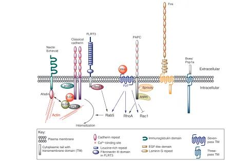
P
p120
β-Cat
Fz7 Classical
cadherin
PAPC
Sprouty
ANR5
α-Cat
EPLIN
RhoA Rac1
Actin
Fmi
Bves/ Pop1a FLRT3
Wnt
Rab5
Internalization Rnd1
Plasma membrane Cadherin repeat Ca2+-binding site
Three-pass TM Seven-pass TM Cytoplasmic tail with
transmembrane domain (TM)
Immunoglobulin domain
Fibronectin III domain in FLRT3
Leucine-rich repeat EGF-like domain Laminin G repeat
Extracellular
Intracellular Nectin
Echinoid
Afadin
[image:5.612.54.498.58.372.2]Key:
Fig. 2. Cell-cell adhesion molecules involved in gastrulation. Classical cadherins are integral membrane proteins characterized by five extracellular (EC) domains that mediate homophilic, trans or cis binding (Pokutta and Weis, 2007). The cytoplasmic domains of all classical
cadherins contain binding sites for β-catenin (β-cat) and the catenin-relative p120, and associate with the actin cytoskeleton, possibly through Eplin. They are regulated by non-canonical Wnt signalling or by the small GTPase Rdn1, which induces cadherin endocytosis in Rab5+vesicles by binding to the cytoplasmic domain of FLRT3. Protocadherins have an additional EC domain and lack cytoplasmic p120 and β-cat binding sites. The cytoplasmic tail of Xenopusparaxial protocadherin C (XPAPC) contains several other binding sites that mediate intracellular signalling and interfere with non-canonical Wnt (PCP) signalling. Flamingo (Fmi) is an atypical seven-pass transmembrane (TM) cadherin-related protein, with eight or nine EC-domains, several EGF and two Laminin G domains, and a cytoplasmic domain that mediates intracellular signalling. Ca2+-independent cell-cell adhesion molecules that are required for gastrulation movements include Bves and Echinoid. Xenopusand DrosophilaBves and Popeye family members have relatively short EC domains, a three-pass TM and a long intracellular domain (Lin et al., 2007; Ripley et al., 2006). Echinoid, a
Drosophilanectin-like immunoglobulin cell-adhesion molecule (Ig-CAM), clusters with classical cadherins via their cytoplasmic binding partners
afadin and α-catenin. ANR5, ankyrin repeats domain protein 5; EGF, epidermal growth factor; Fz7, Frizzled 7.
D
E
V
E
LO
P
M
E
N
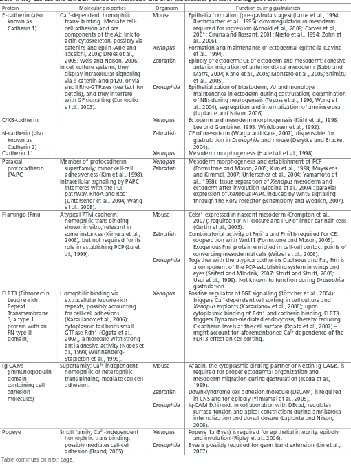
Table 1. Key cell-cell and cell-ECM adhesion molecules and their intracellular partners during gastrulation
Protein Molecular properties Organism Function during gastrulation
E-cadherin (also known as Cadherin 1)
Mouse
Xenopus
Zebrafish
Drosophila
Epithelia formation (pre-gastrula stages) (Larue et al., 1994; Riethmacher et al., 1995); downregulation in mesoderm required for ingression (Arnold et al., 2008; Carver et al., 2001; Ciruna and Rossant, 2001; Nieto et al., 1994; Zohn et al., 2006).
Formation and maintenance of ectodermal epithelia (Levine et al., 1994).
Epiboly of ectoderm; CE of ectoderm and mesoderm; cohesive anterior migration of anterior dorsal mesoderm (Babb and Marrs, 2004; Kane et al., 2005; Montero et al., 2005; Shimizu et al., 2005).
Epithelialization of blastoderm; AJ and monolayer
maintenance in ectoderm during gastrulation; delamination of NBs during neurogenesis (Tepass et al., 1996; Wang et al., 2004); segregation and internalization of amnioserosa (Laplante and Nilson, 2006).
C/XB-cadherin Xenopus Ectoderm and mesoderm morphogenesis (Kühl et al., 1996;
Lee and Gumbiner, 1995; Winklbauer et al., 1992). N-cadherin (also
known as Cadherin 2)
Zebrafish CE of mesoderm (Warga and Kane, 2007); dispensable for
gastrulation in Drosophila and mouse (Derycke and Bracke, 2004).
Cadherin 11
Ca2+-dependent, homophilic
trans- binding. Mediate cell-cell adhesion and are components of the AJ; link to actin cytoskeleton, possibly via catenins and eplin (Abe and Takeichi, 2008; Drees et al., 2005; Weis and Nelson, 2006). In cell culture systems, they
display intracellular signalling via -catenin and p120, or via small Rho-GTPases (see text for details), and they interfere with GF signalling (Comoglio et al., 2003).
Xenopus Mesoderm morphogenesis (Hadeball et al., 1998). Paraxial
protocadherin (PAPC)
Member of protocadherin superfamily; minor cell-cell adhesiveness (Kim et al., 1998). Intracellular signalling by PAPC
interferes with the PCP pathway, RhoA and Rac1 (Unterseher et al., 2004; Wang et al., 2008).
Xenopus
Zebrafish
Mesoderm morphogenesis and establishment of PCP (Formstone and Mason, 2005; Kim et al., 1998; Muyskens and Kimmel, 2007; Unterseher et al., 2004; Yamamoto et al., 1998); tissue separation of Xenopus mesoderm and ectoderm after involution (Medina et al., 2004); paraxial expression of Xenopus PAPC induced by Wnt5 signalling through the Ror2 receptor (Schambony and Wedlich, 2007).
Flamingo (Fmi) Atypical 7TM-cadherin;
homophilic trans binding shown in vitro, relevant in some instances (Kimura et al., 2006), but not required for its role in establishing PCP (Lu et al., 1999).
Mouse
Zebrafish
Drosophila
Celsr1 expressed in nascent mesoderm (Crompton et al., 2007); required for NT closure and PCP of inner ear hair cells (Curtin et al., 2003).
Combinatorial activity of Fmi1a and Fmi1b required for CE; cooperation with Wnt11 (Formstone and Mason, 2005). Exogenous Fmi protein enriched in cell-cell contact points of converging mesodermal cells (Witzel et al., 2006).
Together with the atypical cadherins Dachsous and Fat, Fmi is a component of the PCP-establishing system in wings and eyes (Seifert and Mlodzik, 2007; Strutt and Strutt, 2005; Usui et al., 1999). Not known to function during Drosophila
gastrulation. FLRT3 (Fibronectin
Leucine-rich Repeat Transmembrane 3, a type 1 protein with an FN type III domain)
Homophilic binding via extracellular leucine-rich repeats, possibly accounting for cell-cell adhesions (Karaulanov et al., 2006); cytoplasmic tail binds small GTPase Rdn1 (Ogata et al., 2007), a molecule with strong anti-adhesive activity (Nobes et al., 1998; Wunnenberg-Stapleton et al., 1999).
Xenopus Positive regulator of FGF signalling (Böttcher et al., 2004); triggers Ca2+-dependent cell sorting in cell culture and Xenopus explants (Karaulanov et al., 2006); upon cytoplasmic binding of Rdn1 and cadherin binding, FLRT3 triggers Dynamin-mediated endocytosis, thereby reducing C-cadherin levels at the cell surface (Ogata et al., 2007) – might account for aforementioned Ca2+-dependence of the
FLRT3 effect on cell sorting.
Ig-CAMs
(Immunoglobulin domain-containing cell adhesion molecules)
Superfamily; Ca2+-independent
homophilic or heterophilic trans binding, mediate cell-cell adhesion.
Mouse
Zebrafish
Drosophila
Afadin, the cytoplasmic binding partner of Nectin Ig-CAMs, is required for proper ectodermal organization and
mesoderm migration during gastrulation (Ikeda et al., 1999).
Down-syndrome cell adhesion molecule (DsCAM) is required in CNS and for epiboly (Yimlamai et al., 2005).
Ig-CAM Echinoid, in collaboration with DEcad, regulates surface tension and apical constrictions during amniserosa internalization and dorsal closure (Laplante and Nilson, 2006).
Popeye Small family; Ca2+-independent
homophilic trans binding, possibly mediates cell-cell adhesion (Brand, 2005).
Xenopus
Drosophila
Popeye 1a (Bves) is required for epithelial integrity, epiboly and involution (Ripley et al., 2006).
[image:6.612.52.562.66.740.2]Bves is possibly required for germ band extension (Lin et al., 2007).
Table continues on next page.
D
E
V
E
LO
P
M
E
N
Table 1. Continued
Protein Molecular properties Organism Function during gastrulation
Integrins TM, heteromeric receptor for
ECM proteins, such as FN, collagens, laminins or fibrillins. Integrins mediate cell-ECM adhesion; are components of focal adhesions; their cytoplasmic domains bind a protein complex, consisting of Talin, Paxillin, Vinculin and -actinin, linking the complex to the actin skeleton. Other complex members are the kinases FAK and ILK, and the GEFs - and -PIX, linking the system to Rho GTPases, regulators of the actin cytoskeleton. Integrins also interfere with GF signalling (Comoglio et al., 2003).
Mouse
Chick
Xenopus
Zebrafish
Drosophila
1 integrin, ILK and Talin mutants die shortly after implantation, with massive defects in the polarization of epiblast cells and lack of germ layer formation from the ICM (Fässler and Meyer, 1995; Monkley et al., 2000; Sakai et al., 2003; Stephens et al., 1995).
RhoA-dependent destabilization of integrin-based adhesion is required for basement membrane breakdown and ingression of mesodermal cells in the primitive streak (Nakaya et al., 2008).
Xenopus integrins and their FN ligand are required for the migration of anterior dorsal mesoderm on the blastocoel roof (Davidson et al., 2002; Skalski et al., 1998; Winklbauer and Keller, 1996), for radial intercalation driving epiboly (Marsden and DeSimone, 2001), and for mediolateral intercalation driving CE (Iioka et al., 2007; Marsden and DeSimone, 2003). ILK is required for epiboly and CE, Paxillin for CE (Iioka et al., 2007). Fibrillin is required for the ‘capture’ of intercalating cells at the notochord-somite border during CE (Skoglund and Keller, 2007). Zebrafish FN is required for proper convergence of heart
precursor cells to the midline (Trinh and Stainier, 2004); mesendodermal cells isolated from gastrulating zebrafish embryos adhere to FN-coated substrates in a Wnt11dependent manner (Puech et al., 2005); zebrafish ILK and -PIX are required for blood vessel integrity, a laminin-dependent process, but not for gastrulation (Knoll et al., 2007; Liu et al., 2007).
Drosophila integrins are not required for invagination per se (Leptin et al., 1989), but for the later migration of primordial midgut cells after EMT (Martin-Bermudo et al., 1999; Roote and Zusman, 1995).
LTBP Latent TGF -binding protein;
covalently binds proprotein of transforming growth factor (TGF) ; contains integrin-binding motif RGD; ligand of integrin v 1 (Munger et al., 1998).
Xenopus
Mouse
Xenopus LTBP1 is expressed in nascent mesoderm. LTBP1 overexpression interferes with Nodal/Activin signalling and mesoderm induction, rather than morphogenesis,
suggesting that its interaction with integrin is required not for gastrulation movements, but to localize TGF to the cell surface (Altmann et al., 2002).
LTBP1 deficient mice are viable and fertile, with moderate alterations in facial structures (Drews et al., 2008). HSPGs (syndecans,
glypicans)
Cell surface-associated HSPGs; can bind FN, possibly modulating cellular focal adhesiveness (Morgan et al., 2007). Main function is to modulate GF distribution (non-cell autonomous), or as a co-receptor of GF signalling (Fujise et al., 2003; Topczewski et al., 2001).
Xenopus Xenopus Syndecan 4, in cooperation with FN, binds to Wnt receptor Frizzled 7, promoting non-canonical Wnt signalling to regulate CE movements (Munoz et al., 2006). Syndecan 2 is involved in FN matrix assembly on Xenopus
animal cap cells. According to loss-of-function studies, it is required for signalling by the TGF factor Vg1, transmitting left-right information from ectoderm to migrating mesodermal cells, rather than regulating gastrulation movements (Kramer and Yost, 2002).
Hyaluronan Secreted linear polysaccharide of
high molecular weight; ECM component; implicated in cell adhesion, migration and proliferation; structural and signalling function (Lee and Spicer, 2000).
Zebrafish Hyaluronan-synthesizing enzyme Has2 is required for
lamellipodia formation and motility of lateral mesodermal cells during convergence movements (Bakkers et al., 2004); cell-autonomous effect upstream of Rac1, pointing to a role of hyaluronan as an autocrine signal, rather than a structural component/migration substrate of the ECM; during Xenopus somitogenesis, CD44 could be identified as a crucial hyaluronan receptor (Ori et al., 2006); relevant receptor during zebrafish gastrulation is currently unknown.
AJ, adherens junctions; CE, convergence and extension; CNS, central nervous system; ECM, extracellular matrix; EMT, epithelial-mesenchymal transition; FAK, focal adhesion kinase; FGF, fibroblast growth factor; GEF, guanine nucleotide-exchange factor; GF, growth factor; FN, fibronectin; HSPG, heparan sulfate proteoglycan; ICM, inner cell mass; ILK, integrin-linked kinase; NB, neuroblast; NT, neural tube; PIX, p21-activated kinase-interacting exchange factor; PCP, planar cell polarity; TM, transmembrane.
D
E
V
E
LO
P
M
E
N
An important signalling function during gastrulation has been revealed for Xenopusparaxial protocadherin C (XPAPC, see Table 1). XPAPC is required for mesoderm morphogenesis and for CE movements, although its contribution to mesodermal cell-cell adhesion is minor (Kim et al., 1998). Extracellularly, it binds to the transmembrane receptor Frizzled 7 (Fz7), which is involved in non-canonical Wnt signalling (Medina et al., 2004), while its cytoplasmic tail interacts with ANR5, an ankyrin repeat-containing protein that has scaffolding functions in the cytoskeleton (Chung et al., 2007), and with Sprouty (Wang et al., 2008). Sprouty is an antagonist of FGF (fibroblast growth factor) and EGF (epidermal growth factor) signalling that inhibits CE movements during Xenopusgastrulation (Nutt et al., 2001; Mason et al., 2006). Sprouty also inhibits non-canonical Wnt signalling (Wang et al., 2008), and thus XPAPC might partly fulfil its CE-promoting effect by sequestering and inhibiting Sprouty (Wang et al., 2008), thereby allowing non-canonical Wnt signalling and the PCP system to become active and to activate the small GTPase Rho (Unterseher et al., 2004). In parallel, and possibly independently of PCP, XPAPC inhibits Rac1 (Unterseher et al., 2004).
In addition to XPAPC, other cadherins and Ca2+-independent cell-cell adhesion molecules are involved in gastrulation. Their individual functions, however, are less well understood (for an overview, see Fig. 2 and Table 1).
Integrin ligands and receptors
The interaction of gastrulating cells with the ECM is dominated by the ECM component fibronectin and its integrin receptors (Fig. 3). Other known integrin ligands, such as Fibrillin (see below), collagens and laminins, are strongly expressed only at the end of, or after, gastrulation. Integrins are crucial components of focal adhesions, and they regulate cell migration during development and adulthood (Bökel and Brown, 2002; Lock et al., 2008). Through cytoplasmic adapter proteins, such as Talin, they associate with the actin cytoskeleton. The binding of an ECM protein to an integrin induces a conformational change in the integrin receptor that results in the recruitment and/or activation of other associated focal adhesion proteins, such as the focal adhesion and the integrin-linked kinases FAK and ILK, scaffold proteins like Paxillin, and GEFs such as alpha- and beta-PIX (Rosenberger and Kutsche, 2006), which provide a link to the small GTPases Rac1 and Cdc24 (outside-in signalling). In reverse, the cytoplasmic binding of Talin to an integrin receptor results in a slight unclasping of the integrin subunits, which is a prerequisite for the binding of their extracellular domains to ECM proteins (inside-out signalling) (Gumbiner, 2005; Hynes, 2002). Via such a mechanism, the cortical actomyosin system could interfere with cell adhesiveness, providing a way of ‘mechanosensing’ the forces that are being created by the cells themselves (Bershadsky et al., 2006). Such inside-out signalling could, for instance, be important for reducing cell-matrix adhesion when blebs form during cell migration (see above).
Other proteins involved in cell-ECM interaction with reported roles during gastrulation are described in Table 1 and Fig. 3.
How cell adhesion regulates gastrulation movements and how it is regulated itself
How do adhesion molecules regulate the different modes of gastrulation movement? We discuss this below, re-visit concepts such as selective affinity, and address more recently discovered phenomena, such as the dynamic re-modelling of cell adhesiveness and the instructive function of adhesion gradients in determining movement direction.
Cell sorting, selective affinity and differential adhesion Selective affinity is a well-established concept that is involved in cell sorting in multiple developmental processes. It is dependent on different, but interdependent factors, such as differential adhesion and cortical tension (see Introduction) (Lecuit and Lenne, 2007). The initial differential adhesion hypothesis formulated by Steinberg relied on the assumption that homophilic trans interactions of cadherins (e.g. E-E or N-N) at contacting cell surfaces are distinct from heterophilic interactions between different cadherin molecules (e.g. E-N). Indeed, despite their very similar structures, it appears that the β -strand-swapping mechanism of cadherin dimerization accounts for their strong preference for homophilic trans interactions (Chen et al., 2005; Shan et al., 2000). Cadherin concentrations at cell surfaces are also important. In particular cultures, cells sort out when they express different levels of the same cadherin, but not when they express equal levels of different cadherins (Duguay et al., 2003). Although cell sorting during several development processes has been described as being driven by the expression of different cadherins (Cortes et al., 2003; Price et al., 2002), during germ layer formation it appears to be the difference in the levels of one particular cadherin that is important. Thus, despite the switch from E- to N-cadherin expression that occurs in the developing mesoderm of Drosophilaand in chicken and mouse embryos during EMT (Ciruna and Rossant, 2001; Hatta and Takeichi, 1986), it is primarily the loss of E-cadherin that is relevant. Upon loss of the transcription factor Snail (Barrallo-Gimeno and Nieto, 2005), or of other factors required for E-cadherin downregulation in chick or mouse, primitive streak cells retain their epithelial properties and fail to undergo EMT and ingression (Arnold et al., 2008; Carver et al., 2001; Ciruna and Rossant, 2001; Nieto et al., 1994; Zohn et al., 2006). In zebrafish, the downregulation of E-cadherin by Snail1a and Snail1b is not required for the initial steps of mesoderm internalization, but is required for the extension of the dorsal mesoderm (Blanco et al., 2007; Yamashita et al., 2004). After the ingression of cells, E-cadherin expression is specifically regained in prechordal plate cells, where it is required for their coherent movement towards the animal pole (Blanco et al., 2007; Montero et al., 2005). Upon loss of Snail1b function, E-cadherin expression is also re-initiated in more posterior axial regions, leading to a mixing of prechordal and notochordal cells (Blanco et al., 2007). Thus, it appears to be the different levels of E-cadherin that regulate the separation of these two mesodermal cell types.
Differential cadherin levels are also important in preventing mesodermal and ectodermal cells from remixing after their segregation, as for instance in Brachet’s cleft of Xenopusembryos. This phenomenon can be nicely reproduced in an explant assay, where only mesodermal cells, but not ectodermal cells, fail to re-integrate into the non-involuting ectoderm when placed onto the inner surface of an animal cap (Wacker et al., 2000). This differential behaviour is regulated by XPAPC, which is expressed only in the involuting mesoderm, and not in the overlying ectoderm. XPAPC depletion leads to a failure in tissue separation and to defects in Brachet’s cleft formation (Medina et al., 2004). To what extent this effect is due to XPAPC-mediated differential adhesion or to XPAPC signalling remains unclear. However, activation of RhoA and Jun N-terminal kinase (JNK), components of the non-canonical Wnt/PCP pathway, plays a crucial role in tissue separation (Medina et al., 2004). In addition, an alternate non-canonical Wnt signalling pathway is involved, regulating cell adhesion via protein kinase C and Ca2+(Winklbauer et al., 2001).
That germ layer separation is driven by other forces in addition to differential adhesion is clear from recent studies in zebrafish, which reveal that actomyosin-dependent cell-cortex/surface tensions are
D
E
V
E
LO
P
M
E
N
crucial for the sorting out of mesendodermal and ectodermal progenitor cells (Krieg et al., 2008). Ectodermal cells, which display the weakest homophilic adhesion (cohesion), but the strongest cell surface tension, end up in the center of heterotypic aggregates with mesodermal cells, indicative of a higher homophilic affinity (see also Box 1); conversely, inhibiting Myosin II contractility decreases ectodermal cell tension and inhibits sorting from mesodermal cells, without interfering with adhesiveness.
An interplay between cell adhesion and cortical contraction, involving E-cadherin and the immunoglobulin cell-adhesion molecule (Ig-CAM) Echinoid, also occurs on the dorsal side of the Drosophila embryo to promote the segregation and internalization of the amnioserosa during dorsal closure (Laplante and Nilson, 2006). Echinoid and its related vertebrate Ig-CAMs (nectins) associate with cadherins, with components of the epithelial cell polarity system (such as Par3), and with the cortical actin and actomyosin systems, regulating adhesiveness, surface tension and cell sorting (Lecuit, 2005b; Ogita and Takai, 2008). The same system can also trigger apical constrictions at the borders of Echinoid-positive and negative cells, contributing to epithelial bending (Laplante and Nilson, 2006; Lecuit, 2005b). Mice mutant in afadin, the cytoplasmic binding partner of nectins, display impaired mesodermal gastrulation movements, in addition to ectodermal disorganization (Ikeda et al., 1999). This suggests that the cooperation between cadherins, Ig-CAMs and the epithelial polarity system might also regulate the cytoskeletal rearrangements that drive the formation of protrusions in migrating cells.
Effects of cell adhesiveness on cell migration
Like rock climbers, migrating cells need to grip strongly to the substratum they move on. This can be either the ECM or other cells; both act as substrata during gastrulation, sometimes in combination, requiring regulated cell-ECM and cell-cell adhesion.
ECM-cell adhesion and cell migration
In line with the crucial role that focal adhesions play in cell migration during development and adulthood (Bökel and Brown, 2002; Lock et al., 2008), loss-of-function experiments with blocking antibodies or with antisense reagents have shown that integrins, their ligand
fibronectin, and their intracellular adapter and signalling proteins are required for different morphogenetic movements during amphibian gastrulation (for the migration of anterior dorsal mesoderm on the blastocoel roof, for the radial intercalation that drives epiboly, and for the mediolateral intercalation that drives CE). Essential roles of different members of the focal adhesion complex (FAC) have also been demonstrated in mouse, zebrafish and Drosophila(see Table 1). In Xenopusembryos, fibronectin is deposited as a fibrillar matrix that lines the blastocoel cavity, and is also found between migrating mesodermal cells (Marsden and DeSimone, 2001; Winklbauer, 1998). However, it seems that the fibronectin-integrin interaction is not required primarily for mesoderm cell adhesion to the blastocoel roof, but for cell spreading and for the formation of lamellipodia (Winklbauer and Keller, 1996). Similarly, during radial and mediolateral intercalation in Xenopusgastrulation, it is necessary for the proper establishment of cellular polarity (Marsden and DeSimone, 2001) and for the correct orientation of cellular protrusions (Davidson et al., 2006). Together, these data suggest that the prime function of the fibronectin-integrin interaction might not be cell-ECM adhesion per se, but the coordinated establishment of cellular and ECM polarity to facilitate cell movements through, or on the surface of, this matrix. By mechanisms not fully understood, integrin signalling also affects gastrulation movements by modulating cadherin-dependent cell-cell adhesion (Marsden and DeSimone, 2003).
Cell-cell adhesion and migration
Classical cadherins, key mediators of intercellular adhesiveness, are required not only for the maintenance of tissue integrity, but also for tissue remodelling and cell movements during gastrulation, including cell migrations (see Table 1). Strikingly, in chimeric zebrafish embryos, migrating E-cadherin-deficient prechordal plate cells lag behind their wild-type counterparts, indicating that E-cadherin is required in the migrating cells themselves (Montero et al., 2005). It is currently unclear whether this effect is solely due to reduced cohesion, allowing migrating cells to remain together as a group, or to a reduced affinity to cells at the inner surface of the epiblast, the likely migration substratum. A role of the epiblast as a migration substratum is indicated by the multiple contacts that are observed between Actin
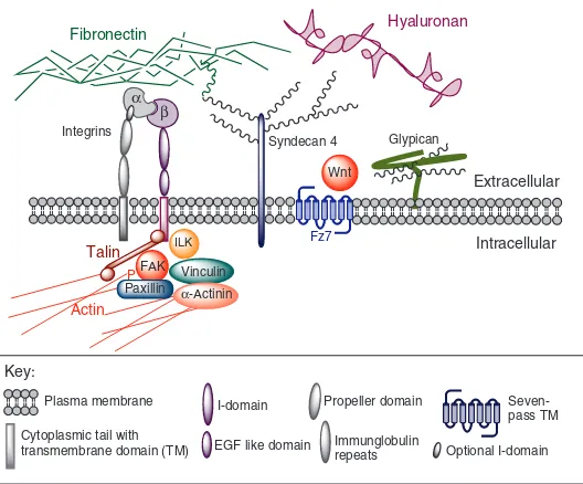
Fz7 α
β Integrins
Syndecan 4 Fibronectin
Glypican
Wnt
Hyaluronan
FAK
Paxillin α-Actinin Vinculin Talin
P ILK
Plasma membrane I-domain
Seven-Cytoplasmic tail with
transmembrane domain (TM) EGF like domain
Extracellular
Intracellular
Propeller domain
Immunglobulin
repeats Optional I-domain
Key:
[image:9.612.52.316.59.278.2]pass TM
Fig. 3. Cell-ECM adhesion molecules involved in gastrulation.
Integrins form heterodimers composed of an αand a βsubunit. The short cytoplasmic domain of the β-subunit binds to the cytoskeletal protein Talin. Integrins link to the actin cytoskeleton and to actin regulators, like Rac1 and Ccd42, via Talin and other cytoplasmic proteins of the focal adhesion complex, including focal adhesion kinase (FAK) or integrin-associated kinase (ILK), the scaffold protein Paxillin, Vinculin, α-actinin and others (Zaidel-Bar et al., 2007). HSPGs are categorized into the subfamilies of transmembrane syndecans, GPI-anchored glypicans and extracellular proteoglycans (Kirn-Safran et al., 2008). Syndecans can bind to fibronectin (FN), possibly modulating cellular focal adhesiveness (Morgan et al., 2007), while interfering with growth factor distribution by modifying the ECM. In reverse, FN might interfere with the growth factor co-receptor function of HSPGs at the cell surface, for instance, modulating signalling through the Wnt receptor Frizzled 7 (Fz7) (Munoz et al., 2006). Hyaluronan is a secreted linear polysaccharide of high molecular weight, but without a polypeptide chain. During zebrafish gastrulation, it seems to act as an autocrine signal, rather than as a migration substrate, activating Rac1 to induce lamellipodia formation (Bakkers et al., 2004). ECM, extracellular matrix; GPI, glycosyl phosphatidylinositol; HSPGs, heparan sulfate proteoglycans.
D
E
V
E
LO
P
M
E
N
mesodermal cell processes and epiblast cells in transmission electron micrographs of gastrulating zebrafish embryos (Montero et al., 2005). The migration of prechordal cells is under the chemotactic control of platelet-derived growth factor (PDGF) (Montero et al., 2003). It remains to be shown whether only the leading edge cells receive the guiding signals, while others follow by cohesion-dependent mechanisms, similar to the situation during chemokine-directed migration of the zebrafish lateral line primordium (Haas and Gilmour, 2006). In sum, these findings show that cadherin-mediated cell-cell adhesiveness is required for coherent migrations and/or migrations on cellular substrates. However, as discussed below, adhesion has to be constantly remodelled to allow these movements to occur.
Dynamic regulation of cell adhesiveness during cell movement
As outlined above, gastrulation movements involve the migration of mesenchymal-like cells, as well as epithelial morphogenesis. Both concepts share crucial features with respect to the remodelling of cell adhesiveness. The regulation of adhesiveness can occur at transcriptional and at post-transcriptional levels, such as by proteolytic cleavage, by the endocytotic trafficking of adhesion molecules, or by interfering with the cytoplasmic components of adhesion complexes and their anchoring to the cytoskeleton.
Remodelling of adhesion complexes during epithelial morphogenesis
During ventral furrow formation in Drosophila embryos, invaginating cells, while undergoing apical constrictions, remain epithelial and in close contact with each other (Fig. 1B). This cell invagination depends on DE-cadherin and its cytoplasmic partner Armadillo, the fly β-catenin homologue (Cox et al., 1996; Wang et al., 2004). Cadherin/Armadillo-containing adherence junctions (AJs) tether the actomyosin system to the apical cell membrane, thereby confining constrictions to the apical side (Dawes-Hoang et al., 2005). A recent study has shown that despite the persistent epithelial organization, a re-distribution of AJs themselves is required to occur (Kölsch et al., 2007). Their disassembly is under the control of the transcriptional repressor Snail (see also above), and apical re-assembly is under the control of the Twist target T48. T48 is a transmembrane protein that is localized to the apical side of ventral furrow cells shortly before invagination. Interestingly, T48 also affects the actomyosin system. It binds RhoGEF2, which in turn, via activation of Rho and Rok, activates Myosin II and the apical constrictions (Fig. 1B). During gastrulation in vertebrates, cells undergoing apical constrictions often display protrusive activities (see Fig. 1M). Interestingly, the RhoGEF2 homologue Xlfc is required for protrusive activity during CE in Xenopus(Kwan and Kirschner, 2005). In epiblast cells of the chick, basally localized Neuroepithelial cell-transforming gene 1 (Net1) protein, another Rho-GEF, is crucial for the maintenance of integrin-based adhesion to the underlying basement membrane; loss of basal Net1 and RhoA activity leads to basement membrane breakdown, which, together with apical constrictions, is a crucial step for subsequent EMT and for the ingression of mesodermal cells in the primitive streak (Levayer and Lecuit, 2008; Nakaya et al., 2008) (Fig. 1M). Together, these findings indicate that the components that are crucial for bringing about the above-described apical constriction of epithelial cells in flies also help to coordinate the more complex cellular events that underlie EMT and the migration of mesenchymal cells during gastrulation in vertebrates.
The progressive re-modelling of AJs within epithelial cells also drives the planar intercalation movements that occur during germ band extension in the Drosophila ectoderm. Here, junctions between two cells shrink, followed by an expansion of junctions with the new neighbours, but without any apparent dissociation of cells (see Fig. 1D). Two factors have been implicated in this process: Myosin II, which is specifically localized at the shrinking junctions (Bertet et al., 2004); and apicobasal polarity regulators, such as Bazooka/Par3, which are at the expanding junctions to assist atypical protein kinase c (aPKC) in balancing AJ symmetry during AJ re-assembly at the medial and lateral sides of the cell (Carthew, 2005; Harris and Peifer, 2007; Zallen and Wieschaus, 2004). Interestingly, Myosin II (Skoglund et al., 2008; Weiser et al., 2007) and components of the apicobasal polarity system are also required for cell intercalations during vertebrate gastrulation, which involve permanent cell dis- and re-associations and highly protrusive cell activities. For instance, the GTPase-activating protein ArfGAP and its physical interaction with aPKC and PAR-6 are required to confine protrusive activity to the mediolateral ends of cells, thereby allowing proper CE during Xenopus gastrulation (Hyodo-Miura et al., 2006).
Recent work has shown that during epithelial morphogenesis in early Drosophila embryos, adhesive bonds are remodelled by modulating the stabilization and immobilization of E-cadherin in the plasma membrane, employing two different actin populations (Cavey et al., 2008). In addition, it has been suggested that membrane traffic is involved (Lecuit, 2005a), similar to the endosomal recycling of E-cadherin during Drosophila wing epithelium morphogenesis (Classen et al., 2005) and during vertebrate gastrulation (see below).
Regulation of cell adhesiveness during vertebrate gastrulation: endocytosis and beyond
Proteolytic cleavage of cadherins, while impinging on cell-cell adhesion in tumour cells (D’Souza-Schorey, 2005; Le et al., 1999; Yap et al., 2007), has not, as yet, been shown to be relevant for gastrulation movements. Another way in which to regulate cadherin function is by its internalization and its trafficking to and from the cell surface (Kamei et al., 1999). Cadherin endocytosis was first shown to be required for gastrulation movements in studies of the GTPase Dynamin, a key regulator of clathrin-mediated endocytosis (Warnock and Schmid, 1996). In this study, a dominant-negative version of Dynamin applied to explanted Xenopusanimal caps caused C-cadherin to accumulate at the cell membrane, while blocking the CE movements that are normally induced in the caps by activin (Jarrett et al., 2002). Elegant recent work (Ogata et al., 2007) implicates two other proteins in Dynamin-dependent C-cadherin endocytosis: the type I transmembrane protein Fibronectin Leucine-rich Repeat Transmembrane 3 (FLRT3) (Lacy et al., 1999), and the small GTPase Rnd1 (Nobes et al., 1998; Wunnenberg-Stapleton et al., 1999). FLRT3 and Rnd1 are both induced by activin in involuting mesodermal cells, and form a complex required for the internalization of C-cadherin in Rab5-positive endosomes during XenopusCE. By this mechanism, cells can undergo mediolateral intercalations and can ‘slide’ past one another without sacrificing tissue integrity. Similarly, the small GTPase Rab5c is required for E-cadherin endocytosis and for the dynamic regulation of cohesion during the anterior migration of prechordal plate cells in the zebrafish embryo (Ulrich et al., 2005). In this case, endocytosis depends on the non-canonical Wnt11 signal, consistent with the involvement of the PCP system in regulating E-cadherin recycling
in the Drosophilawing (Classen et al., 2005).
D
E
V
E
LO
P
M
E
N
It remains unclear how the endocytosis of cadherins is triggered. It is also unclear how the homophilic interactions with cadherins on adjacent cells are weakened, a necessary step for endocytosis-mediated internalization to occur. In cultured epithelial cells, E-cadherin endocytosis is triggered by its ubiquitination via the E3 ubiquitin ligase Hakai (Fujita et al., 2002), and in tumour cells, the disassembly of E-cadherin adhesion complexes is obtained via tyrosine phosphorylation of catenins through integrin signalling via focal adhesion kinase (FAK) (Imamichi and Menke, 2007). This latter mechanism could underlie the observed modulation of C-cadherin-mediated cell adhesiveness by integrins during Xenopus CE (Marsden and DeSimone, 2003). However, to date, no tyrosine phosphorylation of cadherins or catenins has been reported during gastrulation. In addition to post-translational modifications, less-adhesive cadherin conformations could be obtained by interactions with other transmembrane proteins, for example, with FLRT3 itself (see above) (Ogata et al., 2007), or with the protocadherin XPAPC (Chen and Gumbiner, 2006). In this way, loss of XPAPC could cause elevated C-cadherin activity and delayed blastopore closure during Xenopus gastrulation, a defect that can be rescued by the simultaneous partial inactivation of C-cadherin.
Although it is clear that focal adhesion function can be downregulated via the proteolytic degradation of integrin ligands or of the cytoplasmic components of the FAC, it has yet to be demonstrated that gastrulating embryos display a similar internalization of integrins. In migrating cultured cells, integrins do undergo, and serve as crucial regulators of, caveolin-dependent endocytosis (Echarri et al., 2007; Jones et al., 2006). Integrin endocytosis could also regulate the availability of associated growth factors or their inhibitors (Larrain et al., 2003). Indirect evidence for the importance of the proteolysis of integrin ligands for gastrulation movements comes from recent knockdown studies of Mmp14, a membrane-anchored matrix metalloprotease known to degrade Fibronectin, among other ECM substrates. Zebrafish mmp14morphant embryos display compromised cell polarity and cell migration during CE (Coyle et al., 2008). In cell culture systems, downregulation of integrin-dependent focal adhesion can also be obtained by post-translational modifications of its associated kinase FAK or of the scaffold protein Paxillin (Broussard et al., 2008; Zaidel-Bar et al., 2007). Similar regulatory processes also seem to be relevant for gastrulation movements. In Xenopusembryos, non-canonical Wnt signalling induces the ubiquitination and proteasomal degradation of Paxillin in lamellipodia of intercalating cells, thereby interfering with cell polarization and CE (Iioka et al., 2007).
Cell adhesion in determining movement direction
During gastrulation, migrating cells strictly follow navigation cues. But how is this information displayed to the cells? Chemotaxis (Dormann and Weijer, 2006) and adhesion gradients serve in directing cells during organogenesis and in adult life. For example, gradients of atypical cadherins in Drosophilafunction to orient cells during wing and eye development (Seifert and Mlodzik, 2007; Strutt and Strutt, 2005). Here, we propose that comparable events occur in gastrulation, to set up cell polarity and to determine the direction of cell movements. Furthermore, we speculate that adhesion gradients might employ similar intracellular signalling pathways to those that are later used by chemokines to reinforce the direction of cell movements.
The involvement of adhesion gradients in determining the direction of cell movements first came to light in studies of radial intercalations in the zebrafish epiblast, the main driving force of
epiboly (Kane et al., 2005; Warga and Kimmel, 1990). In order to lead to a productive spreading and thinning of the tissue, these radial intercalations have to be unidirectional, and, indeed, cells normally move only from inner to outer layers (Kane et al., 2005). In E-cadherin zebrafish mutants, however, radial intercalations occur in both directions, leading to early embryonic arrest in epiboly. This, together with the higher E-cadherin mRNA levels in outer layers, indicates that stronger E-cadherin-mediated intercellular adhesion within the outer layers of the epiblast (Kane et al., 2005), and between outer epiblast cells and the superficial cells of the enveloping layer (EVL) (Shimizu et al., 2005), is required to keep cells in the other layers and to make the movement unidirectional.
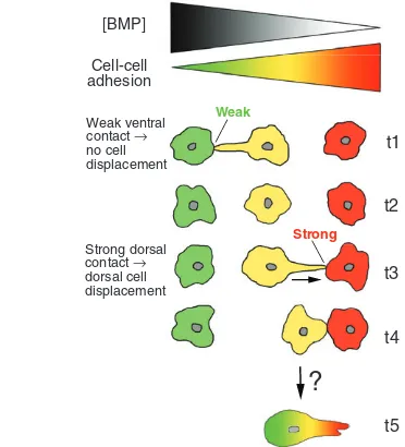
More recent data indicate that a similar mechanism directs active cell migrations during zebrafish dorsal convergence. Different mesodermal cell behaviours driving CE movements can be distinguished in lateral and dorsal regions of the gastrula embryo. In ventral-most regions of the embryo, cells do not move dorsally at all, whereas in lateral regions, they display a rather undirectional migration and a slow net dorsal displacement. Only in paraxial regions does migration occur in straight ventral-to-dorsal paths, and dorsal convergence is fast (Solnica-Krezel, 2006). This spatial pattern also reflects the temporal course of movements of individual cells on their way from lateral regions into the dorsal axis. Interestingly, zebrafish mutant for various PCP pathway components display defects in fast and directed convergence in more dorsal regions, whereas the slow convergence in lateral regions occurs rather normally. In these lateral regions, cells seem to receive their first directional instructions by a bone morphogenetic protein (BMP) signalling gradient. BMPs are well known for their role in determining differential cell fates along the dorsoventral (DV) axis of fish and amphibian embryos (Hammerschmidt and Mullins, 2002). In addition, the BMP gradient has an independent effect on CE (Myers et al., 2002a; Myers et al., 2002b), determining the direction of movements by establishing a reverse gradient of adhesiveness, which progressively increases towards the dorsal midline (von der Hardt et al., 2007; Wallingford and Harland, 2007) (Fig. 4). But why should such a gradient direct the migration of rather loosely organized cells? Time-lapse studies with fluorescently labelled cells have revealed that the cells form transient adhesive bonds, primarily via their lamellipodial protrusions (Fig. 1J). BMP signalling and cadherin-dependent adhesiveness do not affect the polarity or stability of these lamellipodia, but rather their ability to convert subsequent lamellipodial retractions into a productive displacement of the cell. Because of the higher adhesiveness of a dorsal, compared with a ventral, neighbour of a cell, contacts made with dorsal cells are stronger. These stronger contacts might provide a better grip, and/or might induce stronger intracellular signalling and cytoskeletal activities that drive directed migration, thereby causing a net dorsal displacement (see Fig. 4). A similar mechanism might be at play during mediolateral intercalation in Xenopus. Consistent with such a possibility, DV differences in adhesiveness have been seen within the coherent mesodermal sheets of the Xenopusembryo (Reintsch and Hausen, 2001). Here, the adhesive gradient might be set up under the control of a gradient of TGFβ/activin family members, which would not only induce different anterodorsal versus posteroventral mesodermal fates (Green et al., 1992), but also regulate C-cadherin-mediated adhesiveness (Brieher and Gumbiner, 1994), thereby linking embryonic patterning with appropriate morphogenesis through directed CE movements (Howard and Smith, 1993; Ninomiya et al.,
2004).
![Fig. 1. Gastrulation movements in Drosophilathe ventral surface of the blastoderm of a stage 3c chick embryo [reprinted, with permission, from Lawson and Schoenwolf (Lawson andSchoenwolf, 2001)]; arrowhead points to Hensen’s node, arrows indicate primitive](https://thumb-us.123doks.com/thumbv2/123dok_us/8887574.949148/3.612.62.510.59.470/gastrulation-drosophilathe-blastoderm-reprinted-permission-schoenwolf-andschoenwolf-arrowhead.webp)