IJPSR (2013), Vol. 4, Issue 11 (Review Article)
Received on 02 June, 2013; received in revised form, 29 July, 2013; accepted, 26 October, 2013; published 01 November, 2013 GOLD NANOPARTICLES: PROMISING AND POTENTIAL NANOMATERIAL
Madhuri Shringirishi*1, S.K. Prajapati 1, Alok Mahor 1, Shashi Alok 1, Poonam Yadav 1 and Amita Verma2 Department of Pharmaceutics, Institute of Pharmacy, Bundelkhand University 1, Jhansi, Uttar Pradesh, India Department of Pharmaceutical Sciences, Faculty of Health Sciences, Sam Higginbottom Institute of Agriculture, Technology and Sciences-Deemed University 2, Allahabad, Uttar Pradesh, India
ABSTRACT: Gold nanoparticles (AuNPs) have appeared as an attractive candidate for delivery of various drug molecules or considered as extraordinary molecular carriers for the targeting, intracellular trafficking and delivery of a huge array of biomolecules including DNA, RNA, proteins, peptides, drugs, genes and other molecules of therapeutic significance. Particularly gold nanoparticles have attracted intensive interest, because they are easily prepared, have low toxicity and can be readily attached to molecules of biological interest. More and more research shows that AuNPs-based technologies are becoming promising approaches in drug and gene delivery, liver targeting, brain targeting, cancer research and AIDS treatment. The present review focuses on synthesis and functionalization methods of GNPs, the past researchs and reviews about GNPs, their emerging applications and uses and their future prospects.
INTRODUCTION: Nanotechnology has
dynamically developed as an important field of modern research with potential effects in electronic and medicine 1, 3. Nanotechnology can be defined as a research for the design, synthesis, and manipulation of structure of particles with dimension smaller than 100nm 4.
Nanoparticles (NPs), i.e. particles with the dimensions in the range of units to hundreds of nanometers, recently attract an extensive attention in various fields of chemistry, physics, material science, medicine and photonics, due to their unique physical and chemical properties 5, 10.
QUICK RESPONSE CODE
DOI:
10.13040/IJPSR.0975-8232.4(11).4068-82
Article can be accessed online on:
www.ijpsr.com
DOI link: http://dx.doi.org/10.13040/IJPSR.0975-8232.4(11).4068-82
Gold nanoparticles are used in pharmaceutical formulations and biological fluids as gold nanoparticles modified ITO (Au/ITO) electrode is described for the detection of dopamine and serotonin in the presence of a high concentration of ascorbic acid 11.
Nanoparticles are not only excellent materials by structural features but also by functional features. It can be provided changing of functional properties by using various methods beside bulky properties. Electronic 12, optic 13 and catalytic 14 properties of nanoparticles have grown out of their quantum levelled size.
Nanosized particles have gained very importance in modern technology systems due to their high surface area, different optic and optical properties. These nanoparticles can be produced by basic systems like microemulsion, reverse micelle formation, electro deposition etc. These nanoparticles can be replaced to substrate surfaces Keywords:
Gold nanoparticles, Nanomaterial, Nanosized
Correspondence to Author:
Madhuri Shringirishi
Department of Pharmaceutics, Institute of Pharmacy, Bundelkhand University, Jhansi, Uttar Pradesh, India
like Au, Al, glass, silica etc. by physical, chemical or replacement by themselves methods to formation of regular nano orderly structures 15.
Gold (Au) is unique compared to other metals because of its resistance to tarnishing. According to the earliest records, use of Au for medical purposes can be traced back to the Chinese civilization in 2500 BC and after that, several ancient cultures have utilized Au-based materials for medicinal purpose for the treatment of a variety of diseases such as smallpox, skin ulcers, Measles and Syphilis
16
.
In today’s era of nanotechnology, gold nanoparticles (AuNPs) have been used for the treatment of diseases like rheumatoid arthritis and so forth, while considerable research is currently going on for unveiling potential anticancer and antimicrobial and biodiagnostic applications of Au -based materials and compounds for clinical applications 17.
Gold nanoparticles (AuNPs) provide non-toxic carriers for drug and gene delivery applications. With these systems, the gold core imparts stability to the assembly, while the monolayer allows tuning of surface properties such as charge and hydrophobicity. An additional attractive feature of AuNPs is their interaction with thiols, providing an effective and selective means of controlled intracellular release 17.
Synthesis and Functionalization of Gold Nanoparticles: GNPs are the colloidal suspension of gold particles of nanometer sizes. GNPs have been synthesized by following methods;
1. The reduction of chloroauric acid in the presence of a stabilizing agent.
2. The citrate synthesis method includes reduction of chloroauric acid using trisodium citrate resulting into the formation of GNPs. The size of GNPs is determined mainly by the salt concentration, temperature and rate of addition of reactants resulting in size range of 10–25 nm.
3. Widely used method by using toluene with tetra-octanyl ammonium bromide as a phase transfer reagent 18.
4. Chemical reduction using L-Tryptophane as a reducing agent for ionic gold and polyethylene glycol was used to produce AuCl4 − ions to provide higher stability and
uniformity in size, shape, and particle distribution 19.
5. Method using methanol extract of medicinal plants as reducing agent to produce the “GREEN” or environmental friendly GNPs
20
. In another procedure, an amino acid derivative of serrapeptase has been used as stabilizing and reducing agent to synthesize stable “eco-friendly” GNPs 21
. Besides the usual spherical shape, GNPs have been synthesized in various other shapes affecting their physical and biochemical properties.
For example, hexagon and boot shaped GNPs show different surface enhanced Raman scattering (SERs) which in turn can be used to detect molecules conjugated to GNPs such as avidin, thereby making these functionalized GNPs (fGNPs) useful for biolabelling, bioassay, clinical diagnosis and therapy 22. Gold nanocages of six and eight facets have also been synthesized 23.
Similarly, gold nanorods have been synthesized which find usage in biomedical applications for cancer imaging and photothermal therapy 24. In yet another recent study, the GNPs were grown in a lysozyme crystal which could be useful as a bifunctional molecule for specific catalytic activity 25.
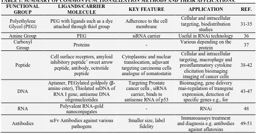
TABLE 1: SUMMARY OF COMMON FUNCTIONALIZATION METHODS AND THEIR APPLICATIONS. FUNCTIONAL
GROUP
LIGANDS/CARRIER
MOLECULE KEY FEATURE APPLICATION REF.
Polyethylene Glycol (PEG)
PEG with ligands such as a dye attached through thiol group
Adherence to the cell membrane
Cellular and intracellular targeting, biodistribution
studies
31-35 Amine Group PEG siRNA carrier Useful in RNAi technology 36
Carboxyl
Group Proteins -
Various depending on the
protein 37
Peptide
Cell surface receptors, amyloid inhibitory peptide+ sweet arrow peptide, antibody, octrotide
peptide
Cytoplasmic and nuclear translocation, adjuvant targeting carcinoma cells analogue of somatostatin
Cellular and intracellular targeting, macrophage and proinflammatory cytokine
elicitation bioimaging imaging of cancer cells
38-42
DNA
Aptamer, PEGylated goldpoly (β-amino ester), Thiolated ssDNA of
RNA I gene, antisense DNA oligonucleotides
Targeting Prostate cancer cells , siRNA
carrier, binds to antisense RNA of p53
Bioimaging, gene delivery rnai-regulation of transgene
expression, detection of specific genes e.g., for
43-47
RNA Polyvalent RNA-gold
nanoconjugates - RNAi 48
Antibodies scFv Antibodies against various pathogens
Smaller size, label fidelity
Immunoassays treatment and diagnosis e.g. antibodies
against aflatoxins
49-51
Methods of Functionalization of Gold Nanoparticles:
1. PEGylation: PEGylation is one of the most commonly used functionalization methods for GNPs. GNPs are coated with a layer of PEG alone or in conjunction with other molecules such as biotin, peptides or oligonucleotides, thereby helping the internalization of these GNPs to the target cells. Due to their ability to bind the cell membrane, these functionalized GNPs can serve as good drug-carriers. PEGylated GNPs functionalized with biomolecules such as lectin, lactose and biotin have been synthesized.
PEGylated GNPs are useful in cellular and intracellular targeting of biological materials. Hetero-bifunctional PEGylated GNPs were synthesized in which GNPs were functionalized with thiol group on one end and coumarin, a fluorescent dye on the other. These fGNPs could make their way into the cells which could be tracked easily because of the attached dye. The stability and functional integrity of PEGylated GNPs is of concern as it is affected by factors such as the molecular weight of PEG, the attached functional groups, the ligand and the size of the GNPs used. The efficacy of one of such group of PEGylated GNPs in the ablation of tumors was tested in mice using
thioctic acid anchored PEGylated GNPs. The internalization of these fGNPs was dependent on the size of the nanoparticles, molecular weight of the PEG and the ligands used for PEGylation. Also, the distribution of these GNPs into various cells was dependent on their physiochemical properties.
2. Peptide/Amino Acid Conjugation:
Functionalization of nanoparticles with amino acids and peptides has been another effective way to enhance specificity and efficacy of nanoparticle based delivery systems. GNPs functionalized with amino acids such as lysine, polylysine and glycine bind DNA with higher efficiency for gene delivery without toxicity. Primary ammonium groups of these amino acids contributed to a higher binding capacity to the cationic groups on DNA. Also lysine dendrons were found superior to polylysine for expression of the reporter β-galatosidase gene.
However, conformational changes were observed in the protein after attachment to the GNPs. GNPs functionalized with peptides has also been used as effective cell-targeting agents. The peptide CALNN and its derivative CALNNR8 were used to functionalize GNPs for targeting intracellular components. Distribution of these fGNPs was dependent on the concentration of the peptide as well as on the size of the GNPs. GNPs (30 nm size) was able to cross cell membrane efficiently by endocytosis and micropinocytosis and showed higher affinity for DNA, RNA and endoplasmic reticulum in the cell.
When in mixture both CALNN and its derivative CALNNR8 could make their way to the nucleus whereas the CALNNR8 was mostly trapped into the endoplasmic reticulum due to the higher affinity of the ER for arginine rich signal peptides. The cell viability could be attributed to the extent of fGNP internalization. Similarly, a sensor for the detection of the interaction between β-amyloid peptide with metallic ions Zn2+ and Ca2+ was designed using GNPs functionalized with β-amyloid peptide-CALNNGK (biotin) G, using standard biotin-streptavidin chemistry.
Time dependent study of the interaction between the fGNP was used to suggest the levels of expression of β-amyloid peptide related genes in a simple colorimetric based assay using the optical changes occurring in the absorption spectra of the fGNPs before and after interaction with the peptides. Peptide functionalized GNPs are also known to activate macrophages, holding promise to be used as adjuvants for vaccine delivery. This is possible due to their ability to bind different biomolecules and expose smaller molecules to the immune system, which are otherwise unrecognizable by the macrophages.
The GNPs functionalized with an amyloid growth inhibitory peptide (AGIP) associated with Alzheimer’s disease were found useful for intracellular drug delivery. They can selectively target the β-amyloid fibers and sweet arrow peptide (SAP) which could be recognized by the bone marrow derived macrophages.
These fGNPs were recognized by the macrophages due to TLR-4, a pattern recognition receptor. These fGNPs further activated the pro-inflammatory cytokines TNF-α, IL-1β and IL-6; thus, stopping macrophage proliferation. These fGNPs were then internalized by the macrophages and processed.
GNPs can therefore be conjugated to adjuvants, cofactors or adaptor proteins for an effective immune response. Cellular and subcellular targeting of fGNPs depends on the peptide used for conjugation and the type of cells in question. PEGylated GNPs (30 nm) functionalized with Arg-Gly-Asp (RGD) peptide and a nuclear localization signal peptide lysine-lysine-lysine-arginine-lysine was found to target specifically the nucleus of cancer cells. Likewise, GNPs functionalized with the peptide conantokin-G were internalized by HER293 cells through selective binding to N -methyl-D-aspartate receptors.
In another study, GNPs functionalized with protein transduction domains (PTDs) from HIV Tat proteins were used to follow their intracellular path. PTDs are peptides that can translocate to cell and nuclear membranes in a temperature and receptor independent manner. fGNPs were shown to make their way either into the nucleus (if nuclear localization signal peptide is used) or to the cytoplasm of the target cells through an endosomal path. Peptide sequence thus regulates the entry of the conjugated GNPs.
Peptide-conjugated GNPs are also being used to devise a protein kinase assay using secondary-ion mass spectrometric imaging. This method uses the change in the mass of the peptide substrate after kinase action and is much simpler as opposed to traditional methods using radioactive or fluorescent labels.
Similarly, bioconjugated gold nanorods have been employed as probes for imaging. A mouse monoclonal antibody specific to human epidermal growth factor receptor 2 (HER2), over-expressed in SKBR3 breast carcinoma cells, was conjugated to either GNPs or nanorods which can be used for biomedical imaging of the carcinoma cells. GNPs functionalized with Bombesin peptides, can be used for imaging of cancer cells as Bombesin has high affinity to gastrin releasing peptides that are over-expressed in cancer cells.
GNPs coated with polyelectrolytes were found to restructure the 3D constructs made of collagen and cardiac fibroblasts, reduced contraction and altered the expression of β-actin, α-smooth muscle actin and collagen type I, suggesting the potential applications for anti-fibrotic therapies. Likewise, GNPs were also found to enhance cross linking of collagen fibrils as well as sites to deliver signaling compounds that direct self-assembly and reduce inflammation.
3. Oligonucleotide Functionalized Nano-particles: Several research groups have devised methods to functionalize gold and other nanoparticles using oligonucleotides either alone or with some modifications. DNA conjugated nanostructures can be synthesized in a controlled manner, either by attaching a specific number of single stranded DNA molecules through thiol caps or by saturating the surface of the GNPs by single stranded DNA molecules.
Kinetic and thermodynamic studies on DNA hybridized to GNPs have shown that ssDNA first adheres to the GNPs and then slowly diffuses on its surface. Secondary structure of a DNA hairpin inhibits interaction between GNPs and DNA thereby increasing the stability of adhered DNA. Aptamer-GNP conjugation has been exploited to target prostate cancer cells. This was achieved by attaching GNPs with an oligonucleotide complementary to the sequence of the anti-PSMA (prostate specific membrane
antigen), thus facilitating the attachment of PSMA-GNPs to anti-PSMA antibody. These results show a promising role of such fGNPs in the detection and imaging of cancerous cells. In another novel study, DNA functionalized GNPs were employed to design a chip based DNA bio bar code sensor to detect target DNA sequences. Here, the bio bar code amplification of the target DNA is assessed using a complementary DNA attached to GNPs and subsequent detection of the amplified DNA instead of the original target DNA.
4. Other Common Functionalization Methods: Apart from DNA and proteins, various other molecules have also been used effectively for functionalization of GNPs for various applications. GNPs functionalized using goat anti-human IgG were used to formulate a bioassay to detect human IgG in serum samples. GNPs modified with carboxyl and alcoholic groups were functionalized using antibodies for the detection of E. coli O157:H7. GNPs have also been employed in the immobilization of enzymes to offer an inert and biocompatible system.
The enzyme glucose oxidase has been immobilized on chitosan-GNPs for the quantitative detection of glucose. This method helps the enzyme retain its activity at higher temperature and extreme conditions. GNPs were used to detect 5-fluorouracil (an anti-leukemic drug) due to the quenching effects of GNPs against the fluorescence of 5-fluorouracil. Also, this conjugate has been shown to have antifungal and antibacterial activity.
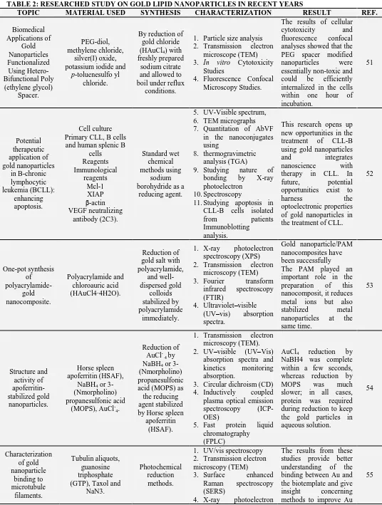
TABLE 2: RESEARCHED STUDY ON GOLD LIPID NANOPARTICLES IN RECENT YEARS
TOPIC MATERIAL USED SYNTHESIS CHARACTERIZATION RESULT REF.
Biomedical Applications of Gold Nanoparticles Functionalized Using Hetero-Bifunctional Poly (ethylene glycol) Spacer. PEG-diol, methylene chloride, silver(I) oxide, potassium iodide and
p-toluenesulfo yl chloride.
By reduction of gold chloride (HAuCl4) with
freshly prepared sodium citrate and allowed to boil under reflux
conditions.
1. Particle size analysis 2. Transmission electron
microscope (TEM)
3. In vitro Cytotoxicity
Studies
4. Fluorescence Confocal Microscopy Studies.
The results of cellular cytotoxicity and fluorescence confocal analyses showed that the PEG spacer modified nanoparticles were essentially non-toxic and could be efficiently internalized in the cells within one hour of incubation. 51 Potential therapeutic application of gold nanoparticles in B-chronic lymphocytic leukemia (BCLL): enhancing apoptosis. Cell culture Primary CLL, B cells and human splenic B
cells Reagents Immunological reagents Mcl-1 XIAP β-actin VEGF neutralizing antibody (2C3). Standard wet chemical methods using sodium borohydride as a
reducing agent.
5. UV-Visible spectrum, 6. TEM micrographs 7. Quantitation of AbVF
in the nanoconjugates using
8. thermogravimetric analysis (TGA)
9. Studying nature of bonding by X-ray photoelectron
10.Spectroscopy
11.Studying apoptosis in CLL-B cells isolated from patients Immunoblotting analysis.
This research opens up new opportunities in the treatment of CLL-B using gold nanoparticles and integrates nanoscience with therapy in CLL. In future, potential opportunities exist to harness the optoelectronic properties of gold nanoparticles in the treatment of CLL.
52 One-pot synthesis of polyacrylamide-gold nanocomposite. Polyacrylamide and chloroauric acid (HAuCl4·4H2O). Reduction of gold salt with polyacrylamide, and well-dispersed gold colloids stabilized by polyacrylamide immediately.
1. X-ray photoelectron spectroscopy (XPS) 2. Transmission electron
microscopy (TEM) 3. Fourier transform
infrared spectroscopy (FTIR)
4. Ultraviolet–visible (UV–vis) absorption spectra.
Gold nanoparticle/PAM nanocomposites have been successfully The PAM played an important role in the preparation of this nanocomposit, it reduces metal ions but also stabilized metal nanoparticles at the same time. 53 Structure and activity of apoferritin-stabilized gold nanoparticles. Horse spleen apoferritin (HSAF),
NaBH4 or
3-(Nmorpholino) propanesulfonic acid
(MOPS), AuCl-4.
Reduction of AuCl- 4 by
NaBH4 or
3-(Nmorpholino) propanesulfonic acid (MOPS) as the reducing agent stabilized by Horse spleen
apoferritin (HSAF).
1. Transmission electron microscopy (TEM). 2. UV–visible (UV–Vis)
absorption spectra and kinetics monitoring absorption.
3. Circular dichroism (CD) 4. Inductively coupled plasma optical emission spectroscopy (ICP-OES)
5. Fast protein liquid chromatography (FPLC)
AuCl4 reduction by
NaBH4 was complete within a few seconds, whereas reduction by MOPS was much slower; in all cases, protein was required during reduction to keep the gold particles in aqueous solution. 54 Characterization of gold nanoparticle binding to microtubule filaments. Tubulin aliquots, guanosine triphosphate (GTP), Taxol and
NaN3.
Photochemical reduction methods.
1. UV/vis spectroscopy 2. Transmission electron microscopy (TEM)
3. Surface enhanced Raman spectroscopy (SERS)
4. X-ray photoelectron
The results from these studies provide better understanding of the binding between Au and the biotemplate and give insight concerning methods to improve Au
spectroscopy (XPS) coverage for MT-templated Au nanowires. Gold Nanoparticle
Synthesis and Characterisation.
Gold chloroauric asid salt
(H[AuCl4]), Trisodium citrate (Na3C6H5O7.2H2O),
By reduction reaction between
tetrachloroauric acid and sodium
citrate
1. Zeta sizer (size and size distribution of the synthesized gold nanoparticles).
The applications of gold nanoparticles are highly extensive as conductive material in sensors and/or biosensors.
56
Stabilized and size-tunable gold
nanoparticles formed in a
quaternary ammonium-based room-temperature ionic liquid under
gamma- irradiation.
Cholin chloride, ZnCl2, Aqueous
hydrogen tetrachloroaurate
(HAuCl4).
Gold nanoparticles under gamma-radiation in a quaternary ammonium-based ionic liquid (QAIL) [Me3NC2H4OH+
[ZnCl5]-.
1. UV/vis spectroscopy 2. Transmission electron microscopy (TEM)
3. Photon cross correction spectroscopy (SERS) 4. X-ray photoelectron
spectroscopy (XPS)
The QAIL surrounding the nanoparticles surface can act as an effective stabilizer of gold nanoparticles, and the nanoparticles size and uniformity are affected by the interactions between the QAIL and clusters during the formation the nanoparticles.
[image:7.612.37.569.210.749.2]57
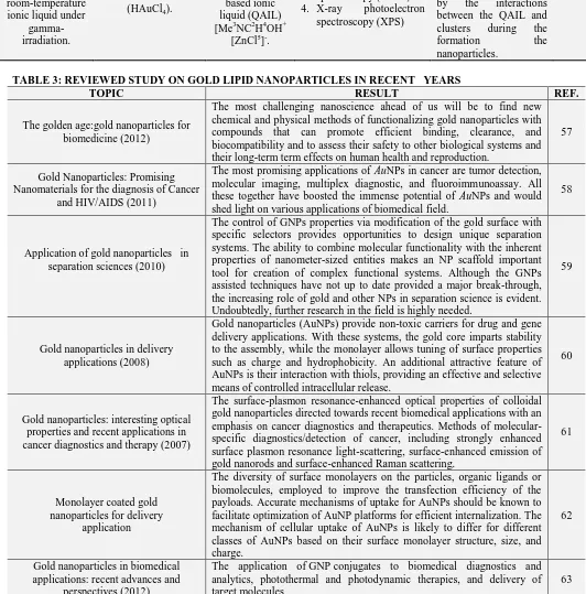
TABLE 3: REVIEWED STUDY ON GOLD LIPID NANOPARTICLES IN RECENT YEARS
TOPIC RESULT REF.
The golden age:gold nanoparticles for biomedicine (2012)
The most challenging nanoscience ahead of us will be to find new chemical and physical methods of functionalizing gold nanoparticles with compounds that can promote efficient binding, clearance, and biocompatibility and to assess their safety to other biological systems and their long-term term effects on human health and reproduction.
57
Gold Nanoparticles: Promising Nanomaterials for the diagnosis of Cancer
and HIV/AIDS (2011)
The most promising applications of AuNPs in cancer are tumor detection, molecular imaging, multiplex diagnostic, and fluoroimmunoassay. All these together have boosted the immense potential of AuNPs and would shed light on various applications of biomedical field.
58
Application of gold nanoparticles in separation sciences (2010)
The control of GNPs properties via modification of the gold surface with specific selectors provides opportunities to design unique separation systems. The ability to combine molecular functionality with the inherent properties of nanometer-sized entities makes an NP scaffold important tool for creation of complex functional systems. Although the GNPs assisted techniques have not up to date provided a major break-through, the increasing role of gold and other NPs in separation science is evident. Undoubtedly, further research in the field is highly needed.
59
Gold nanoparticles in delivery applications (2008)
Gold nanoparticles (AuNPs) provide non-toxic carriers for drug and gene delivery applications. With these systems, the gold core imparts stability to the assembly, while the monolayer allows tuning of surface properties such as charge and hydrophobicity. An additional attractive feature of AuNPs is their interaction with thiols, providing an effective and selective means of controlled intracellular release.
60
Gold nanoparticles: interesting optical properties and recent applications in cancer diagnostics and therapy (2007)
The surface-plasmon resonance-enhanced optical properties of colloidal gold nanoparticles directed towards recent biomedical applications with an emphasis on cancer diagnostics and therapeutics. Methods of molecular-specific diagnostics/detection of cancer, including strongly enhanced surface plasmon resonance light-scattering, surface-enhanced emission of gold nanorods and surface-enhanced Raman scattering.
61
Monolayer coated gold nanoparticles for delivery
application
The diversity of surface monolayers on the particles, organic ligands or biomolecules, employed to improve the transfection efficiency of the payloads. Accurate mechanisms of uptake for AuNPs should be known to facilitate optimization of AuNP platforms for efficient internalization. The mechanism of cellular uptake of AuNPs is likely to differ for different classes of AuNPs based on their surface monolayer structure, size, and charge.
62
Gold nanoparticles in biomedical applications: recent advances and
perspectives (2012)
The application of GNP conjugates to biomedical diagnostics and analytics, photothermal and photodynamic therapies, and delivery of target molecules
Surface Plasmon Resonance Scattering and Absorption of anti-EGFR Antibody Conjugated Gold Nanoparticles in Cancer Diagnostics: Applications in Oral Cancer
(2005)
SPR scattering imaging or SPR absorption spectroscopy generated from antibody conjugated gold nanoparticles can be useful in molecular biosensor techniques for the diagnosis and investigation of oral epithelial living cancer cells in vivo and in vitro.
64
The forthcoming applications of gold nanoparticles in drug and gene delivery
systems (2011)
The attractive features of gold nanoparticles include their surface plasmon resonance, the controlled manner in which they interact with thiol groups, and their non-toxic nature. These attributes can be exploited to provide an effective and selective platform to obtain a targeted intracellular release of some substance. The use of gold nanoparticles can also increase the stability of the payload.
65
Use of Gold Nanoparticles in Diagnostics, Surgery and Medicine: a
Review (2012)
Particularly gold nanoparticles have attracted intensive interest, because they are easily prepared, have low toxicity and can be readily attached to molecules of biological interest. The gold nanoparticles have become more precious than pretty gold because of their wide use and applications.
66
Functionalized gold nanoparticles and their biomedical applications (2011)
GNPs can be considered as extraordinary molecular carriers for the targeting, intracellular trafficking and delivery of a huge array of biomolecules including DNA, RNA, proteins, peptides, drugs, genes and other molecules of therapeutic significance. They do not cause significant cytotoxicity due to their physiochemical properties. Despite these preliminary studies, efforts need to be taken for designing GNPs to enhance the bioavailability of these fGNPs with less immunogenicity and cytotoxicity to be used in vivo.
67
Gold nanoparticles from nanomedicine to nanosensing.
AuNPs-based therapeutic agents could overcome the barriers presented by the human immune and circulatory systems to achieve delivery at diseased sites without uptake by healthy tissues. In principle, such improved targeted delivery could make other AuNPs-based experimental therapeutic techniques, such as photothermal therapy, practicable. With the “right” combination of delivery agents and particle size, AuNPs-based therapeutics could effectively kill the diseased cells while eliminating the horrendous side effects of the conventional chemotherapeutic agents. Special attention should be given to gaining comprehensive insights on the effects of nanoparticle size, ligand conjugation and conjugation chemistry on AuNPs physiological properties. Additionally, the potential for cumulative toxicity upon repeated exposure to AuNPs-based agents must be rigorously investigated. Nanotoxicity may not be a small matter after all! Results from these and related studies will prove informative in further refinement of the design of AuNPs for use in various nanotechnology applications.
49
Emerging applications and uses of Gold Nanoparticles:
1. In diagnosis of Cancer and HIV/AIDS: A number of techniques applying with AuNPs such as surface chemistry mainly by conjugating them with biological molecules have been playing a great role in 21th century for the diagnosis and treatment of various diseases like cancer and HIV. Surface plasmon resonance (SPR) is significant for the absorption and scattering properties of AuNPs. The last 20 years have seen tremendous development of SPR and its use in biomedical applications related to diagnostics and therapy. This technique applies not only for measuring
microarray detection strategies and Protein Chip System for parallel analysis of multitumor markers and its application in cancer detection have also been developed 59.
Monoclonal antibodies (mAbs) can recognize a specific cancer cell. When such monoclonal antibodies attached to gold nanoparticles or nanorods and then expose it to near IR radiation "heating phenomenon" results which can be used in cancer detection. This acoustic signal gives valuable information about the presence ofcancer cells 68.
2. In Surgery: The biological inertness of gold was found to be important in this application and goldplated stents have been found to produce the least number of macroscopic changes in surrounding intravascular tissue. A new surgical procedure for prostate cancer involves the insertion of three gold grains into the prostate. The position of the gold grains can receptorbe detected using X-rays, allowing doctors to target the position of the prostate within one or two millimeters and thereby allow a more precise dose of radiation to be administered to a more targeted area for the treatment of the tumour 67.
3. In Brain Targeted Drug Delivery System: Gold nanoparticles have promising utilities for drug delivery as well as for the diagnosis and treatment of several diseases related to the CNS. AuNPs are retained in a number of organs, such as liver and spleen due to their negative charge and/or processes of Opsonization and hence decreasing their delivery to the brain. It is crucial to alter the nanoparticles’ surface to obtain desired characteristics for achieving enhanced delivery to the brain. Conjugation of 12 nm GNPs with the amphipathic peptide CLPFFD increases the in vivo penetration of these particles to the rat brain. The C (GNP)-LPFFD did not alter the integrity of the blood-brain barrier, and had no effect on cell viability 69.
4. In Liver Targeted Drug Delivery System: Nitric oxide (NO) plays an important role in inhibiting the progress of hepatic fibrosis and as a result complication of portal hypertension by inhibiting human hepatic stellate cell (HSC)
activation. Gold nanoparticles based drug delivery system containing NO donors was developed which was of potential therapeutic application in chronic liver disease. HSC proliferation and the vascular tube formation ability, manifestations of their activation, were significantly weakened by the NO released from these nanoparticles. This study indicates that gold nanoparticles mediated drug delivery systems for introducing NO could be used as a strategy for the treatment of hepatic fibrosis or chronic liver diseases 70.
5. In Magnetic Drug Targeting: Magnetic drug targeting, using core-shell magnetic carrier particles loaded with anti-cancer drugs, is an emerging and significant method of cancer treatment. Kayal et al. synthesized Gold shell-iron core nanoparticles (Fe@Au) by the reverse micelle method with aqueous reactants, surfactant, co-surfactant and oil phase. From Magnetic measurements it was found that the particles were superparamagnetic at room temperature. The anti-cancer drug doxorubicin was loaded onto these Fe@Au nanoparticle carriers. It was found that the amine (-NH2) group of DOX binds to the gold shell. The present studies show that DOX loaded gold coated iron nanoparticles are promising for magnetically targeted drug delivery 71.
6. In Photodynamic Therapy (PDT):
Photodynamic therapy (PDT) is a promising technique for treating various cancers. It involves light,photosensitizers, and tissue oxygen. Most photosensitizing agents, such as porphyrins and are hydrophobic and locate preferentially in apolar sites such as the lipid bilayer membranes of cells. After intravenous injection and accumulation in the target tissue, the photosensitizers, can be excited by light of an appropriate wavelength, can transfer energy to surrounding tissue oxygen which leads to generation of highly reactive oxygen species (e.g., singlet oxygen), and induce apoptpsis or necrosis directly 72.
organelles. But lack of solubility under physiological conditions raises a significant problem for intravenous PDT drug delivery. The administration of such photosensitizers takes long time to reach the maximum accumulation in the tumor sites. This poses an additional risk for toxicity and side effects. Therefore, one needs a delivery carrier that can bestow hydrophilicity during the drug delivery without destroying the hydrophobic characteristics of the drug itself. There are several delivery strategies known to stabilize PDT drugs in aqueous systems. Conjugated polymer nanoparticles are one of them. PEGylated gold nanoparticles (AuNPs) hold the promise to be a highly efficient PDT drug delivery scaffold.
Au NPs are well known for their chemical inertness and have minimum toxicity. By using water-soluble polyethylene glycol (PEG), NPs can be stabilized by steric repulsion to inhibit colloid aggregation in physiological conditions. Drugs on the NPs could be shielded from being uptaken by the reticuloendothelial system (RES). Such drug vectors can preferentially accumulate in tumor sites through the leaky tumor neovasculature and do not return to the circulation 72-73.
7. As Insulin Carrier: Gold nanoparticles synthesized with chitosan were tested as a carrier for insulin. The nanoparticles showed good insulin loading and long-term stability in terms of aggregation.
The use of chitosan served dual purpose by acting as a reducing agent in the synthesis of gold nanoparticles and also promoting the penetration and uptake of insulin across the oral and nasal mucosa in diabetic rats. The study concluded that oral and nasal administration of insulin-loaded, chitosan-reduced gold nano-particles improved pharmacodynamic activity of insulin74.
8. In the delivery of anti HIV agents: Gold nanoparticles coated with multiple copies of an amphiphilic sulfate-ended ligand are able to bind the HIV glycoprotein gp120 as measured by surface plasmon resonance (SPR) and inhibit
in vitro the HIV infection of T-cells. It is
possible to attach both sulfated ligands and other anti-HIV molecules on the same gold cluster and so one can design multifunctional anti-HIV systems 75.
9. In the delivery if Anticancer agents: The anticancer drug, doxorubicin (DOX), was loaded onto DNA-capped gold nanoparticles (AuNPs) designed for specific DOX intercalation 76 To improve selectivity and antitumour activity functionalization of gold nanoparticles (AuNPs) with both a targeting peptide and a drug peptide ligand was attempted. Enhanced activity and selectivity of the peptide multifunctionalized gold nanoparticle conjugates was observed 77.
Gold nanoparticles (AuNPs) were functionalized with an anticancer drug, doxorubicin. As discussed earlier, Doxorubicin was assembled on gold via amino group. The reaction proceeded under mild acidic conditions.
Au NPs could not be adsorbed on doxorubicin in alkaline solution because amino group was not protonated. Under acidic conditions, protonation created a positively charged amino group thus adsorption was easier. The interaction between Au colloids and doxorubicin is believed to be electrostatic 78.
Multidrug resistance (MDR) is a major hurdle to the success of cancer chemotherapy. Doxorubicin was tethered onto the surface of gold nanoparticles with a poly (ethylene glycol) spacer via an acid-labile linkage (DOX-Hyd@AuNPs), it was demonstrated that multidrug resistance in cancer cells can be significantly overcome by a combination of efficient cellular entry and a responsive intracellular release of doxorubicin from the gold nanoparticles in acidic organelles. It released doxorubicin in response to the pH of acidic organelles following endocytosis 79.
methotrexate conjugated to goldnanoparticles in Lewis lung carcinoma cells is about seven times higher than that of free methotrexate 80.
10.In Gene Delivery: The use of nucleic acids to treat and control diseases is termed ‘gene therapy’. Gold nanoparticle conjugates can prepare which protect DNA from degradation81. The light-triggered release of deoxyribonucleic acid (DNA) from gold nanoparticle-based, plasmon resonant vectors, such as nanoshells, shows great promise for gene delivery in living cells. Intracellular light-triggered release can be performed on molecules that associate with the DNA in a DNA host-guest complex bound to nanoshells. The light exposure required for molecular release do not compromise cell viability. This highly controlled co-release of nonbiological molecules accompanying the oligonucleotides may have vast applications in the study of cellular processes and in the development of intracellular targeted therapies
81
.
PEGylated GNPs are one of the most commonly used nanoparticles for gene delivery. A PEGylated GNP based delivery system was evaluated for its transfection efficiency using a plasmid DNA mediated through electro-poration. Gene expression was enhanced to about 100-fold with DNA-PEGylated GNPs compared to naked DNA after intravenous injection69.
11.In Percutaneous Drug Delivery: Co administration of protein drugs with gold nanoparticles enables percutaneous delivery of them. The Au-NPs with a mean size of 5 nm are skin permeable, probably due to the nano-bio interaction with skin lipids and the transient and reversible openings on the stratum corneum. When simultaneously applied with Au-NPs, the protein drugs were also granted to penetrate the skin barrier and migrate into the deep layers. This indicated that co-administration with the skin-permeable Au-NPs could mediate proteins across the skin barrier.
Such co-delivery effect can lead to a simple yet effective method for overcoming the skin barrier for percutaneous protein drug delivery. Employing this strategy, a non-invasive vaccine
delivery system was developed and by topically co-administrating antigens with Au-NPs immune response was elicited in the tested animals.
The results conferred the promise for achieving a needleless transcutaneous vaccination 82.
12.In Photothermal Therapy: Gold nanorods (which are rod-shaped gold nanoparticles) show a surface plasmon band in the near-infrared region. They have been proposed as heating devices for photothermal therapy. Polyethylene glycol-modified gold nanorods systemically administrated. After intravenous injection of gold nanorods followed by near-infrared laser irradiation, significant tumor damage triggered by the photothermal effect was observed 84.
13.As Biosensors: GNPs have been studied and exploited in the development of an assortment of biosensors to detect specific biomolecules significant in disease etiology. Determination of choline in various human samples is clinically important and is usually assayed through the estimation of the enzyme choline esterase. A biosensor developed by combining choline oxidase (ChOx), multi-wall carbon nanotubes (MWCNTs), GNPs and poly-diallyl dimethyl ammonium chloride (PDDA) for the specific detection of choline provided an alternative, significantly sensitive, rapid and efficient approach of detection.
Similarly, uric acid (UA) detection was facilitated using GNPs. UA is an important end product of purine metabolism abnormal levels of which are associated with various metabolic diseases such as gout, hyperuricaemia, pneumonia, kidney damage, cardiovascular diseases and Lesch-Nyhan syndrome. Several methods including colorimetric, enzymatic and electrochemical methods are available for the determination of UA concentration in human fluids.
Here, the absorption maximum of the scattered light by individual nanoparticles was related to the number of molecules of a given analyte bound to individual nanoparticles.
The biosensor model was able to predict the molecular detection limits (minimum no. of detectable molecules) and dynamic range (maximum no. of analyte molecules bound to a nanoparticle) depending on the geometry of the nanoparticles and other parameters of the system 67.
H2O2 biosensor showed a high affinity and
good catalytic activity to H2O2 with good
reproducibility and stability. Hydrogen peroxide is not only an essential mediator in food, clinical, environmental and many other fields, but also a by-product of various enzymatic reactions. Therefore, the as-prepared sensor could be used in the detection of H2O2 in
the detection of antigen or antibody in real serum samples based on the peroxidase labeled immunoassay 85.
14.In Detection: GNPs are also being used for detection of various biological molecules including proteins, enzymes, DNA, antigens and antibodies, etc.
a. Detection of Biological Molecules: GNPs have been used for the detection of proteins, based on their characteristic surface plasmons. For this, GNPs have been functionalized using bifunctional molecules which were conjugated on one side to the GNPs through their thiol group and on the other side to the electron-rich aromatic sidechains of proteins through a diazonium moiety. The model was tested using thrombin as the protein.
The vibrations of the diazo-bond formed between the bifunctional molecule and the target protein tends to enhance due to the conjugation of GNPs constituting the Raman marker. After the functionalized GNPs interact with antithrombin as a sensitive recognition element, immobilized on a substrate, thrombin can be detected through surface enhance Raman Spectroscopy.
GNPs have been used for detection of other biological compounds such as antioxidants which have been studied for their roles in diseases such as cancer, atherosclerosis etc. in suppressing the free radicals. Vitamin E (α-tocopherol) is well known for its antioxidant activity. GNPs functionalized with Trolox, an analogue of vitamin E have been synthesized using self-assembly of thiol ligand, and were evaluated for the free radical scavenging activity. The antioxidant capacity of the GNPs functionalized with Trolox was observed to be higher than that of Trolox alone, showing a promise of these antioxidant-functionalized GNPs in the treatment of various diseases.
b. Detection of Microorganisms: Detection of microorganisms can be achieved by several biochemicals, microbiological and molecular methods. Recent advances in the field of nanotechnology have made it possible to detect microorganisms by using nanoparticles functionalized with oligonucleotides complementary to the gene tags of the microorganisms. In one such study, oligonucleotides complementary to the unique sequences of the heat shock protein 70 (HSP 70) of Cryptosporidium parvum was used to functionalize GNPs, which could be used to detect the oocytes of Cryptosporidium
in a colorimetric assay, offering a simple and robust method of molecular detection.
6-hexanedithiol, GNPs and IgG and the changes in the electron transfer resistance were correlated to the deposition of functionalized GNPs. The increments in the amplified impedance showed good correlation with the protein detection limits 69.
FUTURE PROSPECTS: Gold nanoparticles are promising new vehicle for drug and gene delivery. Control, place and timing of release of the candidate are vital in drug and gene delivery systems. The poor stability of conventional drugs and genes in biological fluids, enzymatic degradation, and difficulties in assuring their penetration through some barrier or nucleus of cells are some of the unfavorable attributes of the existing technologies.
The conjugation of gold nanoparticles with drugs or genes offers greater control and enhanced therapeutic efficacy. In particular, the combination of gold nanoparticles and laser irradiation to control the release of drugs and genes has the potential to provide useful therapeutic benefits. Nevertheless, it is early days in this field and one can achieve a lot.
Most promising application of gold nanoparticles is organ targeting. Liver targeting, brain targeting and tumour targeting is achieved precisely with gold nanoparticles. Magnetic targeting in combination with iron nanoparticles achieved with gold nanoparticles. Advantages of this technique are that Gold nanoparticles are not toxic to human cells. A similar technique with QDs uses semiconductor crystals to mark cancer cells, but the semiconductor material is potentially toxic to the cells and humans.It does not require expensive high-powered microscopes or lasers to view the results. All it takes is a simple, inexpensive microscope and white light. The results are instantaneous. If a cancerous tissue is sprayed with gold nanoparticles containing the antibody, the results can be seen immediately. The scattering is so strong that a single particle can be detected.
ACKNOWLEDGEMENT: The authors are
thankful to the authorities of Bundelkhand University, Jhansi, for providing support to the study and other necessary facility like internet surfing, library and other technical support to write a review article.
REFERENCES:
1. Glomm RW: Functionalized nanoparticles for application in biotechnology. J.Dispersion Sci. Technology 2005;26: 389-314.
2. Chan WCW: Bionanotechnology progress and advances. Biology Blood Marrow Transplantation 2006; 12: 87-91. 3. Boisselier E, Astruc D: Gold nanoparticles in
nanomedicine: preparation, imaging, diagnostics, therapies and toxicity. Chem. Soc. Rev 2009; 38: 1759-1782. 4. Ahmad A, Mukherjee P, Senapati S, Mandal D, Khan M.,
Kumar R, Sastry M. Extracellular biosynthesis of silver nanoparticles using the fungus, Fusarium oxysporum. Colloids Surfaces, B: Biointerfaces 2003; 27: 313-318. 5. Daniel MC, Astruc D. Chem. Rev2004; 104: 293–346. 6. Guo SJ, Wang EK:.Anal. Chim. Acta2007; 598: 181–192. 7. Boisselier E, Astruc D: Chem. Soc. Rev2009; 38: 1759–
1782.
8. Radwan SH, Azzazy HME. Exp. Rev. Mol. Diagn2009; 9: 511–524.
9. Algar WR, Massey M and Krull UJ. Trends Anal. Chem 2009; 28: 292–306.
10. Bawarski WE, Chidlowsky E, Bharali DJ and Mousa SA. Nanomed. Nanotechnol. Biol. Med2008; 4: 273–282. 11. Goyal NR, Gupta KV, Oyama M and Bachheti N: Gold
nanoparticles modified indium tin oxide electrode for the simultaneous determination of dopamine and serotonin: Application in pharmaceutical formulations and biological fluids. Talanta 2007 72; 976–983.
12. Khairutdinov RF: Physical chemistry of nanocrystalline semiconductors. Colloid J. 59 1997; 5: 535-548.
13. Mulvaney P: Surface Plasmon Spectroscopy of Nanosized Metal Particles. Langmuir 1996; 12: 788-800.
14. Meldrum FC, Flath J and Knoll W: Chemical Deposition of PbS on a Series of w Functionalized Self-Assembled Monolayers. J. Mater. Chem 1999; 9: 711-723.
15. Link S: Size and Temperature Dependence of the Plasmon Absorption of Colloidal Gold Nanoparticles. J. Phys. Chem.B 1999; 103: 4212-4217.
16. Daniel MC, Astruc D: Gold Nanoparticles: assembly, supramolecular chemistry, quantum-size-related properties, and applications toward biology, catalysis, and nanotechnology. Chemical Reviews 2004; 104: 293–346. 17. Chen CPo, Mwakwari CS and Oyelere KA: Gold
nanoparticles: From nanomedicine to nanosensing. Nanotechnology, Science and Applications 2008; 1: 45– 66.
18. Ghosh P, Han G, De M, Kim KC and Rotello MV: Gold nanoparticles in delivery applications. Advanced Drug Delivery Reviews2008; 60(11): 1307-1315.
19. Tiwari MP, Vig K, Dennis AV and Singh RS: Functionalized Gold Nanoparticles and Their Biomedical Applications. Nanomaterials2011; 1: 31-63.
20. Mandal S, Selvakannan PR, Phadtare S, Pasricha R and Sastry M: Synthesis of a stable gold hydrosol by the reduction of chloroaurate ions by the amino acid, aspartic acid. Proc. Indian Acad. Sci. Chem. Sci2002; 114: 513– 520.
21. Hung L, Leel AP: Microfluidic devices for the synthesis of nanoparticles and biomaterials. J. Med. Biol. Eng 2007; 27: 1–6.
22. Bhattacharya S, Srivastava A: Synthesis of gold nanoparticles stabilised by metal-chelator and the controlled formation of close-packed aggregates by them. Proc. Indian Acad. Sci. Chem. Sci 2003; 115: 613–619. 23. Akbarzadeh A, Zare D, Farhangi A, Mehrabi MR,
nanoparticles by tryptophane. Am. J. Appl. Sci 2009; 6: 691–695.
24. Ramezani N, Ehsanfar N, Shamsa F, Amin G, Shahverdi HR, Esfahani HM et al: Screening of medicinal plant methanol extracts for the synthesis of gold nanoparticles by their reducing potential. Z. Naturforsch 2008; 63b: 903–908.
25. Ravindra P: Protein-mediated synthesis of gold nanoparticles. Mater. Sci. Eng. B2009; 163: 93–98. 26. Hu M, Qian L, Brinas RP, Lymar ES, Kuznetsova L,
Hainfeld JF: Gold nanoparticle-protein arrays improve resolution for cryo-electron microscopy. J. Struct. Biol 2008; 161: 83–91.
27. Sun Y, Xia Y: Shape-controlled synthesis of gold and silver nanoparticles. Science2002; 298: 2176–2179. 28. Huang Y, Yu F, Park YS, Wang J, Shin MC, Chung H S,
et al: Co-administration of protein drugs with gold nanoparticles to enable percutaneous delivery. Biomaterials2010; 31: 9086–9091.
29. Vekilov PG: Gold nanoparticles: Grown in a crystal. Nat. Nanotech2011; 6: 82–83.
30. Lipka J, Semmler-Behnke M, Sperling RA, Wenk A, Takenaka S, Schleh C, et al: Biodistribution of PEG-modified gold nanoparticles following intratracheal instillation and intravenous injection. Biomaterials 2010; 31: 6574–6581.
31. Cho WS, Cho M, Jeong J, Choi M, Han BS, Shin HS, et al: Size-dependent tissue kinetics of PEG-coated gold nanoparticles. Toxicol. Appl. Pharmacol 2010; 245: 116– 123.
32. Takae S, Akiyama Y, Otsuka H, Nakamura T, Nagasaki Y, Kataoka K: Ligand density effect on biorecognition by PEGylated gold nanoparticles: Regulated Interaction of RCA (120) lectin with lactose installed to the distal end of tethered PEG strands on gold surface. Biomacromolecules 2005;6: 818–824.
33. Ishii T, Otsuka H, Kataoka K and Nagasaki Y: Preparation of functionally PEGylated gold nanoparticles with narrow distribution through autoreduction of auric cation by alpha-biotinyl- PEG-block-[poly(2-N,N -dimethylamino)ethyl methacrylate)]. Langmuir 2004; 20: 561–564.
34. Khalil H, Mahajan D, Rafailovich M, Gelfer M and Pandya K: Synthesis of zerovalent nanophase metal particles stabilized with poly (ethylene glycol). Langmuir 2004; 20: 6896–6903.
35. Lee SH, Bae KH, Kim SH, Lee KR and Park TG: Amine functionalized gold nanoparticles as non-cytotoxic and efficient intracellular siRNA delivery carriers. Int. J. Pharm2008; 364: 94–101.
36. Wangoo N, Bhasin KK, Mehta SK, Suri CR: Synthesis and capping of water-dispersed gold nanoparticles by an amino acid: Bioconjugation and binding studies. J. Colloid Interface Sci2008; 323: 247–254.
37. Sun L, Liu D, Wang Z. Funtional gold nanoparticle-peptide complexes as cell targeting agentsLangmuir2008; 24: 10293–10297.
38. Tkachenko AG, Xie H, Liu Y, Coleman D, Ryan J, Glomm WR, et al: Cellular trajectories of peptide-modified gold particle complexes: Comparison of nuclear localization signals and peptide transduction domains. Bioconjugate Chem 2004; 15: 482–490.
39. Bastis NG, Sanchez-Tillo E, Pujals S, Farrera C, Kogan MJ, Giralt E, et al: Peptides conjugated to gold nanoparticles induces macrophage activation. Mol. Immunol2009; 46: 743–748.
40. Rayavarrapu RG, Peterson W, Ungureanu C, Post JN, van Leeuwen TG and Manohar S: Synthesis and
bioconjugation of gold nanoparticles as potential molecular probes for light-based imaging techniques. Int. J. Biomed. Imaging 2007: 29817:1–29817:10.
41. Surujpaul PP, Gutiérrez-Wing C, Ocampo-García B, Ramírez Fde M, Arteaga de Murphy C, Pedraza-López M, et al: Gold nanoparticles conjugated to [Tyr3]Octreotide peptide. Biophys. Chem2008; 138: 83–90.
42. Javier DJ, Nitin N, Levy M, Ellington A, Richards-Kortum R: Aptamer-targeted gold nanoparticles as molecular specific contrast agents for refelectance imaging. Bioconjugate Chem2008; 19: 1309–1312.
43. Lee JS, Green JJ, Love KT, Sunshine J, Langer R, Anderson DG: Gold, poly(β-amino ester) nanoparticles for small interfering RNA delivery. Nano Lett 2009; 9: 2402– 2406.
44. Kim JH, Jang HH, Ryou SM, Kim S, Bae J, Lee K, Han MS: A functionalized gold nanoparticles-assisted universal carrier for antisense DNA. Chem. Commun 2010; 46: 4151–4153.
45. Rink JS, McMahon KM, Chen X, Mirkin CA, Thaxton CS, Kaufman DB: Transfection of pancreatic islets using polyvalent DNA-functionalized gold nanoparticles. Surgery2010; 148: 335–345.
46. Javier DJ, Castellanos-Gonzalez A, Weigum SE, White AC, Richards-Kortum R: Oligonucleotide-gold nanoparticle networks for detection of Cryptosporidium parvum heat shock protein 70 mRNA. J.Clin. Microbiol 2009; 47, 4060–4066.
47. Giljohann DA, Seferos DS, Prigodich AE, Patel PC, Mirkin CA: Gene regulation with polyvalent siRNA-nanoparticle conjugates. J. Am. Chem. Soc 2009; 131: 2072–2073.
48. Liu Y, Liu Y, Mernaugh RL and Zeng X: Single chain fragment variable recombinant antibody functionalized gold nanoparticles for a highly sensitive colorimetric immunoassay. Biosens. Bioelectron2009; 24: 2853–2857. 49. Shen Z, Yan H, Zhang Y, Mernaugh RL and Zeng X:
Engineering peptide linkers for scFv immunosensors. Anal. Chem 2008; 80: 1910–1917.
50. Sharma A, Matharu Z, Sumana G, Solanki PR, Kim CG and Malhotra BD: Antibody immobilized cysteamine functionalized-gold nanoparticles for aflatoxin detection. Thin Solid Films2010; 519: 1213–1218.
51. Fu W, Shenoy D, Li J, Crasto C, Jones G, Dimarzio C, et al: Biomedical Applications of Gold Nanoparticles Functionalized Using Hetero-Bifunctional Poly(ethylene glycol) Spacer. Materials Research Society 2005; 845. 52. Mukherjee P, Bhattacharya R, Bone N, Lee KY, Patra
Ranjan C, Wang S, et al: Potential therapeutic application of gold nanoparticles in B-chronic lymphocytic leukemia (BCLL): enhancing apoptosis. Journal of Nanobiotechnology2007; 5:4.
53. Bai J, Li Y, Du J, Wang S, Zheng J, Yang Q, et al: One-pot synthesis of polyacrylamide-gold nanocomposite. Materials Chemistry and Physics 2007; 106: 412–415. 54. Zhang L, Swift J, Butts AC, Yerubandi V and Ivan J:
Dmochowski.Structure and activity of apoferritin-stabilized gold nanoparticles. Journal of Inorganic Biochemistry 2007; 101: 1719–1729.
55. Zhou CJ, Wang X, Xue M, Xu Z, Hamasaki T, Yang Y, et al: Characterization of gold nanoparticle binding to microtubule filaments. Materials Science and Engineering C 2009; 30: 20–26.
56. Tabrizi A, Ayhan F and Ayhan H: Gold Nanoparticle Synthesis and Characterisation. Hacettepe J. Biol. & Chem 2009; 37 (3): 217-226.
room-temperature ionic liquid under gamma- irradiation. Nanotechnology2005; 16: 2360-2364.
58. Dreaden CE, Alkilany MA, Huang X, Murphy JC and El-Sayed AM: The golden age: gold nanoparticles for biomedicine. Chem. Soc. Rev 2012; 41: 2740-2779. 59. Kumar A, Boruah MB and Liang JX: Gold Nanoparticles:
Promising Nanomaterials for the Diagnosis of Cancer and HIV/AIDS, Journal of Nanomaterials 2011.
60. Sykora D, Kasicka V, Miksık I, Rezanka P, Zaruba K, Matejka P et al: Application of gold nanoparticles in separation sciences, J. Sep. Sci 2010; 33: 372–387. 61. Huang X, Jain KP, El-Sayed HI and El-SayedAM: Gold
nanoparticles: interesting optical properties and recent applications in cancer diagnostics and therapy. Nanomedicine2007; 2: 681-693.
62. Rana S, Bajaj A, Mout R and RotelloMV: Monolayer coated gold nanoparticles for delivery applications. Adv Drug Deliv Rev 2012; 64(2): 200–216.
63. Dykman L, Khlebtsov N: Gold nanoparticles in biomedical applications: recent advances and perspectives. Chem. Soc. Rev 2012; 41: 2256-2282.
64. El-Sayed HI, El-Sayed AM and Huang X: Surface Plasmon Resonance Scattering and Absorption of anti-EGFR Antibody Conjugated Gold Nanoparticles in Cancer Diagnostics: Applications in Oral Cancer. Nano Lett 2005; 5 (5): 829–834.
65. Pissuwan D, Niidome T and Cortie MB: The forthcoming applications of gold nanoparticles in drug and gene delivery systems. Journal of Controlled Release: Official Journal of the Controlled Release Society 2011; 149(1):65-71.
66. Giasuddin ASM, Jhuma KA and Mujibul Haq AM: Use of Gold Nanoparticles in Diagnostics, Surgery and Medicine: A Review. Bangladesh J Med Biochem 2012; 5(2): 56-60. 67. Tiwari MP, Vig K, Dennis AV and Singh R S:
Functionalized Gold Nanoparticles and Their Biomedical Applications. Nanomaterials2011; 1: 31-63.
68. Kim D, Jeong YY and Jon S: A drug-loaded aptamer-gold nanoparticle bioconjugate for combined CT imaging and therapy of prostate cancer. ACS Nano 2010; 4(7): 3689-3696.
69. Kim C, Ghosh P and Rotello VM: Multimodal drug delivery using gold nanoparticles. Nanoscale2009; 1: 61-67.
70. Das A, Mukherjee P, Singla SK, Guturu P, Frost MC, Mukhopadhyay D, et al: Fabrication and characterization of an inorganic gold and silica nanoparticle mediated drug delivery system for nitric oxide. Nanotechnology 2010; 3: 30.
71. Kayal S, Ramanujan RV: Anti-cancer drug loaded iron-gold core-shell nanoparticles (Fe@Au) for magnetic drug targeting. J Nanosci Nanotechnol 2010; 10 (9):5527-5539. 72. Prajapati BG: Nanoparticles as Platforms For Targeted
Drug Delivery System In Cancer Therapy. The Internet Journal of Nanotechnology 2011:3910.
73. Cheng Y, Sámi AC, Meyers JD, Panagopoulos I, Fei B and Burda C: Highly Efficient Drug Delivery with Gold Nanoparticle Vectors for in Vivo Photodynamic Therapy of Cancer. J Am Chem Soc 2008 August 13; 130(32): 10643–10647.
74. Sona PS: nanoparticulate drug delivery systems for the treatment of diabetes, Digest Journal of Nanomaterials and Biostructures 2010; 5 (2): 411 – 418.
75. Gianvincenzo DP, Marradi M, Martínez-Avila OM, Bedoya LM, Alcamí J and Penadés S: Gold nanoparticles capped with sulfate-ended ligands as anti-HIV agents, Bioorg Med Chem Let 2010; 20(9):2718-2721.
76. Alexander CM, Maye MM and Dabrowiak JC: DNA-capped nanoparticles designed for doxorubicin drug delivery. Chem Commun (Camb) 2011 Mar 28;47 (12):3418-20
77. Hosta-Rigau L, Olmedo l, Arbiol J, Cruz LJ, Kogan MJ and Albiricio F: Multifunctionalized gold nanoparticles with peptides targeted to gastrin-releasing peptide receptor of a tumor cell line, Bioconjug. Chem 2010; 21 (6): 1070-1078.
78. Qin G, Li Z, Xia R, Li F, O'Neill BE, Goodwin JT, et al: Partially polymerized liposomes: stable against leakage yet capable of instantaneous release for remote controlled drug delivery. Nanotechnology 2011; 22(15): 155605.
79. Wang F, Wang YC, Dou S, Xiong MH, Sun TM and Wang J: Doxorubicin-Tethered Responsive Gold Nanoparticles Facilitate Intracellular Drug Delivery for Overcoming Multidrug Resistance in Cancer Cells, ACS Nano 2011.
80. Chen YH, Tsai CY, Huang PY, Chang MY, Cheng PC, Chou CH, et al: Methotrexate conjugated to gold nanoparticles Inhibits tumor growth in a syngeneic lung tumor model, Mol. Pharm 2007; 4 (5): 713–722.
81. Pissuwan D, Niidome T and Cortie MB: The forthcoming applications of gold nanoparticles in drug and gene delivery system, Journal of controlled release 2011; 149: 65-71.
82. Huschka R, Neumann O, Barhoumi A and Halas NJ: Visualizing light-triggered release of molecules inside living cells, Nano Lett 2010; 10(10):4117-22.
83. Huang Y, Yu F, Park YS, Wang J, Shin MC, Chung HS, et al: Co- administration of protein drugs with gold nanoparticles to enable percutaneous delivery, Biomaterials 2010; 31(34): 9086-9091.
84. Niidome T, Shiotani A, Akiyama Y, Ohga A, Nose K, Pissuwan D, et al: Theragnostic approaches using gold nanorods and near infrared light, Yakugaku Zasshi 2010; 130(12):1671-1677.
85. Xuan J, Jia D X, Jiang P L, Abdel-Halim S E and Zhu J J. Gold nanoparticle-assembled capsules and their application as hydrogen peroxide biosensor based on hemoglobin. Bioelectrochemistry 2012; 84: 32–37.
All © 2013 are reserved by International Journal of Pharmaceutical Sciences and Research. This Journal licensed under a Creative Commons Attribution-NonCommercial-ShareAlike 3.0 Unported License.
This article can be downloaded to ANDROID OS based mobile. Scan QR Code using Code/Bar Scanner from your mobile. (Scanners are available on Google Playstore)
How to cite this article: